Click image to see more details
-

-
-
-
-
+6
Product Info Summary
| SKU: | A04899-1 |
|---|---|
| Size: | 0.1 mg |
| Reactive Species: | Human, Rat |
| Host: | Rabbit |
| Application: | ELISA, IF, IHC-P, WB |
Customers Who Bought This Also Bought

Product info
Product Name
Anti-PUMA BBC3 Antibody
SKU/Catalog Number
A04899-1
Size
0.1 mg
Form
Liquid
Description
Boster Bio Anti-PUMA BBC3 Antibody (Catalog # A04899-1). Tested in ELISA, WB, IHC-P, IF applications. This antibody reacts with Human, Rat.
Storage & Handling
PUMA antibody can be stored at 4°C for three months and -20°C, stable for up to one year. Avoid repeated freeze-thaw cycles. Antibodies should not be exposed to prolonged high temperatures.
Cite This Product
Anti-PUMA BBC3 Antibody (Boster Biological Technology, Pleasanton CA, USA, Catalog # A04899-1)
Host
Rabbit
Contents
PUMA Antibody is supplied in PBS containing 0.02% sodium azide.
Clonality
Polyclonal
Isotype
IgG
Immunogen
Anti-PUMA antibody was raised against a peptide corresponding to 14 amino acids near the amino terminus of human PUMA isoform 1. The immunogen is located within the first 50 amino acids of PUMA.
Reactive Species
A04899-1 is reactive to BBC3 in Human, Rat
Observed Molecular Weight
68 kDa
Calculated molecular weight
26.5 kDa
Background of BBC3
Apoptosis is related to many diseases and development. The p53 tumor-suppressor protein induces apoptosis through transcriptional activation of several genes. A novel p53 inducible pro-apoptotic gene was identified recently and designated PUMA (for p53 upregulated modulator of apoptosis) and bbc3 (for Bcl-2 binding component 3) in human and mouse. PUMA/bbc3 is one of the pro-apoptotic Bcl-2 family members including Bax and Noxa, which are also transcriptional targets of p53 (1). The PUMA gene encodes two BH3 domain-containing proteins termed PUMA-alpha and PUMA-beta (2). PUMA proteins bind Bcl-2, localize to the mitochondria, and induce cytochrome c release and apoptosis in response to p53. PUMA may be a direct mediator of p53-induced apoptosis.
Antibody Validation
Boster validates all antibodies on WB, IHC, ICC, Immunofluorescence, and ELISA with known positive control and negative samples to ensure specificity and high affinity, including thorough antibody incubations.
Application & Images
Applications
A04899-1 is guaranteed for ELISA, IF, IHC-P, WB Boster Guarantee
Recommend Dilution
WB: 2-3 μg/mL; IF: 10-20 μg/mL; IHC: 2.5-10 μg/mL.
Antibody validated: Western Blot in human samples; Immunohistochemistry in human samples; Immunofluorescence in human samples. All other applications and species not yet tested. Optimal dilutions for each application should be determined by the researcher.
Validation Images & Assay Conditions
Click image to see more details
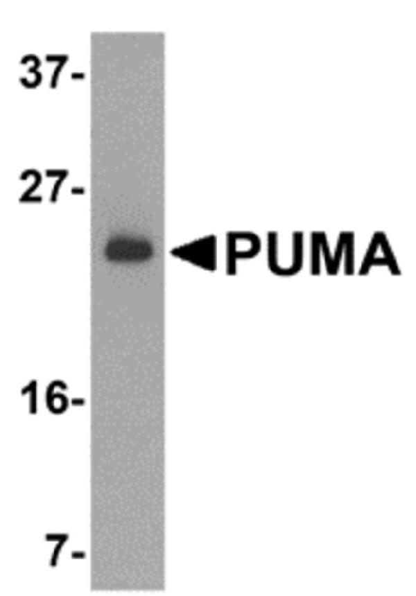
Western Blot Validation of PUMA in K562 Cells
Loading: 15 μg of lysates per lane.
Antibodies: A04899-1 (2 μg/mL), 1 h incubation at RT in 5% NFDM/TBST.
Secondary: Goat anti-rabbit IgG HRP conjugate at 1:10000 dilution

Click image to see more details
Independent Antibody Validation (IAV) via Protein Expression Profile in Human Cells
Loading: 20 μg of lysates per lane.
Antibodies: 3041 (3 μg/mL), A04899-1 (2 μg/mL), beta-actin (1 μg/mL) and GAPDH (0.02 μg/mL), 1 h incubation at RT in 5% NFDM/TBST.
Secondary: Goat anti-rabbit IgG HRP conjugate at 1:10000 dilution.

Click image to see more details
Immunofluorescence Validation of PUMA in K562 Cells
Immunofluorescent analysis of 4% paraformaldehyde-fixed K562 cells labeling PUMA with A04899-1 at 20 μg/mL, followed by goat anti-rabbit IgG secondary antibody at 1/500 dilution (red) and DAPI staining (blue).

Click image to see more details
Immunohistochemistry Validation of PUMA in Human Breast Carcinoma
Immunohistochemical analysis of paraffin-embedded human breast carcinoma tissue using anti-PUMA antibody (A04899-1) at 10 μg/ml. Tissue was fixed with formaldehyde and blocked with 10% serum for 1 h at RT; antigen retrieval was by heat mediation with a citrate buffer (pH6). Samples were incubated with primary antibody overnight at 4˚C. A goat anti-rabbit IgG H&L (HRP) at 1/250 was used as secondary. Counter stained with Hematoxylin.

Click image to see more details
Immunohistochemistry Validation of PUMA in Human Breast Tissue
Immunohistochemical analysis of paraffin-embedded human breast tissue using anti-PUMA antibody (A04899-1) at 2.5 μg/ml. Tissue was fixed with formaldehyde and blocked with 10% serum for 1 h at RT; antigen retrieval was by heat mediation with a citrate buffer (pH6). Samples were incubated with primary antibody overnight at 4˚C. A goat anti-rabbit IgG H&L (HRP) at 1/250 was used as secondary. Counter stained with Hematoxylin.

Click image to see more details
Immunofluorescence Validation of PUMA in K562
Immunofluorescent analysis of 4% paraformaldehyde-fixed K562 cells labeling PUMA with A04899-1 at 10 μg/mL, followed by goat anti-rabbit IgG secondary antibody at 1/500 dilution (red). Image showing cytosol staining on K562 cells

Click image to see more details
Induced Expression of PUMA in MCF7 cells (Wade et al., 2008)
Western analysis of MCF7 treated with the indicated dose of Nutlin-3a or
ABT-737 for 24h. Note that Puma is induced following Nutlin-3a treatment in these cells and PUMA expression was detected by anti-PUMA antibodies (A04899-1)

Click image to see more details
KO Validation of PUMA in Mouse Thymocytes (Michalak et al., 2008)
Western blot analysis of
thymocytes from wt, noxa knockout, puma knockout and noxa/puma double knockout mice cultured for 7 h in the presence or absence of 2.5 Gy g-irradiation.
PUMA expression was not detected in puma KO and double KO mice with anti-PUMA antibodies (A04899-1).

Click image to see more details
KO Validation of PUMA in Mouse Cerebellar Neurons (Ambacher et al., 2012)
Puma expression is induced by potassium withdrawal in cerebellar granule neurons. After 7 days in culture CGNs were either maintained in media containing 25 mM potassium (K25) or switched to low potassium medium containing 5 mM potassium (K5). PUMA protein levels were analyzed by western blot with anti-PUMA antibodies (A04899-1). PUMA expression was not detected in PUMA KO mice and was increased after treatment in WT.

Click image to see more details
Immunofluorescence Validation of PUMA in Rat Retina (Wakabayashi et al., 2012)
PUMA expression in the rat retina detected by anti-PUMA antibodies (A04899-1). The specimens were counterstained with Hoechst 33258 to visualize nuclei (+DNA). GCL, ganglion cell layer; INL, inner nuclear layer; IPL, inner plexiform layer; ONL, outer nuclear layer; OPL, outer plexiform layer; P, postnatal day.
Specific Publications For Anti-PUMA BBC3 Antibody (A04899-1)
Loading publications
Recommended Resources
Here are featured tools and databases that you might find useful.
- Boster's Pathways Library
- Protein Databases
- Bioscience Research Protocol Resources
- Data Processing & Analysis Software
- Photo Editing Software
- Scientific Literature Resources
- Research Paper Management Tools
- Molecular Biology Software
- Primer Design Tools
- Bioinformatics Tools
- Phylogenetic Tree Analysis
Customer Reviews
Have you used Anti-PUMA BBC3 Antibody?
Share your experimental results or join a short interview to earn up to $1,000 in product credits or other rewards.
0 Reviews For Anti-PUMA BBC3 Antibody
Customer Q&As
Have a question?
Find answers in Q&As, reviews.
Can't find your answer?
Submit your question
