BCAP31 / B-cell receptor-associated protein 31 · IHC design guide

Design Immunohistochemistry for BCAP31

This guide uses cytoplasmic tissue staining (HPA tissue IHC) and ER membrane localisation (UniProt) to plan BCAP31 paraffin IHC. It pairs a catalog antibody starting range of 2–5 μg/mL (datasheet A03767-4) with high-staining lung macrophages and unstained cardiomyocytes as reference populations (HPA tissue IHC).

Evidence assembled Oct 2026 · For research use; verify linked source records and product datasheet before use
Immunohistochemistry protocol sheet for BCAP31 (IHC for BCAP31): expected localisation Tissue: cytoplasm (HPA tissue IHC); protein: ER membrane (UniProt), antibody A03767-4, validated IHC image, and IHC protocol steps
Printable BCAP31 IHC protocol sheet — expected localisation Tissue: cytoplasm (HPA tissue IHC); protein: ER membrane (UniProt), antibody A03767-4, controls and protocol steps. Open the full BCAP31 IHC guide →

BCAP31 Immunohistochemistry Experimental Design Guide

Expected localisation, validated protocols, controls and antibodies — the at-a-glance facts below, then the full design guide.

Must know before staining
Expected localisation Tissue: cytoplasm (HPA tissue IHC); protein: ER membrane (UniProt)
Staining pattern Widespread cytoplasmic staining; high in lung macrophages (HPA tissue IHC)
Antigen retrieval EDTA pH 8.0 HIER, heat-mediated (datasheet A03767-4)
Positive control ⓘ Cerebellum+4 more · see all
Negative control ⓘ Heart muscle+4 more · see all
Important caveats
Reasons your staining may differ from the expected pattern.
Fixation Keep fixation consistent across sections (standard IHC practice; not target-specific)
Caveat Presumed off-target binding may confound staining (HPA tissue IHC)
Regulation ER stress redistributes BCAP31 (UniProt)
Isoform / epitope 2 isoforms; epitope differences unspecified (UniProt)
Section 1

Recommended BCAP31 IHC & IF Protocols

The catalog antibody’s IHC-P protocol is followed by 3 published BCAP31 IHC protocols with usable staining details (PMC11683684; PMC6771250; PMC7055246).

Recommended immunohistochemistry (IHC-P) protocol parameters
SampleParaffin-embedded human colon cancer tissue; fixative not specified (datasheet A03767-4)
FixationImage fixative and duration unreported (datasheet A03767-4); verify before use.
Sectioning4–5 µm sections on charged slides (standard)
DeparaffinisationXylene, graded ethanol series to water (standard)
Antigen retrievalHeat retrieval: EDTA pH 8.0 (datasheet A03767-4); 20 min, 95–100 °C (standard)
Peroxidase block3% H2O2, 10 min, room temperature (standard)
Blocking10% goat serum (datasheet A03767-4)
Primary antibodyRabbit anti-BCAP31, 2-5 μg/ml (datasheet A03767-4)
Primary incubationOvernight at 4 °C (datasheet A03767-4)
DetectionHRP-conjugated secondary, DAB chromogen (datasheet A03767-4)
CounterstainHematoxylin, blue, dehydrate and mount (standard)
Expected resultBCAP31-positive staining in bergmann glia - cytoplasm/membrane of cerebellum (HPA tissue IHC: High). HPA tissue profile: Ubiquitous cytoplasmic expression. No signal in the no-primary control.
💡Decision noteStart with heat-mediated EDTA at pH 8.0 (datasheet: A03767-4); citrate retrieval is a published alternative (PMC11683684; PMC6771250).
Section 2

What Is the Expected BCAP31 Staining Pattern?

BCAP31 is an endoplasmic reticulum (ER) membrane protein with three transmembrane segments (UniProt P51572 topology). In paraffin-section IHC, expect mainly cytoplasmic or membrane-associated staining, particularly in cerebellar Bergmann glia and lung macrophages (HPA: High in both). Expression is widespread, but intensity varies by cell type (HPA: ubiquitous cytoplasmic profile). Treat the reference pattern as a guide: HPA rates its tissue IHC evidence Supported, with medium consistency and presumed off-target binding observed (HPA: tissue IHC reliability).

What am I looking at on my slide?
Cytoplasmic/membrane staining in Bergmann glia or lung macrophages, with little nuclear signal.This fits the reported High staining in those cells (HPA: tissue IHC) and BCAP31’s ER membrane location (UniProt P51572). Judge the pattern within the identified cells; section-wide color alone cannot establish specific staining (general IHC practice).
Predominantly nuclear staining, or a pattern confined to an unexpected compartment.A nuclear-dominant result conflicts with the ER localisation reported by UniProt and ICC-IF (UniProt P51572; HPA: subcellular ER). Review morphology and controls before interpreting it as BCAP31; HPA reports presumed off-target binding in its tissue assessment (HPA: tissue IHC reliability).
Strong color in cells listed as Not detected, such as cardiomyocytes or skeletal myocytes.That differs from the HPA reference for those specific cells (HPA: Not detected in heart and skeletal muscle). Consider cross-reactivity or endogenous detection activity, then check controls (general IHC practice). An HPA Not detected call is an observed staining result, not proof that every specimen must be negative.
Diffuse color across cells and surrounding tissue, without a discernible cytoplasmic pattern.Treat this as possible background until controls support a cell-associated signal (general IHC practice). The reference is ubiquitous cytoplasmic expression, not uniform staining of every part of a section (HPA: tissue IHC profile); evaluate background separately from genuine low-level staining.
No discernible signal in Bergmann glia or lung macrophages.This disagrees with the reported High IHC staining in those cells (HPA: tissue IHC). First confirm that the relevant cells are present and identifiable; then assess the staining run and antibody conditions (general IHC practice). A failed reference section cannot by itself establish BCAP31 absence in test tissue.
💡Expected BCAP31 appearanceCall a result consistent when identifiable Bergmann glia or lung macrophages show clear cytoplasmic/membrane-associated staining, corresponding to HPA High staining and ER localisation; nuclear-dominant or cell-independent color warrants investigation (HPA: tissue IHC and subcellular ER; UniProt P51572).
How each factor affects the staining
Compartment and topologyBCAP31 occupies the ER and ER–Golgi intermediate compartment membranes, with three transmembrane segments (UniProt P51572). A cytoplasmic or membrane-associated tissue pattern is therefore plausible; the record does not specify an IHC epitope or retrieval condition.
Cell-type variationHPA reports High staining in Bergmann glia and lung macrophages, Medium in several listed cell types, and Not detected in certain others (HPA: tissue IHC). Compare like cells across sections; a low or negative reference call does not define every cell in an organ.
Antibody evidenceThe three listed antibodies, HPA003906, CAB015350 and CAB015424, each have Supported IHC status (HPA: antibody validation). HPA also notes medium tissue IHC consistency and presumed off-target binding, so use staining distribution and controls together (HPA: tissue IHC reliability).
Isoforms and protein processingTwo isoforms are recorded, with no signal peptide or propeptide and no annotated glycosylation sites or modified residues (UniProt P51572). The supplied record gives no isoform-specific tissue pattern or antibody epitope; do not assign a staining difference to a particular isoform.
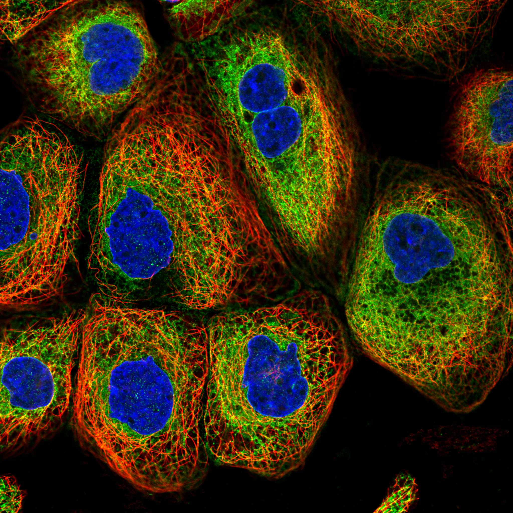
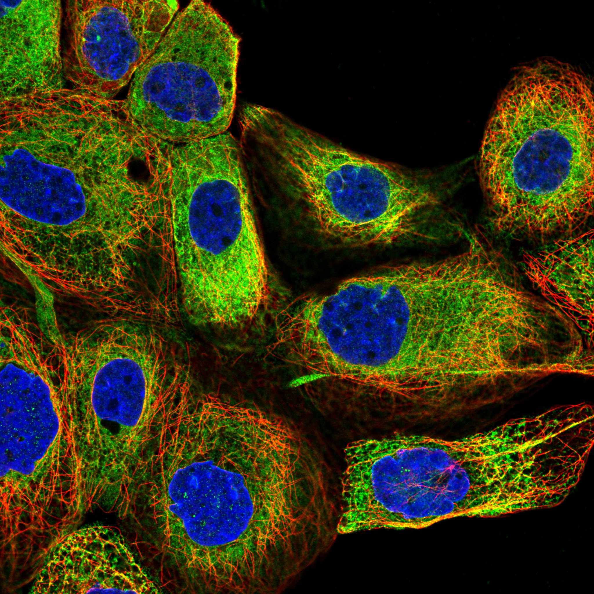
IF/ICC Q&A: What pattern can help assess localisation?HPA reports enhanced ER localisation in ICC-IF, with images from A-431, U-251MG and U2OS (HPA: subcellular). That supports an ER-pattern comparison, while the tissue IHC profile remains the reference for this paraffin-section guide (HPA: tissue IHC).
Why is my staining missing, weak or wrong?
SituationLikely causeNext action
The positive reference section is blank.The run may have failed, the expected cells may be absent from the section, or the signal may be below detection (general IHC practice).Confirm Bergmann glia or lung macrophages are present, then review the documented IHC-P staining conditions and run controls (HPA: High in those cells; general IHC practice).
Reference cells stain weakly despite a clear positive control.Intensity varies across cell types and assay runs; HPA’s High category is an observed reference pattern, not a prescribed color threshold (HPA: tissue IHC).Compare the same cell type under matched staining and counterstain conditions; record whether the cytoplasmic pattern remains discernible (general IHC practice).
Nuclei dominate the apparent signal.This conflicts with reported ER localisation and may reflect nonspecific staining or interpretation of overlapping structures (UniProt P51572; HPA: subcellular ER; general IHC practice).Inspect cell boundaries and the counterstain, compare control sections, and avoid calling nuclear color BCAP31 without independent support (general IHC practice).
Color appears in HPA Not detected cell types.Cross-reactivity or endogenous detection activity can produce misleading chromogenic color (general IHC practice); HPA reports presumed off-target binding (HPA: tissue IHC reliability).Check an appropriate negative reagent control and the detection-system controls; interpret against the exact cell type in the HPA reference (general IHC practice; HPA: tissue IHC).
The entire section has diffuse background.Insufficient blocking, excess detection reagent, or incomplete removal of unbound reagent can obscure localisation (general IHC practice).Review blocking, reagent exposure and washes in the documented IHC-P workflow; require identifiable cell-associated cytoplasmic staining before scoring (general IHC practice; HPA: cytoplasmic profile).
Two antibodies give different cell distributions.HPA lists Supported IHC validation for each listed antibody but reports medium consistency and presumed off-target binding overall (HPA: antibody validation and tissue IHC reliability).Compare the same cells in matched sections, check controls for each antibody, and describe the disagreement rather than assigning either pattern to BCAP31 automatically (general IHC practice).

Sample controls for BCAP31 IHC & IF

🧪Run lung first and score macrophages for staining (HPA: High in lung macrophages). Run heart muscle as the negative tissue and score cardiomyocytes (HPA: Not detected in cardiomyocytes); on the lung slide, neighboring cells without specific staining can show local background, but the supplied HPA rows do not establish any lung cell type as BCAP31-negative (HPA: High in lung macrophages; UniProt P51572 tissue specificity: ubiquitous).
Positive control tissue: Cerebellum (Bergmann glia - cytoplasm/membrane, HPA High)
Negative control tissue: Heart muscle (HPA Not detected)
ICC-IF cell lines (HPA subcellular resource): HPA ICC-IF images show BCAP31 in A-431, U-251MG, U2OS, with annotated localisation: Endoplasmic reticulum (enhanced) (HPA subcellular).
Technical controls: Include a no-primary (secondary-only) slide, a concentration-matched nonimmune rabbit IgG control matched for clonality where applicable, and BCAP31 knockout material as a biological negative (selected-SKU caption: rabbit primary antibody). For chromogenic lung sections, quench endogenous peroxidase and distinguish macrophage pigment from DAB signal (HPA: lung macrophages High; selected-SKU caption: HRP/DAB detection).
⚠️Feasibility: A target-specific fixation window or fixation effect is unreported, and the selected-SKU paraffin-section caption does not state a fixative (selected-SKU caption: fixative not stated). Heat retrieval in EDTA at pH 8.0 was used for the catalog antibody, but the evidence does not establish that retrieval is required or whether frozen sections are easier (selected-SKU caption: heat-mediated EDTA retrieval in a paraffin section). IF/ICC offers an ER localization check in A-431, U-251MG or U2OS cells, while lung macrophage pigment can complicate chromogenic scoring (HPA subcellular: enhanced ER localization and ICC-IF images in those cell lines; HPA: lung macrophages High).

HPA tissue IHC evidence for BCAP31

Comprehensive Human Protein Atlas IHC scoring per tissue (reliability: Supported — Medium consistency between antibody staining and RNA expression data. Presumed off target binding observed and disregarded.). Rows are taken directly from the HPA tissue chart — click any row's HPA link to view the source.

Positive expression · recommended positive controls

TissueCell typeLevelEvidenceSource
Cerebellum Bergmann glia - cytoplasm/membrane High Protein (IHC) HPA →
Lung Macrophages High Protein (IHC) HPA →
Adipose tissue Adipocytes Medium Protein (IHC) HPA →
Adrenal gland Glandular cells Medium Protein (IHC) HPA →
Appendix Glandular cells Medium Protein (IHC) HPA →

Undetected expression · recommended negative controls

TissueCell typeLevelEvidenceSource
Heart muscle Cardiomyocytes Not detected Protein (IHC) HPA →
Oral mucosa Squamous epithelial cells Not detected Protein (IHC) HPA →
Skeletal muscle Myocytes Not detected Protein (IHC) HPA →
Soft tissue Peripheral nerve Not detected Protein (IHC) HPA →
Vagina Squamous epithelial cells Not detected Protein (IHC) HPA →
Section 3

Advanced BCAP31 IHC Tips

These questions focus on interpreting BCAP31 staining in paraffin sections, with one entry covering IF/ICC.

Which retrieval condition should I use first for BCAP31 IHC?
Start with heat-mediated antigen retrieval in EDTA at pH 8.0 for paraffin sections (datasheet A03767-4). Process a known positive alongside study sections, and keep heating, cooling, and section thickness consistent when comparing staining (standard IHC practice). The selected image used human colon cancer, followed by 10% goat serum and 2 μg/ml primary antibody overnight at 4°C; its fixative was unreported (datasheet A03767-4). If staining remains weak, compare another retrieval condition as a controlled fallback, checking whether the expected cytoplasmic pattern improves without diffuse background or damaged morphology (UniProt P51572 localisation; standard IHC practice).
How should I investigate whether fixation caused weak BCAP31 staining?
BCAP31-specific fixation sensitivity is unknown here: the selected paraffin-section caption does not report a fixative, and HPA staining does not establish fixation tolerance (datasheet A03767-4; HPA tissue IHC). For new material, document the fixative, delay to fixation, and fixation duration, then process comparison sections together (standard IHC practice). Hold EDTA retrieval at pH 8.0 and the primary antibody at 2 μg/ml while comparing fixation histories against a positive control (datasheet A03767-4; standard IHC practice). BCAP31 has 3 transmembrane segments and no annotated glycosylation sites, but neither observation establishes a preferred fixation duration (UniProt P51572 topology and PTMs).
What staining pattern is plausible for BCAP31 in paraffin sections?
Assess BCAP31 chiefly as cytoplasmic staining with an ER-associated pattern, rather than scoring nuclear signal as expected expression (HPA tissue IHC; HPA subcellular; UniProt P51572 localisation). BCAP31 occupies the ER membrane and ER–Golgi intermediate compartment membrane and may shuttle toward the cis-Golgi complex (UniProt P51572 localisation). Its association with mitochondria-associated ER membrane through TOMM40 makes limited variation in cytoplasmic distribution biologically plausible, but chromogenic IHC alone cannot resolve those contacts (UniProt P51572 localisation; standard IHC practice). Compare morphology and compartmental staining with a positive section; isolated nuclear or extracellular DAB should prompt a review of specificity and detection background (standard IHC practice).
How can epitope position change my interpretation of BCAP31 IHC?
Check the catalog antibody’s mapped immunogen before comparing staining across preparations, because BCAP31 has 2 annotated isoforms and the supplied evidence does not locate this antibody’s epitope (UniProt P51572 isoforms; datasheet A03767-4). The protein has 3 membrane-spanning segments at residues 7–27, 44–64, and 103–123, with a cytoplasmic region at 124–246 (UniProt P51572 topology). Retrieval can expose epitopes differently in processed sections, so compare conditions on adjacent sections while preserving the same detection settings (standard IHC practice). A change in DAB intensity alone cannot establish isoform abundance without an isoform-discriminating antibody or independent validation (standard IHC practice).
How should I check BCAP31 localisation by IF/ICC alongside IHC?
Use IF/ICC as a separate localisation check: HPA reports enhanced ER localisation and lists images from A-431, U-251MG, and U2OS cells (HPA subcellular). For tissue multiplexing, pair BCAP31 with a validated marker for the cell type under study, such as macrophages when examining lung, and inspect each channel separately (HPA tissue IHC; standard IF practice). Choose a fluorophore channel with low tissue autofluorescence, verify it with an unstained control, and assess spectral bleed-through with single-label controls (standard IF practice). Match permeabilisation to the antibody’s mapped epitope: cytoplasmic residues 124–246 and lumenal loops occupy different sides of the ER membrane (UniProt P51572 topology; standard IF practice).
What should I change when BCAP31 DAB staining is diffuse?
First compare a no-primary control and a positive section to separate detection background from plausible cytoplasmic BCAP31 staining (standard IHC practice; HPA tissue IHC). The selected protocol blocked with 10% goat serum, applied 2 μg/ml rabbit primary overnight at 4°C, and used a peroxidase-linked secondary with DAB (datasheet A03767-4). If the control stains, review endogenous peroxidase blocking, secondary-antibody specificity, washing, and DAB development time before changing retrieval (standard IHC practice). If only the primary-stained section is diffuse, titrate antibody concentration and check morphology; HPA notes presumed off-target binding in its tissue assessment (standard IHC practice; HPA tissue IHC).
How should I score BCAP31 across mixed cell populations? ⚠ ANSWER MARKED FOR VERIFICATION
Define the cell population and cytoplasmic scoring rule before reading slides, then use the same threshold and imaging settings throughout a comparison (HPA tissue IHC; standard IHC practice). Report the percentage of positive cells and, when intensity is reproducible, an H-score from 0–300; for spatial counts, report positive cells per mm² of viable tissue (standard IHC practice). Normalise counts to the relevant cell population or viable tissue area, and record excluded necrosis and section edges (standard IHC practice). Keep cell types separate because HPA reports high staining in lung macrophages and undetected staining in heart cardiomyocytes (HPA tissue IHC).
When is a BCAP31-positive IHC result convincing?
A convincing result has reproducible cytoplasmic staining in morphologically intact cells and fits BCAP31’s ER membrane localisation (HPA tissue IHC; UniProt P51572 localisation; standard IHC practice). Check which cells stain: HPA reports high signal in lung macrophages and Bergmann glia, while cardiomyocytes are reported as undetected (HPA tissue IHC). Treat isolated nuclear signal, section-edge staining, and staining confined to necrotic areas as reasons to recheck the preparation and controls (UniProt P51572 localisation; standard IHC practice). A no-primary control helps identify endogenous enzyme or detection-system signal, and HPA’s Supported rating includes presumed off-target binding that was disregarded (standard IHC practice; HPA tissue IHC).
Boster reagents

Best BCAP31 / B-cell receptor-associated protein 31 IHC Antibodies

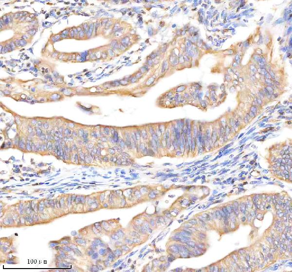
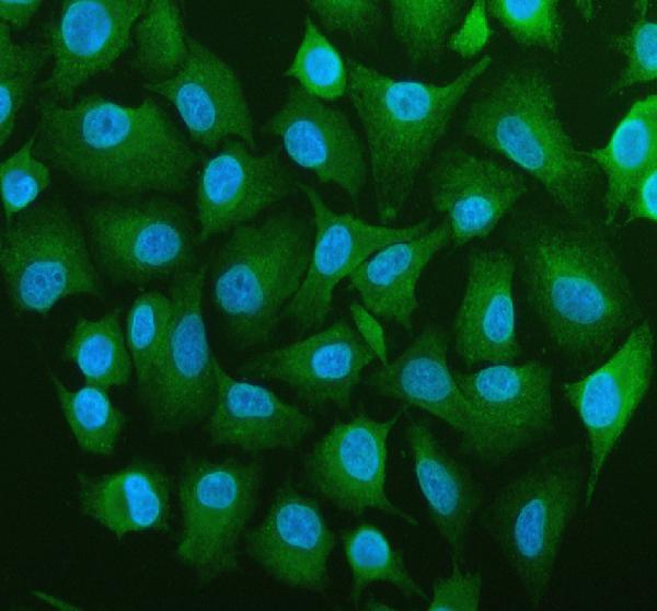
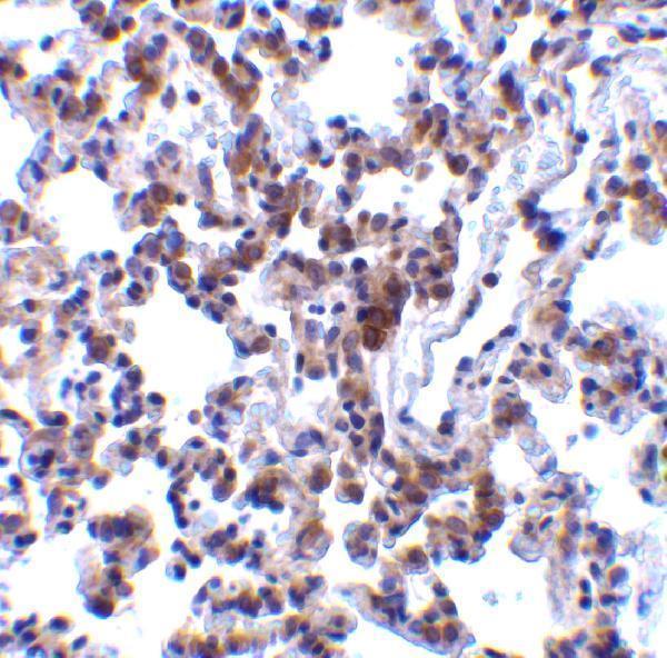
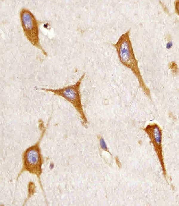
IHC images show BCAP31 staining in human colon cancer, rat lung and human brain sections (catalog IHC captions). IF images show A549 cells and human paraffin sections (A03767-4 IF captions).

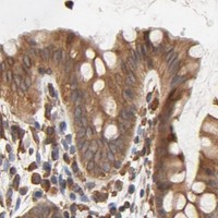
Real IHC data IHC analysis of BAP31/BCAP31 using anti-BAP31/BCAP31 antibody (A03767-4). BAP31/BCAP31 was detected in a paraffin-embedded section of human colon cancer tissue. Heat mediated antigen retrieval was performed in EDTA buffer (pH 8.0, epitope retrieval solution). The tissue section was blocked with 10% goat serum. The tissue section was then incubated with 2 μg/ml rabbit anti-BAP31/BCAP31 Antibody (A03767-4) overnight at 4°C. Peroxidase Conjugated Goat Anti-rabbit IgG was used as secondary antibody and incubated for 30 minutes at 37°C. The tissue section was developed using HRP Conjugated Rabbit IgG Super Vision Assay Kit (Catalog # SV0002) with DAB as the chromogen.
Anti-BAP31/BCAP31 Antibody ®
Cat # A03767-4
Real IHC data Immunohistochemistry Validation of BAP31 in Rat Lung Tissue Immunohistochemical analysis of paraffin-embedded rat lung tissue using anti-BAP31 antibody (A03767) at 2 μg/ml. Tissue was fixed with formaldehyde and blocked with 10% serum for 1 h at RT; antigen retrieval was by heat mediation with a citrate buffer (pH6). Samples were incubated with primary antibody overnight at 4˚C. A goat anti-rabbit IgG H&L (HRP) at 1/250 was used as secondary. Counter stained with Hematoxylin.
Anti-BAP31 BCAP31 Antibody
Cat # A03767
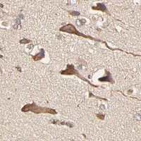
Real IHC data M03767 staining BAP31 in human brain sections by Immunohistochemistry (IHC-P -paraformaldehyde-fixed, paraffin-embedded sections). Tissue was fixed with formaldehyde and blocked with 3% BSA for 0. 5 hour at room temperature; antigen retrieval was by heat mediation with a citrate buffer (pH6). Samples were incubated with primary antibody (1/25) for 1 hours at 37°C. A undiluted biotinylated goat polyvalent antibody was used as the secondary antibody.
Anti-BAP31 Antibody
Cat # M03767

A03767-4 shows IHC in human colon cancer paraffin sections and IF in A549 cells and human paraffin sections (A03767-4 image captions); A03767 shows IHC in formaldehyde-fixed rat lung paraffin sections (A03767 IHC caption). M03767 shows IHC in formaldehyde-fixed human brain paraffin sections (M03767 IHC caption).

Which to pick: For human tissue IHC, choose A03767-4 for paraffin sections at 2–5 μg/mL with EDTA pH 8.0 retrieval; its IHC caption does not report a fixative (A03767-4 datasheet and IHC caption). M03767 is a mouse monoclonal option for paraffin-section human brain paraffin sections at 1:25 with citrate pH 6 retrieval (M03767 catalog and IHC caption); for IF/ICC, choose A03767-4 at 5 μg/mL, supported by images of A549 cells and human paraffin sections (A03767-4 datasheet and IF captions). For cross-species planning, A03767 is a rabbit polyclonal reactive with human, mouse and rat, but its IHC image and stated IHC validation cover rat tissue only (A03767 catalog, dilution notes and IHC caption). The selected A03767-4 tissue-IHC caption documents paraffin sections, but does not specify the fixative (selected-SKU IHC image A03767-4).

Each figure is that product's own IHC / IF validation image from its datasheet.

References

  1. UniProt Consortium. UniProt entry P51572 (BAP31_HUMAN, B-cell receptor-associated protein 31).
  2. Human Protein Atlas. BCAP31 tissue IHC expression (reliability: Supported).
  3. Human Protein Atlas. BCAP31 subcellular location (ICC-IF): Localized to the endoplasmic reticulum..
  4. Human Protein Atlas. BCAP31 antibody validation summary (3 antibodies).
  5. Pan-cancer analysis shows that BCAP31 is a potential prognostic and immunotherapeutic biomarker for multiple cancer types. Frontiers in immunology 2024 — PMC11683684.
  6. BCAP31 drives TNBC development by modulating ligand-independent EGFR trafficking and spontaneous EGFR phosphorylation. Theranostics 2019 — PMC6771250.
  7. BCAP31, a cancer/testis antigen-like protein, can act as a probe for non-small-cell lung cancer metastasis. Scientific reports 2020 — PMC7055246.
  8. A novel BCAP31 variant associated with nonsyndromic auditory neuropathy spectrum disorder: mitochondrial dysfunction, cisplatin sensitivity, and amenability to mitochondrial transplantation. Journal of translational medicine 2025 — PMC12131585.
  9. PubMed PMID:7806238 — UniProt-cited evidence.
  10. PubMed PMID:8706661 — UniProt-cited evidence.
  11. PubMed PMID:8612576 — UniProt-cited evidence.