This website uses cookies to ensure you get the best experience on our website.


- Table of Contents
Plan chromogenic BCAS1 IHC-P with the human-reactive catalog antibody, starting at 5 μg/mL (datasheet: IHC-P). Assess cytoplasmic staining in prostate glandular cells (HPA tissue IHC) alongside a negative control (standard IHC practice).

Expected localisation, validated protocols, controls and antibodies — the at-a-glance facts below, then the full design guide.
| Expected localisation | Cytoplasmic staining in GI tract, bladder, prostate and CNS (HPA tissue IHC) | |
| Staining pattern | Goblet/glandular cytoplasm; cerebellar granular cytoplasm/membrane (HPA tissue IHC) | |
| Antigen retrieval | Citrate pH 6.0 HIER, 95–98 °C, 20 min (rule: cytoplasmic / membrane antigen) | |
| Positive control | Appendix+4 more · see all | |
| Negative control | Adipose tissue+4 more · see all |
| Fixation | Keep fixation consistent across paraffin sections. (standard IHC practice; not target-specific) | |
| Caveat | GI positives vary: colon endocrine; small-intestinal goblet cells (HPA tissue IHC) | |
| Regulation | Down-regulated in some colorectal tumors (UniProt) | |
| Isoform / epitope | Two isoforms; no TM segment; check isoform epitope coverage (UniProt) |
The catalog antibody protocol is followed by one published chromogenic IHC protocol for BCAS1 in human multiple sclerosis brain lesions (PMC11772157).
| Sample | Tissue sections; selected-image fixative not specified (standard IHC workflow) |
| Fixation | Image fixative and duration unreported (datasheet A13589); verify before use. |
| Sectioning | 4–5 µm sections on charged slides (standard) |
| Deparaffinisation | Xylene, graded ethanol series to water (standard) |
| Antigen retrieval | Heat-induced epitope retrieval in citrate buffer, pH 6.0, 20 min at 95–98 °C (standard rule: cytoplasmic / membrane antigen) |
| Peroxidase block | 3% H2O2, 10 min, room temperature (standard) |
| Blocking | 10% normal serum of the secondary host, 30 min, room temperature (standard) |
| Primary antibody | Rabbit anti-BCAS1, 5 μg/mL (datasheet A13589) |
| Primary incubation | Overnight at 4 °C (standard) |
| Detection | HRP-polymer secondary, DAB chromogen 5–10 min (standard) |
| Counterstain | Hematoxylin, blue, dehydrate and mount (standard) |
| Expected result | BCAS1-positive staining in goblet cells of appendix (HPA tissue IHC: High). HPA tissue profile: Cytoplasmic expression in the gastrointestinal tract, urinary bladder, prostate and the CNS. No signal in the no-primary control. |
BCAS1 should chiefly appear in the cytoplasm on IHC sections, with strong staining in selected epithelial populations, including prostate glandular cells and intestinal goblet cells (HPA: tissue IHC, High). The protein is cytoplasmic and has no transmembrane segment (UniProt O75363: localisation and topology). HPA rates its tissue staining “Enhanced,” meaning consistency with RNA expression while external verification remains pending (HPA: reliability description).
| Distinct cytoplasmic reaction product in prostate glandular cells or duodenal goblet cells, with limited staining in adjacent cells. | This supports the expected cell-selective pattern: both populations are rated High by tissue IHC (HPA: prostate and duodenum). Assess the stained cell type as well as intensity; HPA reports cytoplasmic expression across several tissues, but its cell-level findings do not imply that every cell in a positive section should stain (HPA: tissue IHC profile). |
| Predominantly nuclear staining, or a broad extracellular deposit, instead of a cell-associated cytoplasmic pattern. | Treat this as discordant with the reported localisation (UniProt O75363: Cytoplasm; HPA: tissue IHC profile). General IHC practice: inspect the counterstain and a negative control before assigning the signal to BCAS1; review detection and wash conditions if the same pattern appears across unrelated structures. |
| Strong staining in adipocytes while expected epithelial cells show little or none. | HPA reports BCAS1 as Not detected in adipose-tissue adipocytes, whereas prostate glandular and duodenal goblet cells are High (HPA: tissue IHC). General IHC practice: consider antibody cross-reactivity or endogenous detection activity; compare a suitable negative control and check whether the signal follows cell morphology. |
| Haze or granular colour spread across the section, obscuring cell borders and the counterstain. | That distribution prevents a reliable call on the selective cytoplasmic pattern reported for BCAS1 (HPA: tissue IHC profile; UniProt O75363: Cytoplasm). General IHC practice: examine reagent-only or primary-omission controls, blocking, washes and development time to distinguish nonspecific background from interpretable cell-associated signal. |
| No convincing signal in the expected cells of a prostate or duodenum section. | An absent result conflicts with HPA's High calls for prostate glandular cells and duodenal goblet cells (HPA: tissue IHC). General IHC practice: first confirm that the relevant cells are present, then check the positive control, primary antibody conditions, detection reagents and antigen retrieval before interpreting the specimen as negative. |
| Choice of reference tissue and cells | Prostate glandular cells, duodenal goblet cells and small-intestine goblet cells are High, while adipose-tissue adipocytes are Not detected (HPA: tissue IHC). Compare the named cell populations, since a whole-section average can conceal the reported distribution. |
| Strength and scope of validation | The tissue profile is rated Enhanced for consistency between staining and RNA expression, with external verification pending (HPA: reliability description). HPA lists Enhanced IHC status for HPA051816, HPA054745 and CAB033558 (HPA: antibody validation); those ratings do not validate an unlisted antibody. |
| Isoform and epitope interpretation | Two BCAS1 isoforms are recorded, but their antibody epitopes are not supplied here (UniProt O75363: isoforms; HPA: supplied antibody data). Do not infer which isoform an observed stain represents or use an apparent difference in staining to assign isoform-specific expression. |
| Does IF/ICC show the same pattern? | HPA approves a vesicular ICC-IF location, while UniProt gives the broader cytoplasmic location (HPA: subcellular ICC-IF; UniProt O75363: Cytoplasm). The vesicular observation concerns ICC-IF images and should not be imposed as the required appearance of chromogenic tissue IHC. |
| Situation | Likely cause | Next action |
|---|---|---|
| Positive tissue is blank. | The expected cells may be absent from the examined area, or an IHC workflow step may have failed; prostate glandular and duodenal goblet cells are High in HPA tissue IHC (HPA: tissue IHC). | Locate the named cells on the counterstained section. As general IHC practice, check a positive control, reagent preparation, retrieval conditions, primary incubation and detection before scoring the specimen. |
| Signal is weak in a tissue expected to be positive. | HPA's High rating applies to specific cells, not every part of that tissue (HPA: tissue IHC). Weak signal may also reflect general workflow conditions; no BCAS1-specific fixation effect is reported in the supplied sources. | Reassess the named cell population. As general IHC practice, compare the positive control and review validated retrieval, antibody incubation and chromogen development conditions. |
| Adipocytes stain strongly. | That finding conflicts with HPA's Not detected call for adipose-tissue adipocytes (HPA: tissue IHC); nonspecific binding or endogenous detection activity is possible under general IHC practice. | Check a primary-omission control and the detection system; reassess blocking and cell identification. Preserve the cell-specific distinction: the HPA breast entry also concerns adipocytes, not every breast cell (HPA: tissue IHC). |
| Signal is mainly nuclear. | Nuclear dominance is inconsistent with the recorded cytoplasmic localisation (UniProt O75363: Cytoplasm; HPA: tissue IHC profile). | Use the counterstain to verify compartment assignment. As general IHC practice, compare control sections and inspect background, antibody conditions and detection before accepting nuclear signal as specific. |
| Diffuse colour masks cell boundaries. | The distribution cannot be matched confidently to HPA's cell-specific cytoplasmic observations (HPA: tissue IHC); general IHC causes include excess background or overdevelopment. | As general IHC practice, compare the negative control, review blocking and washes, and shorten chromogen development where appropriate; then rescore only clearly localised cellular staining. |
| CNS staining seems inconsistent across cell types. | HPA reports High cerebellar granular-cell staining but Low hippocampal and caudate glial-cell staining; UniProt reports high brain expression, particularly in oligodendrocytes (HPA: tissue IHC; UniProt O75363: tissue specificity). | Record region and cell identity before comparing results. Do not convert UniProt's broader brain-expression statement into an expectation of uniformly strong IHC staining in all glial cells. |

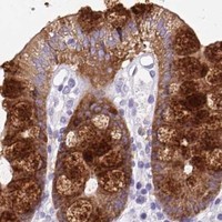


Comprehensive Human Protein Atlas IHC scoring per tissue (reliability: Enhanced — High consistency between antibody staining and RNA expression data. Pending external verification.). Rows are taken directly from the HPA tissue chart — click any row's HPA link to view the source.
| Tissue | Cell type | Level | Evidence | Source |
|---|---|---|---|---|
| Appendix | Goblet cells | High | Protein (IHC) | HPA → |
| Bronchus | Respiratory epithelial cells | High | Protein (IHC) | HPA → |
| Cerebellum | Granular cells - cytoplasm/membrane | High | Protein (IHC) | HPA → |
| Colon | Endocrine cells | High | Protein (IHC) | HPA → |
| Duodenum | Goblet cells | High | Protein (IHC) | HPA → |
| Tissue | Cell type | Level | Evidence | Source |
|---|---|---|---|---|
| Adipose tissue | Adipocytes | Not detected | Protein (IHC) | HPA → |
| Adrenal gland | Glandular cells | Not detected | Protein (IHC) | HPA → |
| Bone marrow | Hematopoietic cells | Not detected | Protein (IHC) | HPA → |
| Breast | Adipocytes | Not detected | Protein (IHC) | HPA → |
| Cervix | Glandular cells | Not detected | Protein (IHC) | HPA → |
Troubleshoot BCAS1 chromogenic IHC in paraffin sections by checking retrieval, compartment, cell identity, controls, and scoring before interpreting staining.
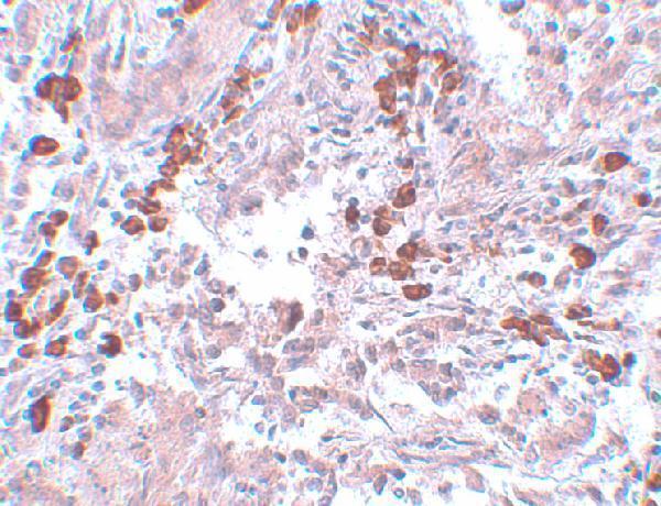
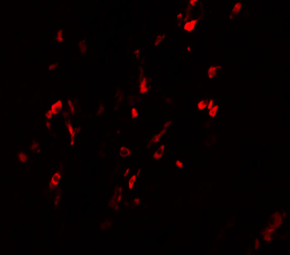
The IHC-validated anti-BCAS1 antibody has human breast carcinoma tissue IHC data (A13589 IHC image caption) and human breast carcinoma cell IF data (A13589 IF image caption).
A13589 is listed for human IHC-P and IF (catalog: applications and reactivity). Its figures show IHC in human breast carcinoma at 5 μg/mL and IF in human breast carcinoma cells at 20 μg/mL (A13589 image captions).
Which to pick: Choose A13589 for paraffin-section IHC (catalog: IHC-P); its IHC figure shows human breast carcinoma, but the fixative is unreported (A13589 IHC image caption). For IF/ICC planning, A13589 has IF data in human breast carcinoma cells; ICC is not separately listed (A13589 IF image caption; catalog: applications). No cross-species option is supported because A13589 lists human reactivity only (catalog: reactivity).