This website uses cookies to ensure you get the best experience on our website.


- Table of Contents
Plan chromogenic BCAS2 IHC-P around nuclear and nucleolar tissue staining (HPA tissue IHC). Testis spermatogonia provide a high-staining reference (HPA tissue IHC); start the IHC-validated antibody A07113 at 5 μg/mL (datasheet: IHC-P).

Expected localisation, validated protocols, controls and antibodies — the at-a-glance facts below, then the full design guide.
| Expected localisation | Nuclear and nucleolar staining (HPA tissue IHC) | |
| Staining pattern | Nuclear and nucleolar staining across tissue cell types (HPA tissue IHC) | |
| Antigen retrieval | Tris-EDTA pH 9.0 HIER, 95–98 °C, 20 min (rule: nuclear antigen) | |
| Positive control | Testis+4 more · see all | |
| Negative control | Adipose tissue |
| Fixation | Keep formalin fixation consistent across sections. (standard IHC practice; not target-specific) | |
| Caveat | Adipocytes may be unstained despite the general tissue pattern (HPA tissue IHC) | |
| Regulation | Ubiquitously expressed (UniProt) | |
| Isoform / epitope | No isoforms; the annotated chain starts at residue 2 (UniProt) |
The catalog antibody protocol is followed by three published BCAS2 IHC protocols for pig testis, esophageal carcinoma tissue, and human prostate specimens (PMC10607209; PMC5783606; PMC4453457).
| Sample | Tissue sections; selected-image fixative not specified (standard IHC workflow) |
| Fixation | Image fixative and duration unreported (datasheet A07113); verify before use. |
| Sectioning | 4–5 µm sections on charged slides (standard) |
| Deparaffinisation | Xylene, graded ethanol series to water (standard) |
| Antigen retrieval | Heat-induced epitope retrieval in Tris-EDTA buffer, pH 9.0, 20 min at 95–98 °C (standard rule: nuclear antigen) |
| Peroxidase block | 3% H2O2, 10 min, room temperature (standard) |
| Blocking | 10% normal serum of the secondary host, 30 min, room temperature (standard) |
| Primary antibody | Rabbit anti-BCAS2, 5 μg/mL (datasheet A07113) |
| Primary incubation | Overnight at 4 °C (standard) |
| Detection | HRP-polymer secondary, DAB chromogen 5–10 min (standard) |
| Counterstain | Hematoxylin, blue, dehydrate and mount (standard) |
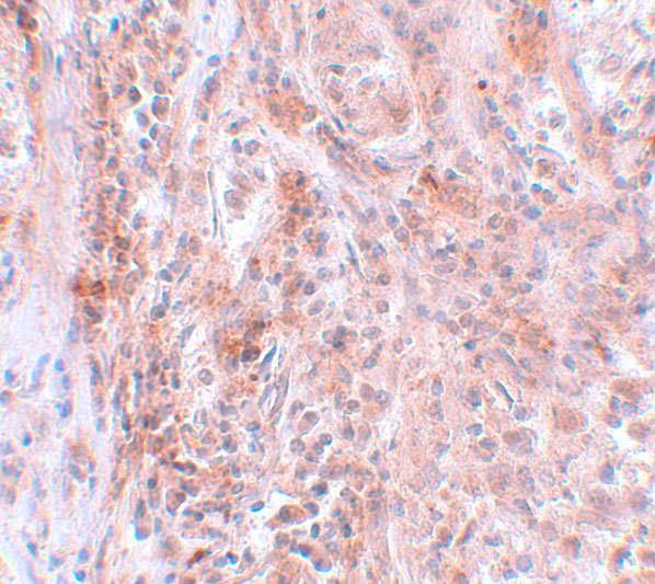
| Expected result | BCAS2-positive staining in spermatogonia cells of testis (HPA tissue IHC: High). HPA tissue profile: General nuclear and nucleolar expression. No signal in the no-primary control. |
BCAS2 is expected in nuclei and nucleoli (UniProt O75934; HPA: general nuclear and nucleolar expression). Testis spermatogonia show high staining, while several other cell populations show medium staining (HPA: tissue IHC). Tissue IHC reliability is Supported, with medium consistency between staining and RNA data (HPA: reliability). BCAS2 has no transmembrane segment (UniProt O75934 topology).
| Distinct nuclear staining, sometimes accenting nucleoli, in spermatogonia or other HPA-positive cells. | This matches the reported compartment and cell distribution (UniProt O75934: nucleus and nucleolus; HPA: general nuclear and nucleolar expression). Spermatogonia are a high-staining reference; adrenal glandular cells and cerebral cortical neurons are medium-staining examples (HPA: tissue IHC). Judge cells within their tissue context rather than expecting every nucleus to have equal intensity. |
| Predominantly cytoplasmic or membranous chromogen with little nuclear signal. | The dominant compartment conflicts with BCAS2’s reported nuclear and nucleolar location (UniProt O75934; HPA: tissue IHC). A membrane pattern also lacks support from its topology (UniProt O75934: no transmembrane segment). Treat it as suspect staining and check localisation in a known-positive tissue before interpreting it as BCAS2. |
| Strong staining in adipocytes, especially if nuclei in a known-positive area remain weak. | HPA reports BCAS2 as not detected in adipocytes (HPA: adipose tissue IHC). This discrepancy can reflect nonspecific antibody binding or endogenous detection activity; it does not by itself identify which occurred. Compare primary-antibody and no-primary controls, and assess whether any chromogen is actually nuclear (General IHC practice). |
| A diffuse haze covers cells, stroma, and empty areas without clear nuclear boundaries. | That appearance cannot support a BCAS2 localisation call because the expected tissue pattern is nuclear and nucleolar (HPA: tissue IHC; UniProt O75934). Excess background can arise from nonspecific binding or detection chemistry (General IHC practice). Score only distinguishable cellular signal after the background has been resolved. |
| No nuclear signal in a section containing identifiable spermatogonia. | This conflicts with the high staining reported for spermatogonia (HPA: testis IHC), but a failed assay is only one possibility. First confirm that the relevant cells are present and interpretable, then review the antibody and detection controls (General IHC practice). HPA’s Supported reliability does not guarantee positivity in every specimen (HPA: reliability). |
| Tissue and cell selection | BCAS2 is described as ubiquitously expressed (UniProt O75934), yet HPA staining varies by cell type: high in spermatogonia, medium in selected glandular, lymphoid, ciliated, glial, neuronal, and squamous cells, and not detected in adipocytes (HPA: tissue IHC). Use the named cell population when comparing signal; tissue-wide averages can obscure the pattern. |
| Evidence strength and antibody choice | The tissue IHC profile is Supported, with medium consistency against RNA data; the listed HPA067881 antibody has Supported IHC validation (HPA: reliability; HPA: antibody validation). These labels support a reference pattern but do not establish identical staining with every IHC-validated antibody or every specimen. |
| Compartment and molecular context | UniProt places BCAS2 in the nucleus and nucleolus and describes a spliceosome role (UniProt O75934: subcellular location and function). It records no transmembrane segment, signal peptide, or propeptide (UniProt O75934: topology and processing). These facts support a nuclear readout; they do not predict an antibody epitope or antigen-retrieval requirement. |
| IF/ICC Q&A: Should punctate nuclear signal be expected? | Yes, nuclear speckles are the supported main IF/ICC location, and centrosome is an approved location (HPA: subcellular ICC-IF). Those cell-image findings provide context for IF/ICC interpretation; they do not require a punctate chromogenic pattern in paraffin tissue, whose reported profile is general nuclear and nucleolar expression (HPA: tissue IHC). |
| Target-specific fixation sensitivity | No target-specific fixation effect is established by the supplied UniProt or HPA records. Select and assess retrieval and detection conditions as general IHC workflow variables (General IHC practice); do not infer a BCAS2 fixation response from its topology, modifications, or tissue staining levels. |
| Situation | Likely cause | Next action |
|---|---|---|
| Weak or absent signal in identifiable spermatogonia. | The result differs from the reported high level in those cells (HPA: testis IHC); the source does not identify a BCAS2-specific cause. | Check cell preservation, primary-antibody application, detection reagents, and a known-positive control; review retrieval as a general IHC variable (General IHC practice). |
| Chromogen is mainly cytoplasmic or membranous. | That distribution conflicts with the nuclear and nucleolar reference and lacks topology support for a membrane pattern (UniProt O75934; HPA: tissue IHC). | Compare with a known-positive section and a no-primary control; only score a cellular nuclear pattern as supportive (General IHC practice; HPA: tissue IHC). |
| Adipocytes appear strongly positive. | They are reported as not detected (HPA: adipose tissue IHC); nonspecific binding or endogenous detection activity is possible (General IHC practice). | Inspect subcellular placement and no-primary controls, then reassess blocking and detection background (General IHC practice). |
| Colour appears in stroma or across the whole section. | Diffuse signal obscures the expected nuclear and nucleolar profile (HPA: tissue IHC); background may arise during antibody or chromogen detection (General IHC practice). | Compare a no-primary control, check washing and blocking, and adjust detection conditions if background persists (General IHC practice). |
| Only some cell types stain within an otherwise interpretable section. | Cell-type variation is compatible with the HPA range from high spermatogonia to medium, low, or undetected populations (HPA: tissue IHC). | Identify the cells before scoring and compare each with its HPA cell-specific reference; do not require equal intensity across a tissue (HPA: tissue IHC). |
| IF/ICC speckles are seen, but the paraffin section lacks discrete puncta. | HPA describes nuclear speckles in ICC-IF and general nuclear and nucleolar expression in tissue IHC (HPA: subcellular ICC-IF; HPA: tissue IHC). | Interpret the paraffin section against its tissue IHC reference: assess nuclear localisation and the named positive cells without requiring resolvable speckles (HPA: tissue IHC). |


Comprehensive Human Protein Atlas IHC scoring per tissue (reliability: Supported — Medium consistency between antibody staining and RNA expression data.). Rows are taken directly from the HPA tissue chart — click any row's HPA link to view the source.
| Tissue | Cell type | Level | Evidence | Source |
|---|---|---|---|---|
| Testis | Spermatogonia cells | High | Protein (IHC) | HPA → |
| Adrenal gland | Glandular cells | Medium | Protein (IHC) | HPA → |
| Appendix | Lymphoid tissue | Medium | Protein (IHC) | HPA → |
| Bronchus | Ciliated cells (cell body) | Medium | Protein (IHC) | HPA → |
| Caudate | Glial cells | Medium | Protein (IHC) | HPA → |
| Tissue | Cell type | Level | Evidence | Source |
|---|---|---|---|---|
| Adipose tissue | Adipocytes | Not detected | Protein (IHC) | HPA → |
Troubleshooting BCAS2 staining in paraffin sections centers on retrieval, nuclear localisation and controls that distinguish signal from tissue artefact.
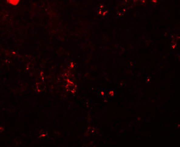
A07113 has human breast carcinoma IHC and IF images (catalog image captions); its listed reactivity includes human, mouse and rat (catalog applications and reactivity).
A07113 is the only SKU rendered, with IHC data from human breast carcinoma at 5 μg/mL (A07113 IHC image caption). Its IF image also shows human breast carcinoma tissue at 20 μg/mL (A07113 IF image caption).
Which to pick: Choose A07113 for paraffin-section IHC because it lists IHC-P and has its own human breast carcinoma IHC image (A07113 applications; A07113 IHC image caption); the fixative is unreported (A07113 IHC image caption). Choose A07113 for tissue IF with an image, or A07113-1 when an ICC-listed antibody is needed; A07113-1 has no IHC image or IHC-P listing (A07113 IF image caption; A07113-1 applications and image captions). Both list human, mouse and rat reactivity, but the supplied IHC and IF images show human tissue only (catalog reactivity; A07113 image captions).