This website uses cookies to ensure you get the best experience on our website.


- Table of Contents
For BCAS4 paraffin IHC, expect general cytoplasmic staining and consider appendix glandular cells as a high-staining reference (HPA tissue IHC). Start the catalog antibody at 5 μg/mL (datasheet), and interpret scoring in light of the uncertain tissue profile (HPA tissue IHC).

Expected localisation, validated protocols, controls and antibodies — the at-a-glance facts below, then the full design guide.
| Expected localisation | General cytoplasmic tissue staining (HPA tissue IHC) | |
| Staining pattern | Cytoplasmic staining in glandular and other cells (HPA tissue IHC) | |
| Antigen retrieval | Citrate pH 6.0 HIER, 95–98 °C, 20 min (rule: cytoplasmic / membrane antigen) | |
| Positive control | Appendix+4 more · see all | |
| Negative control | None in HPA (detected in all 45 tissues); use no-primary + isotype controls |
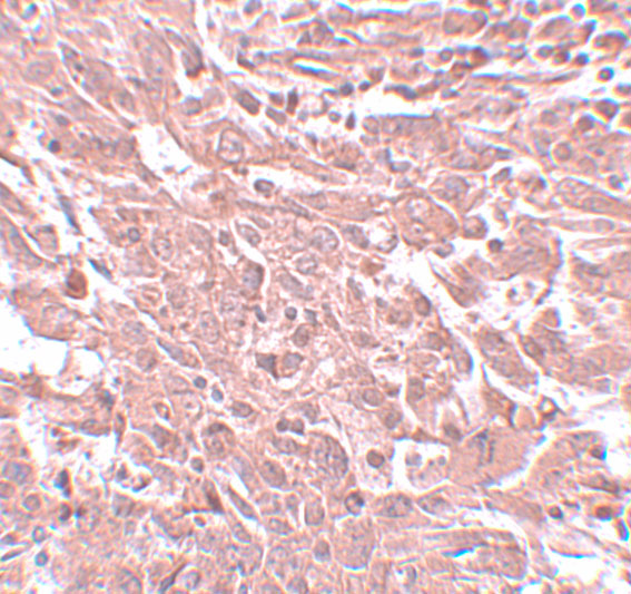
| Fixation | Matched tissue-IHC evidence does not establish the fixation claim. Validate the specimen-specific method before use. (selected-SKU IHC image A13963-1) | |
| Caveat | Low staining–RNA concordance; tissue profile uncertain (HPA tissue IHC) | |
| Regulation | Lymphoid tissue–enhanced RNA (HPA tissue RNA) | |
| Isoform / epitope | 3 isoforms; cytoplasmic epitope coverage unknown (UniProt) |
The catalog antibody’s IHC-P protocol is paired with one published BCAS4 chromogenic IHC protocol (PMC13520631).
| Sample | Tissue sections; selected-image fixative not specified (standard IHC workflow) |
| Fixation | Image fixative and duration unreported (datasheet A13963-1); verify before use. |
| Sectioning | 4–5 µm sections on charged slides (standard) |
| Deparaffinisation | Xylene, graded ethanol series to water (standard) |
| Antigen retrieval | Heat-induced epitope retrieval in citrate buffer, pH 6.0, 20 min at 95–98 °C (standard rule: cytoplasmic / membrane antigen) |
| Peroxidase block | 3% H2O2, 10 min, room temperature (standard) |
| Blocking | 10% normal serum of the secondary host, 30 min, room temperature (standard) |
| Primary antibody | Rabbit anti-BCAS4, 5 μg/mL (datasheet A13963-1) |
| Primary incubation | Overnight at 4 °C (standard) |
| Detection | HRP-polymer secondary, DAB chromogen 5–10 min (standard) |
| Counterstain | Hematoxylin, blue, dehydrate and mount (standard) |
| Expected result | BCAS4-positive staining in glandular cells of appendix (HPA tissue IHC: High). HPA tissue profile: General cytoplasmic expression. No signal in the no-primary control. |
In paraffin-section IHC, expect BCAS4 staining mainly in the cytoplasm (UniProt Q8TDM0: cytoplasm; no transmembrane segment). HPA reports high staining in selected glandular, squamous, respiratory, follicle and cardiac cells (HPA: tissue IHC). Treat the pattern as provisional: HPA rates tissue staining Uncertain because antibody staining and RNA expression show low consistency (HPA: tissue IHC reliability).
| Cytoplasmic chromogen in appendix glandular cells or esophageal squamous epithelial cells. | This fits the reported compartment and cell types (UniProt Q8TDM0: cytoplasm; HPA: High in both). Compare stained cells with tissue morphology and controls before scoring; the HPA tissue IHC result is Uncertain (HPA: reliability). |
| Predominantly nuclear or sharply membranous staining, with little cytoplasmic signal. | This differs from the reported cytoplasmic location and lacks support from the topology record (UniProt Q8TDM0: cytoplasm; no transmembrane segment). Consider nonspecific staining or a detection artefact; review controls before calling it BCAS4. |
| Strong staining in fibroblasts, lymph-node germinal center cells or bone-marrow hematopoietic cells. | HPA reports Low staining in these cells (HPA: tissue IHC). Unexpected strong signal raises concern for cross-reactivity or endogenous detection activity; compare it with the negative control and local background. |
| Uniform chromogen across cells and extracellular spaces, obscuring cell boundaries. | A diffuse deposit is difficult to assign to the reported cytoplasmic pattern (UniProt Q8TDM0: cytoplasm). In standard IHC practice, inspect reagent-only controls and washing, blocking and detection conditions before interpreting intensity. |
| No detectable signal in appendix glandular cells or another HPA high-staining site. | That result conflicts with the reported tissue pattern (HPA: High in appendix glandular cells and other listed cells). First check whether the assay and control slide developed; because HPA rates tissue IHC Uncertain, absence alone does not establish biological loss (HPA: reliability). |
| Compartment and topology | BCAS4 is annotated as cytoplasmic, with no transmembrane segment (UniProt Q8TDM0). Use cell boundaries and counterstain to judge whether chromogen lies in cytoplasm; topology alone does not validate antibody specificity. |
| Tissue and cell selection | HPA reports High staining in several specified cell types and Low staining in fibroblasts, germinal center cells and hematopoietic cells (HPA: tissue IHC). Select reference areas by cell type, not tissue name alone. |
| Isoforms and epitope coverage | UniProt lists 3 isoforms (UniProt Q8TDM0: isoforms 1–3). The supplied record does not map the antibody epitope, so staining cannot be assigned to an isoform or assumed to detect every isoform. |
| Evidence strength | HPA rates tissue IHC Uncertain and HPA038812 IHC Uncertain (HPA: tissue reliability; antibody validation). Report the observed pattern and controls; avoid treating intensity as a validated measure of BCAS4 abundance. |
| IF/ICC cross-check — Q&A | Q: What localisation is reported in IF/ICC? A: Cytosol and intermediate filaments, both approved (HPA: subcellular ICC-IF). This supports a localisation comparison, but does not resolve the uncertain tissue IHC assessment (HPA: tissue reliability). |
| Situation | Likely cause | Next action |
|---|---|---|
| No chromogen in a reported high-staining cell population. | The run may have failed, or signal may be below the chosen assay's detection threshold; biological absence is also possible. HPA tissue IHC remains Uncertain (HPA: reliability). | Check a run control, section integrity and the reagent sequence; then review retrieval, antibody dilution and development as general IHC variables. Do not infer BCAS4-specific fixation sensitivity from this record. |
| Nuclear staining dominates the section. | Nuclear predominance conflicts with the cytoplasmic annotation (UniProt Q8TDM0: cytoplasm). Background or antibody cross-reactivity is possible, but appearance alone cannot identify the cause. | Compare reagent-only and tissue controls, inspect cytoplasmic signal in the same cells, and record nuclear staining separately from any BCAS4-compatible pattern (UniProt Q8TDM0: cytoplasm). |
| Strong signal appears in fibroblasts or germinal center cells. | These are Low-staining populations in the HPA tissue record (HPA: fibroblasts; germinal center cells). Cross-reactivity or endogenous detection activity should be considered. | Confirm cell identity by morphology, compare an appropriate detection control, and reassess blocking or detection conditions as general IHC practice before scoring those cells positive. |
| Chromogen is widespread and obscures cellular detail. | Diffuse background prevents a reliable cytoplasmic call (UniProt Q8TDM0: cytoplasm). Excess detection signal or inadequate washing is a general IHC possibility, not a BCAS4-specific finding. | Inspect the reagent-only control, adjust general washing or development conditions if indicated, and score only signal that can be assigned to intact cells. |
| Adjacent high-staining candidate tissues differ markedly in intensity. | HPA lists several High-staining cell types but rates the aggregate tissue IHC evidence Uncertain (HPA: tissue IHC). Differences cannot be attributed to BCAS4 abundance from staining alone. | Compare equivalent cell populations and matched run controls; document distribution and intensity separately, and avoid ranking tissues by chromogen strength without independent validation. |
| IHC looks membranous while ICC-IF looks filamentous or cytosolic. | ICC-IF reports approved cytosol and intermediate-filament localisation, whereas UniProt annotates cytoplasm and no transmembrane segment (HPA: subcellular ICC-IF; UniProt Q8TDM0: topology). | Review IHC morphology and detection controls, then describe the discordance. The ICC-IF result does not upgrade tissue IHC reliability, which HPA calls Uncertain (HPA: tissue reliability). |


Comprehensive Human Protein Atlas IHC scoring per tissue (reliability: Uncertain — Low consistency between antibody staining and RNA expression data.). Rows are taken directly from the HPA tissue chart — click any row's HPA link to view the source.
| Tissue | Cell type | Level | Evidence | Source |
|---|---|---|---|---|
| Appendix | Glandular cells | High | Protein (IHC) | HPA → |
| Cervix | Glandular cells | High | Protein (IHC) | HPA → |
| Epididymis | Glandular cells | High | Protein (IHC) | HPA → |
| Esophagus | Squamous epithelial cells | High | Protein (IHC) | HPA → |
| Gallbladder | Glandular cells | High | Protein (IHC) | HPA → |
| Tissue | Cell type | Level | Evidence | Source |
|---|---|---|---|---|
| None in HPA: BCAS4 is detected in all 45 scored tissues. Use a no-primary (secondary-only) and an isotype control instead. | ||||
Troubleshoot BCAS4 staining in paraffin sections by checking cytoplasmic localisation, tissue morphology, controls, and the limits of the available antibody evidence.
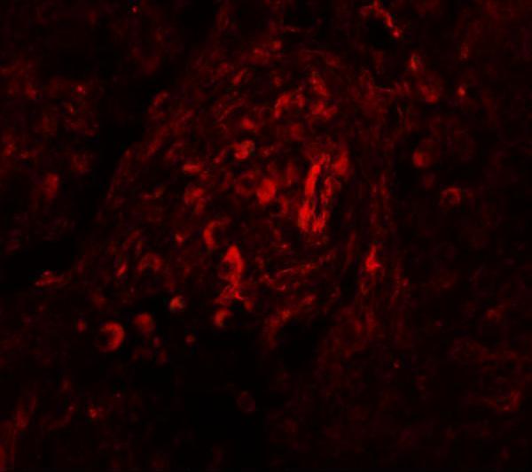
A13963-1 has IHC and IF images from human breast carcinoma tissue (IHC and IF image captions); the catalog lists Human and Mouse reactivity (catalog: reactivity).
A13963-1 is listed for IHC-P and IF, with Human and Mouse reactivity (catalog: applications and reactivity). Its IHC image shows human breast carcinoma at 5 μg/mL, and its IF image shows human breast carcinoma tissue at 20 μg/mL (IHC and IF image captions).
Which to pick: Choose A13963-1 for paraffin-section IHC (catalog: IHC-P application); its human breast carcinoma IHC caption does not report the fixative (IHC image caption). For tissue IF, choose A13963-1 based on its human breast carcinoma IF image; ICC validation is unreported (IF image caption; catalog: applications). For work involving Mouse samples, A13963-1 lists Mouse reactivity, but the supplied IHC and IF images show Human samples only; its clone is unreported (catalog: reactivity and clone; IHC and IF image captions).