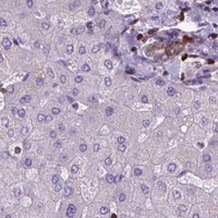
BCL2 / Apoptosis regulator Bcl-2 · IHC design guide

Design Immunohistochemistry for BCL2

Plan BCL2 staining in paraffin sections using an IHC-validated antibody (datasheet: M00040-2). Use the reported cytoplasmic tissue pattern to select controls and interpret staining by cell population (HPA tissue IHC).

Evidence assembled Oct 2026 · For research use; verify linked source records and product datasheet before use
Immunohistochemistry protocol sheet for BCL2 (IHC for BCL2): expected localisation Cytoplasmic tissue staining (HPA tissue IHC), antibody M00040-1, validated IHC image, and IHC protocol steps
Printable BCL2 IHC protocol sheet — expected localisation Cytoplasmic tissue staining (HPA tissue IHC), antibody M00040-1, controls and protocol steps. Open the full BCL2 IHC guide →

BCL2 Immunohistochemistry Experimental Design Guide

Expected localisation, validated protocols, controls and antibodies — the at-a-glance facts below, then the full design guide.

Must know before staining
Expected localisation Cytoplasmic tissue staining (HPA tissue IHC)
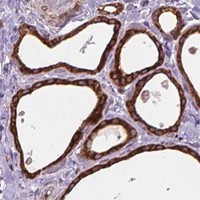
Staining pattern Cytoplasmic staining in thyroid and lymphoid organs (HPA tissue IHC)
Antigen retrieval Tris-EDTA pH 9.0 HIER, 95–98 °C, 20 min (rule: nuclear antigen)
Positive control ⓘ Appendix+4 more · see all
Negative control ⓘ Adrenal gland+4 more · see all
Important caveats
Reasons your staining may differ from the expected pattern.
Fixation Matched tissue-IHC evidence does not establish the fixation claim. Validate the specimen-specific method before use. (selected-SKU IHC image M00040-1)
Caveat Lymphoid staining varies by cell population (HPA tissue IHC)
Regulation Expression varies by tissue and cell type (HPA tissue IHC)
Isoform / epitope 2 isoforms, Alpha and Beta; check epitope coverage (UniProt)
Section 1

Recommended BCL2 IHC & IF Protocols

The catalog antibody's IHC-P protocol is paired with published workflows for breast lesions (PMC9592210) and follicular lymphoma (PMC5930855).

Recommended immunohistochemistry (IHC-P) protocol parameters
SampleParaffin-embedded tissue sections; fixative not specified (datasheet M00040-1; sample unspecified)
FixationImage fixative and duration unreported (datasheet M00040-1); verify before use.
Sectioning4–5 µm sections on charged slides (standard)
DeparaffinisationXylene, graded ethanol series to water (standard)
Antigen retrievalHeat-induced epitope retrieval in Tris-EDTA buffer, pH 9.0, 20 min at 95–98 °C (standard rule: nuclear antigen)
Peroxidase block3% H2O2, 10 min, room temperature (standard)
Blocking10% normal serum of the secondary host, 30 min, room temperature (standard)
Primary antibodyRabbit monoclonal (clone HF-2) anti-BCL2, 1:50-1:100 (datasheet M00040-1)
Primary incubationOvernight at 4 °C (standard)
DetectionHRP-polymer secondary, DAB chromogen 5–10 min (standard)
CounterstainHematoxylin, blue, dehydrate and mount (standard)
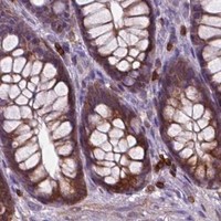
Expected resultBCL2-positive staining in non-germinal center cells of appendix (HPA tissue IHC: High). HPA tissue profile: Cytoplasmic expression in several different tissue types, including thyroid gland and lymphoid organs. No signal in the no-primary control.
💡Decision noteStart with Tris-EDTA pH 9.0 at 95–98 °C for 20 min (page retrieval setting); neither included excerpt specifies retrieval (PMC9592210; PMC5930855).
Section 2

What Is the Expected BCL2 Staining Pattern?

BCL2 is annotated at the mitochondrial outer membrane, ER membrane and nuclear membrane, with a transmembrane segment at residues 212–233 (UniProt P10415 topology and location). In paraffin IHC, expect cytoplasmic staining, including high staining in lymph node non-germinal center cells (HPA tissue IHC). HPA rates tissue IHC reliability Enhanced, with medium consistency between staining and RNA expression (HPA tissue IHC).

What am I looking at on my slide?
Cytoplasmic stain in lymph node non-germinal center cells (HPA: High)Matches the observed tissue pattern (HPA tissue IHC).
Uniform intranuclear stainUnexpected for membrane-associated BCL2; review for artifact (UniProt P10415 location).
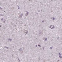
Strong stain in lung alveolar cells (HPA: Not detected)Discordant with HPA; assess cross-reactivity or endogenous activity (HPA tissue IHC; general IHC practice).
Diffuse cell-free brown hazeSuggests background; review blocking, washes and detection controls (general IHC practice).
No stain in lymph node non-germinal center cells (HPA: High)Check retrieval, antibody and detection controls (general IHC practice).
💡Expected BCL2 appearanceA convincing positive is cytoplasmic, high staining in lymph node non-germinal center cells (HPA tissue IHC); uniform intranuclear or cell-free brown signal warrants artifact review (UniProt P10415 location; general IHC practice).
How each factor affects the staining
Membrane topology (UniProt P10415)Residues 212–233 form a transmembrane segment (UniProt P10415 topology).
IHC antibody validation (HPA antibodies)CAB000003 has Enhanced IHC validation (HPA antibodies).
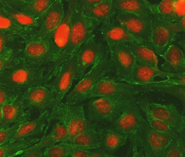
IF/ICC Q: Where is BCL2 seen?A: At mitochondria in SiHa and U2OS images (HPA subcellular ICC-IF).
Why is my staining missing, weak or wrong?
SituationLikely causeNext action
Known-positive lymph node cells are unstained (HPA: High)Retrieval or detection may have failed (general IHC practice).Check retrieval and detection controls (general IHC practice).
Uniform intranuclear signalDiscordant with annotated membrane locations (UniProt P10415 location).Review primary-omission control and counterstain (general IHC practice).
Lung alveolar cells stain strongly (HPA: Not detected)Possible cross-reactivity or endogenous activity (general IHC practice).Check a detection-only control and cell identity (general IHC practice).
Diffuse brown haze across the sectionPossible excess background (general IHC practice).Review blocking, washes and antibody dilution (general IHC practice).
Stain appears in blank areas of the slidePossible detection precipitate (general IHC practice).Inspect reagent handling and detection-only control (general IHC practice).

Sample controls for BCL2 IHC & IF

🧪Run appendix first: non-germinal center cells should stain (HPA: High in appendix non-germinal center cells). Use heart muscle as the negative tissue: cardiomyocytes are listed as not detected (HPA: Not detected in heart muscle cardiomyocytes). On the appendix slide, compare staining with cells outside the scored compartment as an internal background reference; their BCL2 status is not established by the supplied HPA row.
Positive control tissue: Appendix (Non-germinal center cells, HPA High)
Negative control tissue: Adrenal gland (HPA Not detected)
ICC-IF cell lines (HPA subcellular resource): HPA ICC-IF images show BCL2 in SiHa, U2OS, with annotated localisation: Mitochondria (enhanced) (HPA subcellular).
Technical controls: Include a no-primary (secondary-only) slide and an isotype control matched to the primary antibody’s host species and class (standard IHC practice). If available, use BCL2-knockout material or an immunizing-peptide block as a biological specificity control (standard IHC practice). For chromogenic IHC, quench endogenous peroxidase and check for endogenous biotin if using biotin-based detection; for IF, assess tissue autofluorescence (standard IHC/IF practice).
⚠️Feasibility: No supplied target or application evidence reports a BCL2-specific fixation window or establishes whether antigen retrieval is required; optimize retrieval for the IHC-validated antibody (supplied evidence: no fixation-window or retrieval result). The selected M00040-1 image shows paraffin-section lymphoma staining at 1:50, but its fixative is unreported (selected tissue-IHC caption). The supplied evidence does not establish whether frozen sections or IF are easier; in appendix, check blood-rich or inflammatory areas for endogenous peroxidase background during chromogenic detection (standard IHC practice).

HPA tissue IHC evidence for BCL2

Comprehensive Human Protein Atlas IHC scoring per tissue (reliability: Enhanced — Medium consistency between antibody staining and RNA expression data.). Rows are taken directly from the HPA tissue chart — click any row's HPA link to view the source.

Positive expression · recommended positive controls

TissueCell typeLevelEvidenceSource
Appendix Non-germinal center cells High Protein (IHC) HPA →
Bone marrow Hematopoietic cells High Protein (IHC) HPA →
Bronchus Respiratory epithelial cells High Protein (IHC) HPA →
Endometrium Cells in endometrial stroma High Protein (IHC) HPA →
Epididymis Glandular cells High Protein (IHC) HPA →

Undetected expression · recommended negative controls

TissueCell typeLevelEvidenceSource
Adrenal gland Glandular cells Not detected Protein (IHC) HPA →
Caudate Glial cells Not detected Protein (IHC) HPA →
Cerebellum Cells in granular layer Not detected Protein (IHC) HPA →
Cerebral cortex Endothelial cells Not detected Protein (IHC) HPA →
Heart muscle Cardiomyocytes Not detected Protein (IHC) HPA →
Section 3

Advanced BCL2 IHC Tips

Troubleshoot BCL2 staining in paraffin sections by checking retrieval, compartment, cell identity and controls before scoring chromogenic signal.

Which retrieval conditions should I start with for BCL2 in paraffin sections?
Start with Tris-EDTA at pH 9.0 for heat-induced retrieval at 95–98 °C for 20 min (page retrieval setting). If staining is weak, compare adjacent sections with a milder retrieval condition while holding antibody dilution and detection constant; changes in retrieval can alter epitope exposure and tissue integrity (standard IHC practice). Cool sections consistently, then compare signal in non-germinal center cells of lymph node with a no-primary control (HPA tissue IHC: High in lymph node non-germinal center cells; standard IHC practice). Interpret improvement by cellular pattern as well as intensity, because BCL2 is associated with mitochondrial outer, endoplasmic reticulum and nuclear membranes (UniProt P10415 localisation).
How should I troubleshoot variable BCL2 staining after fixation?
Target-specific fixation sensitivity is unknown from the supplied evidence; the selected image establishes staining in paraffin-embedded non-Hodgkin's lymphoma but does not state its fixative (catalog antibody M00040-1 tissue-IHC caption). Record fixative, duration and tissue processing for each case, then compare sections processed together before changing antibody concentration (standard IHC practice). Overfixation or underfixation can change antigen accessibility and morphology in IHC generally, so check preserved cellular detail alongside signal (standard IHC practice). Use the same retrieval and detection run for a known positive control, such as lymph node non-germinal center cells, to distinguish processing variation from a failed stain (HPA tissue IHC: High in lymph node non-germinal center cells; standard IHC practice).
What BCL2 staining pattern is plausible, and when is localisation suspicious?
Assess chromogenic BCL2 chiefly as a cytoplasmic pattern at light-microscope resolution, consistent with its broad tissue-IHC profile (HPA tissue IHC: cytoplasmic expression). Its annotated sites include mitochondrial outer, endoplasmic reticulum and nuclear membranes, while subcellular IF evidence favors mitochondria (UniProt P10415 localisation; HPA subcellular: mitochondria, enhanced). Do not interpret isolated nuclear filling as expected membrane-associated BCL2 without corroboration; inspect a no-primary control and adjacent morphology first (UniProt P10415 localisation; standard IHC practice). Compare the pattern in non-germinal center lymph-node cells with nearby cells, keeping compartment and cell identity separate from overall brown intensity (HPA tissue IHC: High in lymph node non-germinal center cells; standard IHC practice).
Could the chosen antibody miss a BCL2 isoform or modified epitope?
BCL2 has Alpha and Beta isoforms, but the supplied antibody caption does not define the recognized epitope or establish isoform coverage (UniProt P10415 isoforms; catalog antibody M00040-1 tissue-IHC caption). Confirm the immunogen and isoform sequence coverage from the antibody's documentation before treating a negative stain as absence of total BCL2 (standard IHC practice). The record lists phosphorylation at residues 69, 70 and 87; whether any of these changes this antibody's binding is unknown (UniProt P10415 modified residues; catalog antibody M00040-1 tissue-IHC caption). Keep retrieval and detection matched when comparing samples, and use an independently mapped epitope if isoform or modification dependence matters (standard IHC practice).
How can IF help resolve an ambiguous chromogenic BCL2 pattern?
Use IF as a separate corroborating assay: multiplex BCL2 with a marker for the expected cell population, such as CD45 for hematopoietic cells in bone marrow (HPA tissue IHC: High in bone marrow hematopoietic cells; standard IF practice). Choose a fluorophore channel after checking tissue autofluorescence and include single-color controls to assess bleed-through (standard IF practice). Because BCL2 is associated with intracellular membrane surfaces, optimize permeabilisation for access to the antibody's mapped epitope; its membrane-facing side is not supplied here (UniProt P10415 topology: transmembrane segment 212–233; standard IF practice). Compare compartment-level IF with chromogenic IHC cautiously, since HPA subcellular images support mitochondrial localisation but do not validate this catalog antibody for IF (HPA subcellular: mitochondria, enhanced; catalog antibody M00040-1 tissue-IHC caption).
What causes diffuse brown signal despite weak cellular BCL2 staining?
First inspect the no-primary and detection-only controls for secondary-reagent binding or endogenous peroxidase signal, then repeat an effective peroxidase block if needed (standard chromogenic IHC practice). Check whether background follows tissue edges, folds or damaged regions rather than intact cells; these patterns can reflect processing or reagent pooling (standard IHC practice). Titrate the primary antibody around the demonstrated 1:50 image condition while keeping retrieval, incubation and DAB development comparable (catalog antibody M00040-1 tissue-IHC caption; standard IHC practice). Judge improvement by the contrast between expected cytoplasmic staining and background in control cells, including alveolar cells reported as not detected (HPA tissue IHC: cytoplasmic expression; HPA tissue IHC: Not detected in lung alveolar cells).
How should I quantify BCL2 staining across paraffin sections? ⚠ ANSWER MARKED FOR VERIFICATION
Define the cell population and region before scoring, then record the percentage of positive cells and cytoplasmic intensity using a consistent threshold (standard IHC practice; HPA tissue IHC: cytoplasmic expression). An H-score combines intensity categories with their positive-cell percentages; report the scoring scale and threshold used rather than comparing raw brown area alone (standard IHC practice). If counting stained cells, report density per mm² of viable, evaluated tissue and normalize to the relevant cell population when its abundance varies (standard IHC practice). Exclude folds and necrotic areas, and keep retrieval, imaging and DAB development comparable across sections (standard IHC practice).
How do I distinguish true BCL2 positivity from an artefact?
A credible positive shows cell-associated cytoplasmic signal in a plausible population, such as non-germinal center lymph-node cells, with appropriate control staining (HPA tissue IHC: cytoplasmic expression; HPA tissue IHC: High in lymph node non-germinal center cells; standard IHC practice). Check whether a nuclear-only deposit conflicts with BCL2's annotated membrane-associated sites before calling it positive (UniProt P10415 localisation). Edge accentuation, necrotic debris and signal reproduced without primary antibody argue for staining artefact, including residual endogenous enzyme activity (standard chromogenic IHC practice). Interpret a negative result against matched positive tissue and morphology, because tissue expression varies and the selected image alone does not establish fixation conditions (HPA tissue IHC: variable cell-level expression; catalog antibody M00040-1 tissue-IHC caption).
Boster reagents

Best BCL2 / Apoptosis regulator Bcl-2 IHC Antibodies

The catalog includes human IHC examples in paraffin sections and spleen, plus a human ICC/IF example (image captions); listed antibody reactivity spans human, mouse and rat (catalog: reactivity).

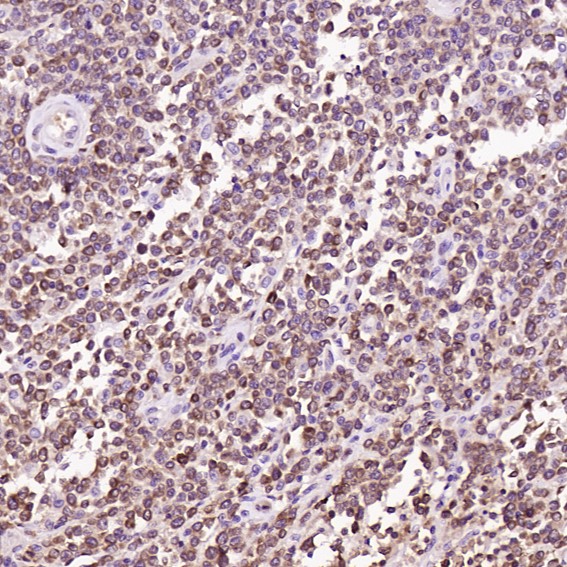
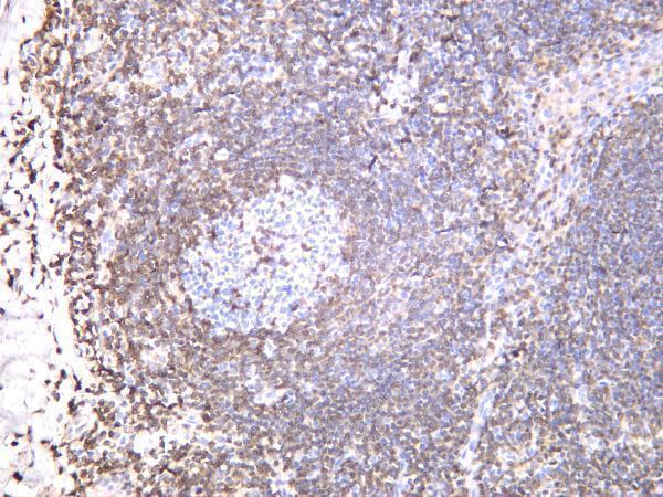
Real IHC data Immunohistochemical analysis of paraffin-embedded Human non-Hodgkin's lymphoma, using the Antibody at 1:50 dilution.
Anti-Bcl-2 Rabbit Monoclonal Antibody
Cat # M00040-1
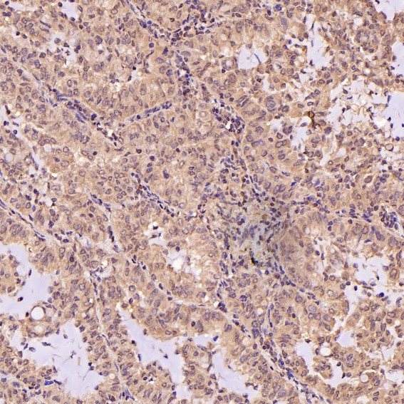
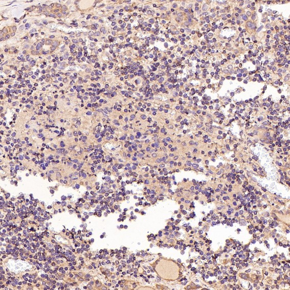
Real IHC data Immunohistochemical analysis of paraffin-embedded Human lung adenocarcinoma, using the Antibody at 1:100 dilution.
Anti-Bcl-2 Rabbit Monoclonal Antibody
Cat # M00040-2
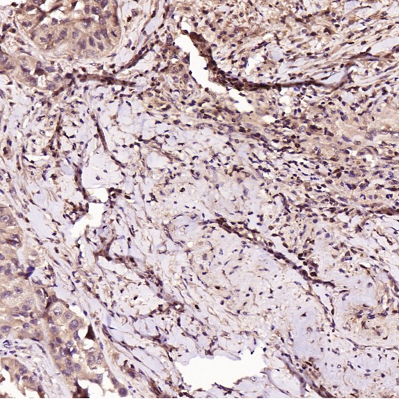
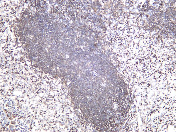
Real IHC data Human spleen was stained with anti-Bcl-2 rabbit antibody
Anti-Bcl-2 Rabbit Monoclonal Antibody
Cat # M00040-4
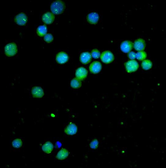
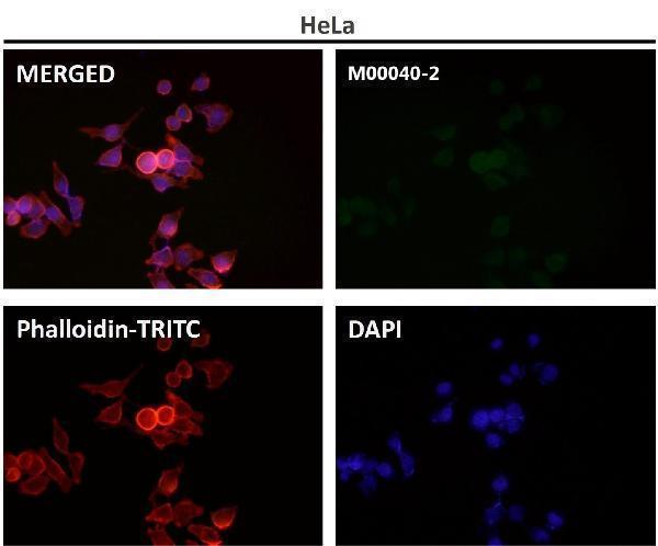
Real IF data IF analysis of BCL2 and Tubulin alpha using anti-BCL2 antibody (A00040-2) and anti-Tubulin alpha antibody (M03989-3). BCL2 and Tubulin alpha were detected in immunocytochemical section of U20S cells. Enzyme antigen retrieval was performed using IHC enzyme antigen retrieval reagent (AR0022) for 15 mins. The cells were blocked with 10% goat serum. And then incubated with 5μg/mL rabbit anti-BCL2 antibody (A00040-2) and mouse anti-Tubulin alpha Antibody (M03989-3) overnight at 4°C. Cy3 Conjugated Goat Anti-Rabbit IgG (BA1032) and DyLight®488 Conjugated Goat Anti-Mouse IgG (BA1126) were used as secondary antibody at 1:100 dilution and incubated for 30 minutes at 37°C. Visualize using a fluorescence microscope and filter sets appropriate for the label used.
Anti-Bcl-2/BCL2 Antibody ®
Cat # A00040-2

M00040-1 shows human non-Hodgkin’s lymphoma in paraffin sections at 1:50; M00040-2 shows human lung adenocarcinoma in paraffin sections at 1:100 (respective IHC image captions). M00040-4 shows human spleen by IHC, while A00040-2 shows BCL2 in U20S cells by ICC/IF at 5 μg/mL (respective image captions).

Which to pick: For paraffin-section IHC, choose M00040-1 or M00040-2: both are rabbit monoclonals with IHC applications and paraffin-section examples; the captions do not report the fixative (catalog: applications and clonality; respective IHC image captions). For ICC/IF, choose A00040-2 at 5 μg/mL; its application list includes ICC and IF but not IHC (catalog: applications and IF dilution). For work across species, consider M00040-4, listed for human, mouse and rat IHC; its card shows human spleen staining without reporting section processing or fixative (catalog: reactivity and applications; M00040-4 IHC image caption).

Each figure is that product's own IHC / IF validation image from its datasheet.

References

  1. UniProt Consortium. UniProt entry P10415 (BCL2_HUMAN, Apoptosis regulator Bcl-2).
  2. Human Protein Atlas. BCL2 tissue IHC expression (reliability: Enhanced).
  3. Human Protein Atlas. BCL2 subcellular location (ICC-IF): Localized to the mitochondria..
  4. Human Protein Atlas. BCL2 antibody validation summary (3 antibodies).
  5. Patterns of Immunohistochemical Expression of P53, BCL2, PTEN, and HER2/neu Tumor Markers in Specific Breast Cancer Lesions. Evidence-based complementary and alternative medicine : eCAM 2022 — PMC9592210.
  6. Double-hit lymphomas: clinical, morphological, immunohistochemical and cytogenetic study in a series of Brazilian patients with high-grade non-Hodgkin lymphoma. Diagnostic pathology 2017 — PMC5219810.
  7. The t(14;18) translocation is absent from endothelial and follicular dendritic cells of follicular lymphoma (FL) and shows heterogeneous presence in preserved FL mantle zones. Diagnostic pathology 2018 — PMC5930855.
  8. Clinical and Pathological Features of Double-Hit and Triple-Hit High-Grade B-Cell Lymphomas: A Retrospective Study from Three Portuguese Tertiary Centers. International journal of hematology-oncology and stem cell research 2022 — PMC9547779.
  9. PubMed PMID:3523487 — UniProt-cited evidence.
  10. PubMed PMID:1508712 — UniProt-cited evidence.
  11. PubMed PMID:2875799 — UniProt-cited evidence.