Click image to see more details
-

-
-
-
-
+13
Product Info Summary
| SKU: | A00040-2 |
|---|---|
| Size: | 100 μg/vial |
| Reactive Species: | Human |
| Host: | Rabbit |
| Application: | ELISA, Flow Cytometry, IF, ICC, WB |
Customers Who Bought This Also Bought

Product info
Product Name
Anti-Bcl-2/BCL2 Antibody Picoband®
SKU/Catalog Number
A00040-2
Size
100 μg/vial
Form
Lyophilized
Description
Boster Bio Anti-Bcl-2/BCL2 Antibody Picoband® catalog # A00040-2. Tested in ELISA, Flow Cytometry, IF, ICC, WB applications. This antibody reacts with Human. The brand Picoband indicates this is a premium antibody that guarantees superior quality, high affinity, and strong signals with minimal background in Western blot applications. Only our best-performing antibodies are designated as Picoband, ensuring unmatched performance.
Storage & Handling
Store at -20˚C for one year from date of receipt. After reconstitution, at 4˚C for one month. It can also be aliquotted and stored frozen at -20˚C for six months. Avoid repeated freeze-thaw cycles.
Cite This Product
Anti-Bcl-2/BCL2 Antibody Picoband® (Boster Biological Technology, Pleasanton CA, USA, Catalog # A00040-2)
Host
Rabbit
Contents
Each vial contains 4 mg Trehalose, 0.9 mg NaCl and 0.2 mg Na2HPO4.
Clonality
Polyclonal
Isotype
Rabbit IgG
Immunogen
E. coli-derived human Bcl-2 recombinant protein (Position: Q118-E165).
Cross-reactivity
No cross-reactivity with other proteins.
Reactive Species
A00040-2 is reactive to BCL2 in Human
Observed Molecular Weight
26 kDa
Calculated molecular weight
26.3 kDa
Background of BCL2
Immunoreactive BCL2 protein in the neoplastic cells of almost all follicular lymphomas whereas no BCL2 protein was detected in follicles affected by nonneoplastic processes or in normal lymphoid tissue. Every tumor with molecular-genetic evidence of t (14;18) translocation expressed detectable levels of BCL2 protein, regardless of whether the breakpoint was located in or at a distance from the BCL2 gene. Overexpression of BCL2 blocks the apoptotic death of a pro-B-lymphocyte cell line.
Antibody Validation
Boster validates all antibodies on WB, IHC, ICC, Immunofluorescence, and ELISA with known positive control and negative samples to ensure specificity and high affinity, including thorough antibody incubations.
Application & Images
Applications
A00040-2 is guaranteed for ELISA, Flow Cytometry, IF, ICC, WB Boster Guarantee
Assay Dilutions Recommendation
The recommendations below provide a starting point for assay optimization. The actual working concentration varies and should be decided by the user.
Western blot, 0.1-0.5μg/ml, Human
Immunocytochemistry/Immunofluorescence, 5 μg/ml, Human
Flow Cytometry (Fixed), 1-3 μg/1x106 cells, Human
Positive Control
WB: human Jurkat whole cell, human HL-60 whole cell, human THP-1 whole cell, huamn OE19 whole cell
ICC/IF: U20S cell
FCM: U20S cell
Validation Images & Assay Conditions
Click image to see more details
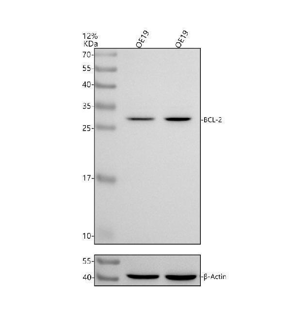
Western blot analysis of Bcl-2 using anti-Bcl-2 antibody (A00040-2).
Electrophoresis was performed on a 10% SDS-PAGE gel at 80V (Stacking gel) / 120V (Resolving gel) for 2 hours. The sample well of each lane was loaded with 30 μg of sample under reducing conditions.
Lane 1: huamn OE19 whole cell lysates,
Lane 2: human OE19 whole cell lysates.
After electrophoresis, proteins were transferred to a nitrocellulose membrane at 150 mA for 50-90 minutes. Blocked the membrane with 5% non-fat milk/TBS for 1.5 hour at RT. The membrane was incubated with rabbit anti-Bcl-2 antigen affinity purified monoclonal antibody (A00040-2) at 0.5 μg/mL overnight at 4°C, then washed with TBS-0.1%Tween-20 3 times with 5 minutes each and probed with a goat anti-rabbit IgG-HRP secondary antibody (Catalog # BA1054) at a dilution of 1:5000 for 1.5 hour at RT. The signal is developed using an ECL Plus Western Blotting Substrate (Catalog # AR1196-200) with Tanon 5200 system. A specific band was detected for Bcl-2 at approximately ~30 kDa. The expected band size for Bcl-2 is at ~26 kDa.

Click image to see more details
Western blot analysis of BCL2 using anti-BCL2 antibody (A00040-2).
Electrophoresis was performed on a 5-20% SDS-PAGE gel at 70V (Stacking gel) / 90V (Resolving gel) for 2-3 hours. The sample well of each lane was loaded with 30 ug of sample under reducing conditions.
Lane 1: human Jurkat whole cell lysates,
Lane 2: human HL-60 whole cell lysates,
Lane 3: human THP-1 whole cell lysates.
After electrophoresis, proteins were transferred to a nitrocellulose membrane at 150 mA for 50-90 minutes. Blocked the membrane with 5% non-fat milk/TBS for 1.5 hour at RT. The membrane was incubated with rabbit anti-BCL2 antigen affinity purified polyclonal antibody (Catalog # A00040-2) at 0.5 μg/mL overnight at 4°C, then washed with TBS-0.1%Tween 3 times with 5 minutes each and probed with a goat anti-rabbit IgG-HRP secondary antibody at a dilution of 1:5000 for 1.5 hour at RT. The signal is developed using an Enhanced Chemiluminescent detection (ECL) kit (Catalog # EK1002) with Tanon 5200 system. A specific band was detected for BCL2 at approximately 26 kDa. The expected band size for BCL2 is at 26 kDa.

Click image to see more details
Flow Cytometry analysis of U20S cells using anti-BCL2 antibody (A00040-2).
Overlay histogram showing U20S cells stained with A00040-2 (Blue line). To facilitate intracellular staining, cells were fixed with 4% paraformaldehyde and permeabilized with permeabilization buffer. The cells were blocked with 10% normal goat serum. And then incubated with rabbit anti-BCL2 Antibody (A00040-2, 1 μg/1x106 cells) for 30 min at 20°C. DyLight®488 conjugated goat anti-rabbit IgG (BA1127, 5-10 μg/1x106 cells) was used as secondary antibody for 30 minutes at 20°C. Isotype control antibody (Green line) was rabbit IgG (1 μg/1x106) used under the same conditions. Unlabelled sample without incubation with primary antibody and secondary antibody (Red line) was used as a blank control.

Click image to see more details
IF analysis of BCL2 and Tubulin alpha using anti-BCL2 antibody (A00040-2) and anti-Tubulin alpha antibody (M03989-3).
BCL2 and Tubulin alpha were detected in immunocytochemical section of U20S cells. Enzyme antigen retrieval was performed using IHC enzyme antigen retrieval reagent (AR0022) for 15 mins. The cells were blocked with 10% goat serum. And then incubated with 5μg/mL rabbit anti-BCL2 antibody (A00040-2) and mouse anti-Tubulin alpha Antibody (M03989-3) overnight at 4°C. Cy3 Conjugated Goat Anti-Rabbit IgG (BA1032) and DyLight®488 Conjugated Goat Anti-Mouse IgG (BA1126) were used as secondary antibody at 1:100 dilution and incubated for 30 minutes at 37°C. Visualize using a fluorescence microscope and filter sets appropriate for the label used.

Click image to see more details
Protective effects of maltol on cisplatin-induced injury in HEK293 cells. ( A ) The cytotoxic effects of cisplatin on HEK293 cells. ( B ) Effect of maltol on the activity of normal cells. ( C ) The viability of HEK293 cells incubated with maltol after cisplatin exposure. Effects of maltol on the protein expression levels of Bcl-2, Bax and caspase 3, 8, 9 as well as GAPDH protein was used as a loading control. ( D ) Cells were used for western blot analysis of indicated proteins (upper panel). Column chart represents relative protein levels compared with the control group after normalization to GAPDH levels (lower panel) Values are expressed as mean ± S.D. n = 8. ** p < 0.01 vs . normal group; # p < 0.05, ## p < 0.01 vs . cisplatin group.
Index in PubMed under a CC BY license. PMID: 30374107

Click image to see more details
Celastrol attenuates ganglion cells apoptosis in the retina of EAE rats. Treatment of celastrol decreased the number of TUNEL-positive cells (A) , upregulated expression of Bcl-2 (B) and downregulated expression of Bax, cleaved-caspase 3 and cleaved-PARP. Scale bar: 100 μm. Data were shown as mean ± SD, n = 5. ∗∗ P < 0.01 versus control group, ## P < 0.01 versus EAE group, †† P < 0.01 versus low dosage of celastrol group.
Index in PubMed under a CC BY license. PMID: 28239352

Click image to see more details
Trimetazidine inhibited EE-induced apoptosis of myocardial cells. (A) The apoptosis in myocardial tissues was evaluated by TUNEL staining. Scale bar is 50 μm. Western blot analysis of the levels of apoptosis-related proteins B-cell lymphoma 2 (Bcl-2) (B) , Bcl-2-associated X protein (Bax) (C) , cleaved caspase-3 (D) , cleaved PARP (E) , and cytoplasmic cytochrome complex (Cytochrome c, Cyto-C) (F) in myocardial tissues. GAPDH was used as a loading control. Each value is shown as mean ± SD ( n = 6). ∗ P < 0.05, ∗∗ P < 0.01, ∗∗∗ P < 0.001, versus the indicated group.
Index in PubMed under a CC BY license. PMID: 30890937

Click image to see more details
Effects of maltol on the levels of inflammation cytokines in cisplatin-induced renal toxicity. ( A ) Effects of maltol on the positive expressions of Bax, Bcl-2, iNOS and COX-2 in renal tissues were examined by IHC in renal tissues (magnification × 200), And the column chart shows stained area, semiquantitative analysis of Bax, Bcl-2, iNOS and COX-2 expression in kidneys to IHC. ( B ) Inflammation cytokines level of TNF-α, IL-1β, iNOS and NF-κB in serum of mice were measured by ELISA kits. All values were expressed as mean ± S.D. * p < 0.05, ** p < 0.01 vs . normal group; # p < 0.05, ## p < 0.01 vs . cisplatin group.
Index in PubMed under a CC BY license. PMID: 30374107

Click image to see more details
Inhibitors of ERK, JNK, and p38 MAPK reversed the effect of TRB3 overexpression on PASMC biological behavior. A The CCK-8 assay was used to evaluated the proliferation of TRB3-overexpressing cells after treating with U0126, SP600125, and SB203580. B – D Western blot analysis of PCNA expression in TRB3 overexpressing cells incubated with U0126, SP600125, or SB203580 for 12 h. E Cell apoptosis induced by U0126, SP600125 and SB203580 in TRB3-overexpressing cells was evaluated using flow cytometry, and the percentage of early apoptotic (Annexin V+/PI−) and late apoptotic (Annexin V+ /PI+) cells was analyzed. F – H Western blot analysis of the protein expression of PARP, BAX, and Bcl2 in TRB3-overexpressing cells incubated with U0126, SP600125, and SB203580 for 12 h. I Crystal violet staining is presented at × 200 magnification for the Transwell assay and the migrated cells were counted and analyzed. J – L Western blot analysis of MMP9 expression in TRB3-overexpressing cells incubated with U0126, SP600125, and SB203580 for 12 h. Data represent the mean ± SEM (n = 4). *P < 0.05 compared to normoxic PASMCs transfected with lv-NC. # P < 0.05 and ## P < 0.01 compared to PASMCs transfected with lv-TRB3. U0126, an ERK signaling inhibitor; SP600125, an JNK signaling inhibitor; SB203580, an p38 MAPK signaling inhibitor
Index in PubMed under a CC BY license. PMID: 34906150

Click image to see more details
TPX2 regulated proliferation, apoptosis, and aerobic glycolysis in glioma cells. a – l LN229 and U251 cells were introduced with si-NC or si-TPX2. a The transfection efficiency of si-TPX2 was checked with RT-qPCR assay in LN229 and U251 cells. b , c The cell viability of LN229 and U251 cells was determined with MTT assay. d The apoptosis rate of transfected LN229 and U251 cells was represented by flow cytometry assay. e The western blot assay was used to assay the expression levels of Bcl-2 and Bax in LN229 and U251 cells. f The activity of caspase-3 was detected with a caspase-3 assay kit. g – i The glucose, lactate, and ATP production levels were shown. j The protein expression levels of HK2 and LDHA were estimated by western blot assay in LN229 and U251 cells. k , l LDHA enzyme activity and ROS content were evaluated in LN229 and U251 cells post-transfection. * P < 0.05
Index in PubMed under a CC BY license. PMID: 32774168

Click image to see more details
The influences of circPOSTN silencing on proliferation, apoptosis and aerobic glycolysis of glioma cells. a – l LN229 and U251 cells were transfected with si-circPOSTN or si-NC. a The interference efficiency of si-circPOSTN was analyzed with RT-qPCR assay in LN229 and U251 cells. b , c Effect of circPOSTN silencing on the cell viability of LN229 and U251 cells was assessed with MTT assay. d The apoptosis rate was computed with flow cytometry assay in transfected LN229 and U251 cells. e The western blot assay showed the expression levels of Bcl-2 and Bax in LN229 and U251 cells. f The caspase-3 activity was measured with a caspase-3 assay kit. g – i The concentration of glucose and lactate in the culture medium, as well as ATP production level were measured with a series of kits, respectively. j The protein expression levels of HK2 and LDHA were determined with western blot assay in transfected LN229 and U251 cells. k – l LDHA enzyme activity and ROS accumulation were evaluated in LN229 and U251 cells post-transfection with lactate dehydrogenase activity detection kit and reactive oxygen species assay kit, respectively. * P < 0.05
Index in PubMed under a CC BY license. PMID: 32774168

Click image to see more details
Knockdown of circPOSTN mediated-effects on proliferation and apoptosis of glioma cells could be eliminated by silencing miR-361-5p. a – j LN229 and U251 cells were transfected with si-NC, si-circPOSTN, si-circPOSTN + anti-miR-NC, or si-circPOSTN + anti-miR-361-5p. a , b The relativity expression level of miR-361-5p was analyzed with RT-qPCR assay in LN229 and U251 cells. c , d MTT assay was administrated to assess cell viability of LN229 and U251 cells after transfection. e , f The apoptosis of transfected LN229 and U251 cells was monitored by flow cytometry. g , h The western blot assay was employed to show the expression levels of Bcl-2 and Bax in LN229 and U251 cells. i , j The caspase-3 activity was examined by caspase-3 assay kit. * P < 0.05
Index in PubMed under a CC BY license. PMID: 32774168

Click image to see more details
CircHIPK3 regulates autophagy and apoptosis via the CircHIPK3/miR-20b-5p/ATG7 axis. A Luciferase reporter assay showed that miR-20b-5p mimics directly binds to the 3′-UTR of ATG7 and inhibits luciferase activity. * P < 0.05 compared with the NC-mimics group. B ATG7 protein expression was detected by western blotting. n = 3. * P < 0.05 compared with the MNC group. # P < 0.05 compared with the INC group. C The protein expression levels of LC3-II, P62, and ATG7 were measured by western blotting. n = 3. D The intracellular ROS level was detected by flow cytometry. n = 3. E Annexin V-FITC/PI flow cytometry was used to evaluate apoptosis under different treatment conditions. n = 3. F Apoptosis-related proteins, including procaspase-3, cleaved caspase-3, Bax, and Bcl-2, were detected by western blotting. n = 3. * P < 0.05 compared with the H/R group. # P < 0.05 compared with the H/R + sicircHIPK3 group.
Index in PubMed under a CC BY license. PMID: 33824287

Click image to see more details
MiR-20b-5p inhibits autophagy and apoptosis of cardiomyocytes under H/R conditions. A Transfection efficacy of miR-20b-5p mimics and miR-20b-5p inhibitors in cardiomyocytes. B Western blot showed the effect of transfection of miR-20b-5p mimics and miR-20b-5p inhibitors on the expression of LC3II and P62 in normal cardiomyocytes. n = 3. C Western blot showed the effect of transfection of miR-20b-5p mimics and miR-20b-5p inhibitors on the expression of apoptosis-related proteins, including procaspase-3, cleaved caspase-3, Bax, and Bcl-2 in normal cardiomyocytes. n = 3. D Western blot showed the effects of miR-20b-5p mimics and miR-20b-5p inhibitor transfection on the expression of LC3II and P62 in cardiomyocytes under H/R conditions. n = 3. E Annexin V-FITC/PI flow cytometry was used to evaluate the effect of miR-20b-5p mimics and miR-20b-5p inhibitors on cardiomyocyte apoptosis under H/R conditions. n = 3. F Western blot analyzed the expression of apoptosis-related proteins, including procaspase-3, cleaved caspase-3, Bax, and Bcl-2 in cardiomyocytes under H/R conditions by miR-20b-5p mimics and miR-20b-5p inhibitors transfection. n = 3. * P < 0.05 compared with the mimics-NC (MNC) group. # P < 0.05 compared with the inhibitors-NC (INC) group.
Index in PubMed under a CC BY license. PMID: 33824287

Click image to see more details
CircHIPK3 promotes H/R-induced cardiomyocyte apoptosis. A The intracellular ROS level was detected by flow cytometry. n = 3. B Annexin V-FITC/PI flow cytometry was used to evaluate the effect of circHIPK3 on cardiomyocyte apoptosis. n = 3. C Apoptosis-related proteins, including procaspase-3, cleaved caspase-3, Bax, and Bcl-2, were detected by western blotting. n = 3. * P < 0.05 compared with the normal group. # P < 0.05 compared with the H/R group.
Index in PubMed under a CC BY license. PMID: 33824287

Click image to see more details
The effect of the combination of alteronol and ADM on protein levels of apoptosis-related molecules in 4T1 cells. (A) The protein levels of Bax and Bcl-2 were measured by western blot. (B) Quantitative analysis of Bax and Bcl-2 protein levels in 4T1 cells after treatment with alteronol and/or ADM. (C) The protein levels of cleaved PARP, cleaved caspase-9, and cleaved caspase-3 were examined by western blot. (D) Quantitative analysis of cleaved PARP, cleaved caspase-9, and cleaved caspase-3 protein levels after the indicated treatments. ∗ P < 0.05, ∗∗ P < 0.01 vs. control group. # P < 0.05, ## P < 0.01 vs. alteronol group. & P < 0.05, && P < 0.01 vs. ADM group. All data are expressed as mean ± SD of three independent experiments.
Index in PubMed under a CC BY license. PMID: 31001113

Click image to see more details
Effect of TRB3 knockdown on proliferation, migration, and apoptosis of hypoxic PASMCs. PASMCs were transfected with lentiviral vectors encoding short hairpin RNAs targeting against rat TRB3(si-TRB3) or negative control(si-NC) for further analysis. A CCK-8 assays were performed to test cell viability after TRB3 knockdown in hypoxic PASMCs. B The EdU proliferation assay was used to measure cell proliferation. All images are × 400 magnification. C EdU‐positive cells were analyzed. D and E The expression of PCNA and Survivin was examined by western blot, and the gray value was quantified by ImageJ software. F Cell apoptosis was evaluated by Hoechst 33342 staining, and the percentage of apoptosis was quantified. G Crystal violet staining of PASMCs is presented at × 200 magnification for the Transwell assay, and the migrated cells were counted manually. H Apoptosis-related protein expression was evaluated and quantification of the analysis is shown. I The expression of MMP9 was evaluated. Data represent the mean ± SEM (n = 4). *P < 0.05 and **P < 0.01 compared to normoxic PASMCs transfected with si-NC. # P < 0.05 and ## P < 0.01 compared to hypoxic PASMCs transfected with si-TRB3. si-NC, lentiviral vectors encoding the negative control sequence; si-TRB3, lentiviral vectors encoding the short-hairpin RNAs against rat TRB3; PCNA, proliferating cell nuclear antigen; TRB3, Tribbles homolog 3; BAX, BCL2 associated X; Bcl2, B cell leukemia/lymphoma 2; MMP9, matrix metallopeptidase 9
Index in PubMed under a CC BY license. PMID: 34906150
Specific Publications For Anti-Bcl-2/BCL2 Antibody Picoband® (A00040-2)
Loading publications
Recommended Resources
Here are featured tools and databases that you might find useful.
- Boster's Pathways Library
- Protein Databases
- Bioscience Research Protocol Resources
- Data Processing & Analysis Software
- Photo Editing Software
- Scientific Literature Resources
- Research Paper Management Tools
- Molecular Biology Software
- Primer Design Tools
- Bioinformatics Tools
- Phylogenetic Tree Analysis
Customer Reviews
Have you used Anti-Bcl-2/BCL2 Antibody Picoband®?
Share your experimental results or join a short interview to earn up to $1,000 in product credits or other rewards.
0 Reviews For Anti-Bcl-2/BCL2 Antibody Picoband®
Customer Q&As
Have a question?
Find answers in Q&As, reviews.
Can't find your answer?
Submit your question
16 Customer Q&As for Anti-Bcl-2/BCL2 Antibody Picoband®
Question
Is a blocking peptide available for product anti-Bcl-2/BCL2 antibody (A00040-2)?
Verified Customer
Verified customer
Asked: 2020-04-21
Answer
We do provide the blocking peptide for product anti-Bcl-2/BCL2 antibody (A00040-2). If you would like to place an order for it please contact support@bosterbio.com and make a special request.
Boster Scientific Support
Answered: 2020-04-21
Question
We have tried in the past anti-Bcl-2/BCL2 antibody for IHC on testis in a previous project. I am using rat, and We intend to use the antibody for ELISA next. My question regards examining testis as well as thyroid gland in our next experiment. Do you have any suggestion on which antibody would work the best for ELISA?
Verified Customer
Verified customer
Asked: 2019-12-19
Answer
I viewed the website and datasheets of our anti-Bcl-2/BCL2 antibody and it appears that A00040-2 has been validated on rat in both IHC and ELISA. Thus A00040-2 should work for your application. Our Boster satisfaction guarantee will cover this product for ELISA in rat even if the specific tissue type has not been validated. We do have a comprehensive range of products for ELISA detection and you can check out our website bosterbio.com to find out more information about them.
Boster Scientific Support
Answered: 2019-12-19
Question
We were well pleased with the WB result of your anti-Bcl-2/BCL2 antibody. However we have observed positive staining in testis mitochondrion outer membrane using this antibody. Is that expected? Could you tell me where is BCL2 supposed to be expressed?
Verified Customer
Verified customer
Asked: 2019-12-19
Answer
Based on literature, testis does express BCL2. Generally BCL2 expresses in mitochondrion outer membrane. Regarding which tissues have BCL2 expression, here are a few articles citing expression in various tissues:
Testis, Pubmed ID: 15489334
Boster Scientific Support
Answered: 2019-12-19
Question
I am interested in to test anti-Bcl-2/BCL2 antibody A00040-2 on rat thyroid gland for research purposes, then I may be interested in using anti-Bcl-2/BCL2 antibody A00040-2 for diagnostic purposes as well. Is the antibody suitable for diagnostic purposes?
Verified Customer
Verified customer
Asked: 2019-10-15
Answer
The products we sell, including anti-Bcl-2/BCL2 antibody A00040-2, are only intended for research use. They would not be suitable for use in diagnostic work. If you have the means to develop a product into diagnostic use, and are interested in collaborating with us and develop our product into an IVD product, please contact us for more discussions.
Boster Scientific Support
Answered: 2019-10-15
Question
I was wanting to use your anti-Bcl-2/BCL2 antibody for ELISA for rat thyroid gland on frozen tissues, but I want to know if it has been validated for this particular application. Has this antibody been validated and is this antibody a good choice for rat thyroid gland identification?
Verified Customer
Verified customer
Asked: 2019-08-06
Answer
You can see on the product datasheet, A00040-2 anti-Bcl-2/BCL2 antibody has been validated for ELISA, IHC, WB on human, mouse, rat tissues. We have an innovator award program that if you test this antibody and show it works in rat thyroid gland in IHC-frozen, you can get your next antibody for free.
Boster Scientific Support
Answered: 2019-08-06
Question
We are currently using anti-Bcl-2/BCL2 antibody A00040-2 for rat tissue, and we are happy with the ELISA results. The species of reactivity given in the datasheet says human, mouse, rat. Is it likely that the antibody can work on primate tissues as well?
Verified Customer
Verified customer
Asked: 2019-07-03
Answer
The anti-Bcl-2/BCL2 antibody (A00040-2) has not been tested for cross reactivity specifically with primate tissues, though there is a good chance of cross reactivity. We have an innovator award program that if you test this antibody and show it works in primate you can get your next antibody for free. Please contact me if I can help you with anything.
Boster Scientific Support
Answered: 2019-07-03
Question
I am interested in using your anti-Bcl-2/BCL2 antibody for post-embryonic development studies. Has this antibody been tested with western blotting on small intestine tissue? We would like to see some validation images before ordering.
Verified Customer
Verified customer
Asked: 2019-06-28
Answer
Thanks for your inquiry. This A00040-2 anti-Bcl-2/BCL2 antibody is validated on small intestine tissue. It is guaranteed to work for ELISA, IHC, WB in human, mouse, rat. Our Boster guarantee will cover your intended experiment even if the sample type has not been be directly tested.
Boster Scientific Support
Answered: 2019-06-28
Question
I have a question about product A00040-2, anti-Bcl-2/BCL2 antibody. I was wondering if it would be possible to conjugate this antibody with biotin. I would need it to be without BSA or sodium azide. I am planning on using a buffer exchange of sodium azide with PBS only. Would there be problems for me to conjugate the antibody and store it in -20 degrees in small aliquots?
G. Carter
Verified customer
Asked: 2019-04-22
Answer
We do not recommend storing this antibody with PBS buffer only in -20 degrees. If you want to store it in -20 degrees it is best to add some cryoprotectant like glycerol. If you want carrier free A00040-2 anti-Bcl-2/BCL2 antibody, we can provide it to you in a special formula with trehalose and/or glycerol. These molecules will not interfere with conjugation chemistry and provide a good level of protection for the antibody from degradation. Please be sure to specify this in your purchase order.
Boster Scientific Support
Answered: 2019-04-22
Question
Is there a BSA free version of anti-Bcl-2/BCL2 antibody A00040-2 available?
Verified Customer
Verified customer
Asked: 2018-05-16
Answer
Thank you for your recent telephone inquiry. I can confirm that some lots of this anti-Bcl-2/BCL2 antibody A00040-2 are BSA free. For now, these lots are available and we can make a BSA free formula for you free of charge. It will take 3 extra days to prepare. If you require this antibody BSA free again in future, please do not hesitate to contact me and I will be pleased to check which lots we have in stock that are BSA free.
Boster Scientific Support
Answered: 2018-05-16
Question
Will anti-Bcl-2/BCL2 antibody A00040-2 work for ELISA with thyroid gland?
Verified Customer
Verified customer
Asked: 2017-08-01
Answer
According to the expression profile of thyroid gland, BCL2 is highly expressed in thyroid gland. So, it is likely that anti-Bcl-2/BCL2 antibody A00040-2 will work for ELISA with thyroid gland.
Boster Scientific Support
Answered: 2017-08-01
Question
Thanks for helping with my inquiry over the phone. Here are the WB image, lot number and protocol we used for thyroid gland using anti-Bcl-2/BCL2 antibody A00040-2. Let me know if you need anything else.
L. Williams
Verified customer
Asked: 2016-05-10
Answer
We appreciate the data. You have provided everything we needed. Our lab team are working to resolve your inquiry as quickly as possible, and we appreciate your patience and understanding! Please let me know if there is anything you need in the meantime.
Boster Scientific Support
Answered: 2016-05-10
Question
I see that the anti-Bcl-2/BCL2 antibody A00040-2 works with ELISA, what is the protocol used to produce the result images on the product page?
Z. Jackson
Verified customer
Asked: 2016-01-19
Answer
You can find protocols for ELISA on the "support/technical resources" section of our navigation menu. If you have any further questions, please send an email to support@bosterbio.com
Boster Scientific Support
Answered: 2016-01-19
Question
Is this A00040-2 anti-Bcl-2/BCL2 antibody reactive to the isotypes of BCL2?
N. Dhar
Verified customer
Asked: 2015-09-28
Answer
The immunogen of A00040-2 anti-Bcl-2/BCL2 antibody is E. coli-derived human Bcl-2 recombinant protein (Position: Q118-E165). Could you tell me which isotype you are interested in so I can help see if the immunogen is part of this isotype?
Boster Scientific Support
Answered: 2015-09-28
Question
I have attached the WB image, lot number and protocol we used for thyroid gland using anti-Bcl-2/BCL2 antibody A00040-2. Please let me know if you require anything else.
G. Banerjee
Verified customer
Asked: 2015-05-05
Answer
Thank you very much for the data. Our lab team are working to resolve this as quickly as possible, and we appreciate your patience and understanding! You have provided everything we needed. Please let me know if there is anything you need in the meantime.
Boster Scientific Support
Answered: 2015-05-05
Question
Does A00040-2 anti-Bcl-2/BCL2 antibody work on parafin embedded sections? If so, which fixation method do you recommend we use (PFA, paraformaldehyde, other)?
M. Jha
Verified customer
Asked: 2014-03-07
Answer
As indicated on the product datasheet, A00040-2 anti-Bcl-2/BCL2 antibody as been tested on ELISA. It is best to use PFA for fixation because it has better tissue penetration ability. PFA needs to be prepared fresh before use. Long term stored PFA turns into formalin, as the PFA molecules congregate and become formalin.
Boster Scientific Support
Answered: 2014-03-07
Question
We have observed staining in mouse testis. Are there any suggestions? Is anti-Bcl-2/BCL2 antibody supposed to stain testis positively?
S. Collins
Verified customer
Asked: 2013-06-14
Answer
Based on literature testis does express BCL2. Based on Uniprot.org, BCL2 is expressed in thyroid gland, testis, among other tissues. Regarding which tissues have BCL2 expression, here are a few articles citing expression in various tissues:
Testis, Pubmed ID: 15489334
Boster Scientific Support
Answered: 2013-06-14
