This website uses cookies to ensure you get the best experience on our website.


- Table of Contents
Plan chromogenic BIRC6 IHC in paraffin sections around the cytoplasmic tissue pattern (HPA tissue IHC). Compare high-staining colon glandular cells with squamous epithelial cells reported as undetected in esophagus, while accounting for medium consistency between antibody staining and RNA expression (HPA tissue IHC).

Expected localisation, validated protocols, controls and antibodies — the at-a-glance facts below, then the full design guide.
| Expected localisation | Cytoplasmic staining in most tissues (HPA tissue IHC) | |
| Staining pattern | Cytoplasmic staining across many tissue cell types (HPA tissue IHC) | |
| Antigen retrieval | Citrate pH 6.0 HIER, 95–98 °C, 20 min (rule: cytoplasmic / membrane antigen) | |
| Positive control | Adrenal gland+4 more · see all | |
| Negative control | Adipose tissue+4 more · see all |
| Fixation | Keep fixation consistent across sections. (standard IHC practice; not target-specific) | |
| Caveat | Antibody staining and RNA show medium consistency (HPA tissue IHC) | |
| Regulation | Staining varies by cell type (HPA tissue IHC) | |
| Isoform / epitope | No annotated isoforms; one chain with no transmembrane segment (UniProt) |
Compare the catalog antibody’s IHC-P protocol (datasheet) with four published BIRC6 IHC protocols (PMC10706016; PMC4196171; PMC4416929; PMC7317768).
| Sample | Tissue sections; selected-image fixative not specified (standard IHC workflow) |
| Fixation | Image fixative and duration unreported (datasheet A01773); verify before use. |
| Sectioning | 4–5 µm sections on charged slides (standard) |
| Deparaffinisation | Xylene, graded ethanol series to water (standard) |
| Antigen retrieval | Heat-induced epitope retrieval in citrate buffer, pH 6.0, 20 min at 95–98 °C (standard rule: cytoplasmic / membrane antigen) |
| Peroxidase block | 3% H2O2, 10 min, room temperature (standard) |
| Blocking | 10% normal serum of the secondary host, 30 min, room temperature (standard) |
| Primary antibody | Rabbit anti-BIRC6, 5 μg/mL (datasheet A01773) |
| Primary incubation | Overnight at 4 °C (standard) |
| Detection | HRP-polymer secondary, DAB chromogen 5–10 min (standard) |
| Counterstain | Hematoxylin, blue, dehydrate and mount (standard) |
| Expected result | BIRC6-positive staining in glandular cells of adrenal gland (HPA tissue IHC: High). HPA tissue profile: Cytoplasmic expression in most tissues. No signal in the no-primary control. |
BIRC6 staining is expected mainly in the cytoplasm of many tissue cell types, with high staining reported in selected glandular, respiratory epithelial, and trophoblastic cells (HPA: tissue IHC). Its Golgi and endosomal associations and cell cycle dependent redistribution can shape localisation (UniProt Q9NR09: subcellular location). BIRC6 has no transmembrane segment (UniProt Q9NR09: topology). HPA rates tissue IHC reliability Approved, with medium consistency between staining and RNA data (HPA: tissue IHC).
| Cytoplasmic staining in colon glandular cells or bronchial respiratory epithelial cells. | This matches reported high staining in those cell types (HPA: tissue IHC). Compare cells within the section; high describes the HPA observation, not a required intensity for every run (HPA: tissue IHC). |
| A sharply nuclear dominant pattern, with little cytoplasmic staining. | Treat this as discordant with the reported cytoplasmic tissue pattern (HPA: tissue IHC) and Golgi, endosomal, and cytoskeletal locations (UniProt Q9NR09: subcellular location). Check whether counterstain or nonspecific detection is being mistaken for target signal (general IHC practice). |
| Strong staining confined to adipocytes, while expected positive glandular cells are unstained. | Adipocytes are reported as not detected, whereas colon glandular cells are high (HPA: tissue IHC). Consider cross-reactivity or endogenous chromogen generating activity, but first verify tissue identity and assay controls (general IHC practice). |
| Uniform haze over cells, stroma, and empty areas. | A field wide deposit is difficult to reconcile with the reported cell associated cytoplasmic pattern (HPA: tissue IHC). It suggests nonspecific background or detection reagent deposition; assess a no primary control (general IHC practice). |
| No staining in a section expected to contain high expressing cells. | Absence in colon glandular cells or placental trophoblastic cells conflicts with their reported high staining (HPA: tissue IHC). Check section quality, retrieval, antibody application, and detection before concluding that the sample lacks BIRC6 (general IHC practice). |
| Which tissue and cell type should anchor interpretation? | Use a reported high group such as colon glandular cells or bronchial respiratory epithelial cells (HPA: tissue IHC). A negative comparator must match the specified cell type: adipocytes, not every cell in adipose tissue, are listed as not detected (HPA: tissue IHC). |
| How much confidence does tissue IHC validation provide? | Tissue staining is Approved but has medium consistency with RNA data (HPA: tissue IHC). HPA074409 is IHC Approved; HPA074738 has ICC approval but no IHC status in the supplied record (HPA: antibodies). Treat unexpected patterns as findings to verify, not proof of a new localisation. |
| Does subcellular location vary through division? | BIRC6 is associated with a pericentriolar compartment in interphase, spindle poles in metaphase, and the midbody ring during cytokinesis (UniProt Q9NR09: subcellular location). A rare division associated focus may therefore be compatible with the record; score the prevalent cytoplasmic pattern separately. |
| Does membrane topology imply surface staining? | No transmembrane segment is reported (UniProt Q9NR09: topology). Its trans Golgi network membrane and endosomal localisation does not establish plasma membrane staining as the expected tissue IHC pattern (UniProt Q9NR09: subcellular location; HPA: tissue IHC). |
| IF/ICC: what pattern should appear? | Golgi apparatus and vesicles are the supported main locations, with an additional supported mitotic spindle location (HPA: subcellular ICC-IF). These resolved IF structures provide context for localisation; routine chromogenic sections are assessed against the broader cytoplasmic tissue pattern (HPA: tissue IHC). |
| What is known about antigen retrieval sensitivity? | No target specific retrieval or fixation effect is established by the supplied UniProt and HPA records. Retrieval is a general paraffin IHC optimisation variable; compare conditions with tissue and reagent controls before attributing a weak result to BIRC6 epitope masking (general IHC practice). |
| Situation | Likely cause | Next action |
|---|---|---|
| A reported high cell type is blank. | The result conflicts with the reported high staining in colon glandular cells or placental trophoblastic cells (HPA: tissue IHC); assay failure is one possibility (general IHC practice). | Confirm the intended cells are present, then review primary antibody use, retrieval, chromogen development, and a concurrently stained positive control (general IHC practice). |
| All sections show uniform brown background. | Diffuse deposition is inconsistent with the reported cytoplasmic cell pattern (HPA: tissue IHC); nonspecific binding or detection background may contribute (general IHC practice). | Inspect a no primary control, blocking and washes, and the detection reagents. Adjust those general IHC conditions before scoring cellular staining (general IHC practice). |
| Signal appears only where endogenous detection activity is likely. | Endogenous activity can create chromogenic signal independent of primary antibody binding (general IHC practice). Such signal cannot by itself establish BIRC6 expression. | Run a no primary control and apply the detection system's appropriate endogenous activity block; then reassess cell specific cytoplasmic staining (general IHC practice). |
| Staining is nuclear dominant. | That pattern differs from HPA's cytoplasmic tissue profile (HPA: tissue IHC) and the supplied UniProt locations (UniProt Q9NR09: subcellular location). | Check counterstain and no primary controls, then compare with a reported high cytoplasmic cell type before accepting the signal as BIRC6 (HPA: tissue IHC; general IHC practice). |
| A low staining tissue is used as the sole negative control. | Thyroid glandular cells and duodenal glandular cells are listed as low, while BIRC6 has low tissue specificity overall (HPA: tissue IHC). Weak signal there need not indicate assay background. | Use a cell type reported as not detected, such as adipocytes, and retain a no primary control to distinguish tissue signal from reagent background (HPA: tissue IHC; general IHC practice). |
| An ICC approved antibody is assumed suitable for paraffin IHC. | HPA074738 has ICC approval but no IHC status in the supplied antibody record; HPA074409 has IHC approval (HPA: antibodies). | Base paraffin tissue expectations on the IHC approved antibody and the HPA tissue pattern. Do not transfer ICC validation or its resolved Golgi and vesicle appearance into an IHC performance claim (HPA: antibodies; HPA: tissue IHC; HPA: subcellular ICC-IF). |
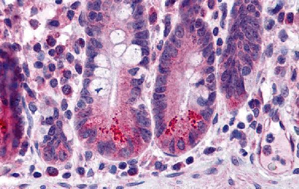
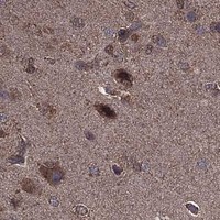


Comprehensive Human Protein Atlas IHC scoring per tissue (reliability: Approved — Medium consistency between antibody staining and RNA expression data.). Rows are taken directly from the HPA tissue chart — click any row's HPA link to view the source.
| Tissue | Cell type | Level | Evidence | Source |
|---|---|---|---|---|
| Adrenal gland | Glandular cells | High | Protein (IHC) | HPA → |
| Breast | Glandular cells | High | Protein (IHC) | HPA → |
| Bronchus | Respiratory epithelial cells | High | Protein (IHC) | HPA → |
| Colon | Glandular cells | High | Protein (IHC) | HPA → |
| Fallopian tube | Glandular cells | High | Protein (IHC) | HPA → |
| Tissue | Cell type | Level | Evidence | Source |
|---|---|---|---|---|
| Adipose tissue | Adipocytes | Not detected | Protein (IHC) | HPA → |
| Caudate | Glial cells | Not detected | Protein (IHC) | HPA → |
| Esophagus | Squamous epithelial cells | Not detected | Protein (IHC) | HPA → |
| Lymph node | Non-germinal center cells | Not detected | Protein (IHC) | HPA → |
| Oral mucosa | Squamous epithelial cells | Not detected | Protein (IHC) | HPA → |
Troubleshoot BIRC6 staining in paraffin sections by checking retrieval, tissue controls, compartment patterns, and scoring before interpreting chromogenic signal.
The IHC-validated BIRC6 antibody has a human small-intestine IHC image (A01773 image caption). IF/ICC data are not listed (catalog: applications).
A01773 is listed for human IHC-P (catalog: applications and reactivity). Its own image shows human small-intestine staining at 5 μg/mL (A01773 image caption).
Which to pick: Choose A01773, a rabbit polyclonal antibody, for human paraffin-section IHC (catalog: host, clonality, reactivity, IHC-P; A01773 image caption: human small intestine). Its image caption does not report the fixative (A01773 image caption). No IF/ICC or cross-species option is listed (catalog: A01773 applications and human-only reactivity).