This website uses cookies to ensure you get the best experience on our website.
- Table of Contents
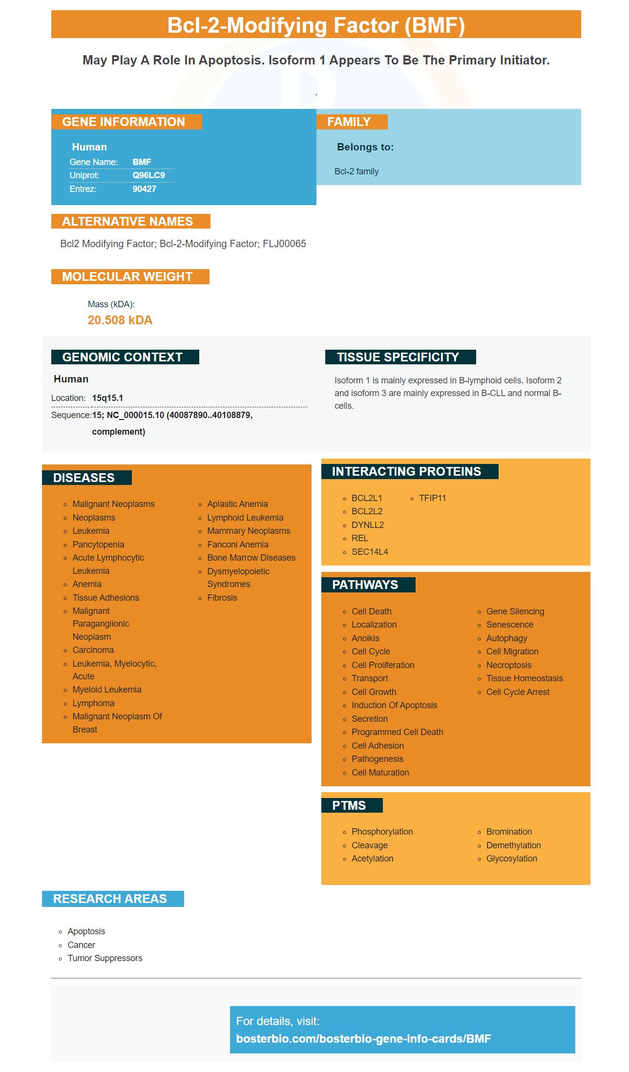
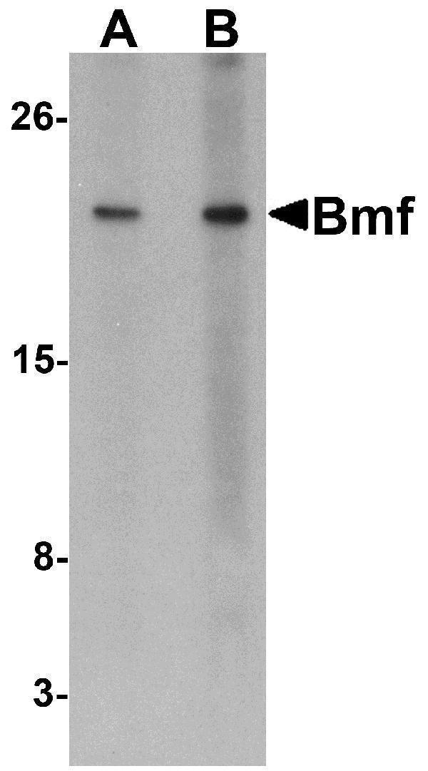
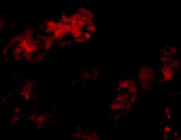
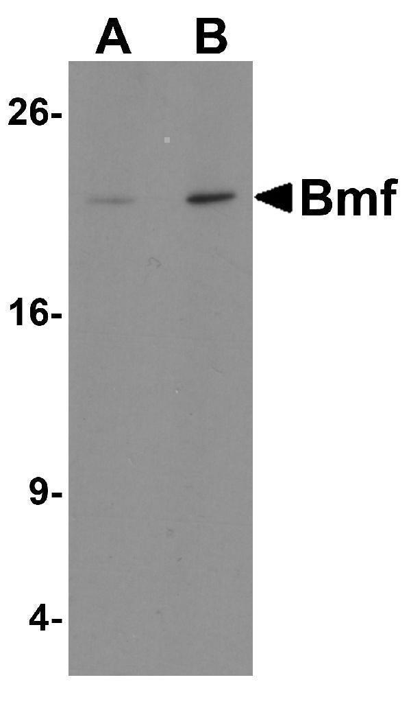
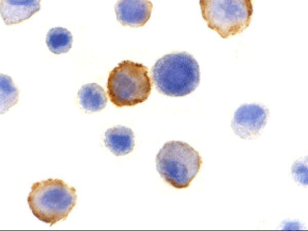
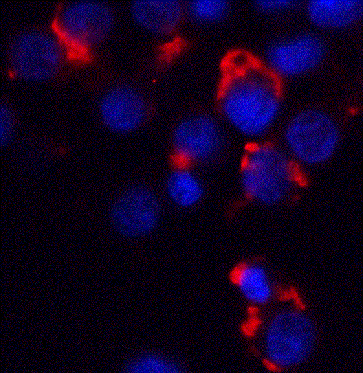
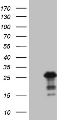
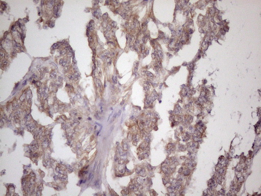
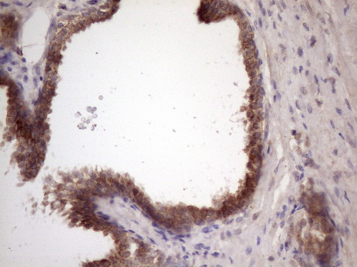
Facts about Bcl-2-modifying factor.
.
Human | |
---|---|
Gene Name: | BMF |
Uniprot: | Q96LC9 |
Entrez: | 90427 |
Belongs to: |
---|
Bcl-2 family |
Bcl2 modifying factor; bcl-2-modifying factor; FLJ00065
Mass (kDA):
20.508 kDA
Human | |
---|---|
Location: | 15q15.1 |
Sequence: | 15; NC_000015.10 (40087890..40108879, complement) |
Isoform 1 is mainly expressed in B-lymphoid cells. Isoform 2 and isoform 3 are mainly expressed in B-CLL and normal B-cells.
The Boster Bio BMF Marker - This is a useful tool for genetic analysis. This marker was developed by Steven Boster (a biotechnology veteran) and can detect mutations in the BMF Protein. It can also induce phenotypic change. The BMF marker is validated by Western blotting and immunohistochemistry. Boster bio is a marker that can be used to identify the benefits and uses of this marker.
The BMF/MDS gene sequence assay covers all exons, and twenty base pairs with intronic sequence that flank each exon. The assay also sequences the regulatory and intronic regions of the gene. The median coverage of the 383kb targeted area was 1887X for 81 patients who were referred between June 2015 and July 2016. This coverage was sufficient to identify all classes mutations.
The assay is able to detect all classes of mutations, and copy number variations. In the study, the assay accurately identified all classes of mutations, including copy number variants and genomic rearrangements. Blinded analysis of genomic data from 14 patients with known and unknown mutations validated the assay. It identified copy number variants, as well other abnormalities in subsets of patients.
BMF/MDS patients can be analyzed genomically to reveal previously unknown phenotypes and genes. This information can be used to aid in the management of BMF/MDS cases. It is possible to find additional genes responsible for the disease by sequencing whole exomes and genomes. It is important to remember that only four of the patients with constitutional damaging mutations had a positive family history. Research is ongoing.
In the current study, we have demonstrated that boster bio's BMF marker induces a phenotypic change in the cell line GC-h2. We used tissue microarrays that contain 17 primary GC tissues, 12 adjacent normal tissues, 16 metastatic lesions, and five normal gastric mucosa tissues. The BMF marker induces cell phenotypic changes by detecting specific proteins being expressed in these cells.
Generally, antibodies are validated on Western blotting by demonstrating specificity and selectivity in complex samples. Validated antibodies have high band intensities in the overexpressed cell line and low or no bands in the control. This indicates that they are specific. Experiments must be reported in detail to minimize this error. Many peer-reviewed journals encourage detailed reporting. This article will cover the validation of antibodies using Western blotting.
This study was carried out using the XCell Surelock Electrophoresis Device and iBlot Dry Blotting Machine from Invitrogen. The lysates were probed with an alpha (a)-tubulin mouse monoclonal antibody, Cat. No. 236-10501, and a goat anti-mouse HRP conjugate. The standard curve used for computing the protein concentration was calculated by assaying diluted human recombinant peptides in the same westernblotting membrane.
PTM analyses require validation of antibody specificity. Cross-reactive blots must be removed from Westernblotting because modified and unmodified versions of a target can often migrate together. This is to avoid incorrect interpretations. PTM-directed antibodies should be specific to the modification and not cross-reacting with the unmodified target. If this is true, the antibody must be validated using a primary or secondary antibody.
It is possible to also test the efficiency of the transfer by protein staining. It can confirm if the protein has moved to the membrane, but does not guarantee that the antibodies have been bound. The stain should be removed. Ponceau S Stain is the most common stain. However it is very sensitive and difficult to photograph. It also ages quickly so documentation is difficult.
Steven Boster founded the Boster Biotech Company. He was known as "he who converts scientific research in the lavatory." He has also developed hundreds of primary antibodies, and the PicoKine(tm), an ELISA platform. To create high-sensitivity ELISA kit kits, he uses trade secrets. In this article, we will discuss the BMF marker's uses in immunohistochemistry.
Boster Bio, an antibody manufacturer, specializes in picogram sensitive ELISA kits and IHC optimized polyclonal antibodies. Boster Bio has been in operation since 1993 and focuses on the development of antibodies that have high specificity and sensitivity. Its products were validated for IHC/WB and Flow cytometry. They are cited more than 29,000 times in scientific publications. Boster Bio offers a variety of antibodies in a variety format including WB and ELISA as well as Flow cytometry.
ELISA is a convenient, fast, and accurate method for measuring specific analytes. It uses high-affinity antibodies which can wash out non-specific binding materials. Boster Bio has been a leader in the industry's development of high-affinity antibacterial coatings for ELISA plates. This makes them the gold standard for biomedical and scientific research. Scientists worldwide trust Boster Bio's BMF marker.
ELISA is performed using a microplate containing an antigen and a capture antibodies. Antibodies are used to detect protein molecules in a target sample. ELISA tests use reagents that are immobilized on polystyrene micelles. To avoid false positives, the microplates should have a binding capacity of 400 ng/cm2. ELISAs can either be performed with one or more types of signals. Matching pairs is recommended.
Boster Bio's antibody labeled with biotinylated antibodies was used to measure the MPO/NAG activities in an ELISA. The results of these experiments were expressed as a percentage of the total antibody load. This means that the BMF markers can provide highly reproducible results for your research. This is the best way to determine if a protein is present.
PMID: 11546872 by Puthalakath H., et al. Bmf: a proapoptotic BH3-only protein regulated by interaction with the myosin V actin motor complex, activated by anoikis.
PMID: 14574334 by Morales A.A., et al. Expression and transcriptional regulation of functionally distinct Bmf isoforms in B-chronic lymphocytic leukemia cells.