This website uses cookies to ensure you get the best experience on our website.


- Table of Contents
Real validated BMP6 Western blot protocols, expected-band and isoform facts, troubleshooting for weak or shifted signal, and recommended anti-BMP6 WB antibodies. Everything you need to plan the experiment before you commit precious samples.
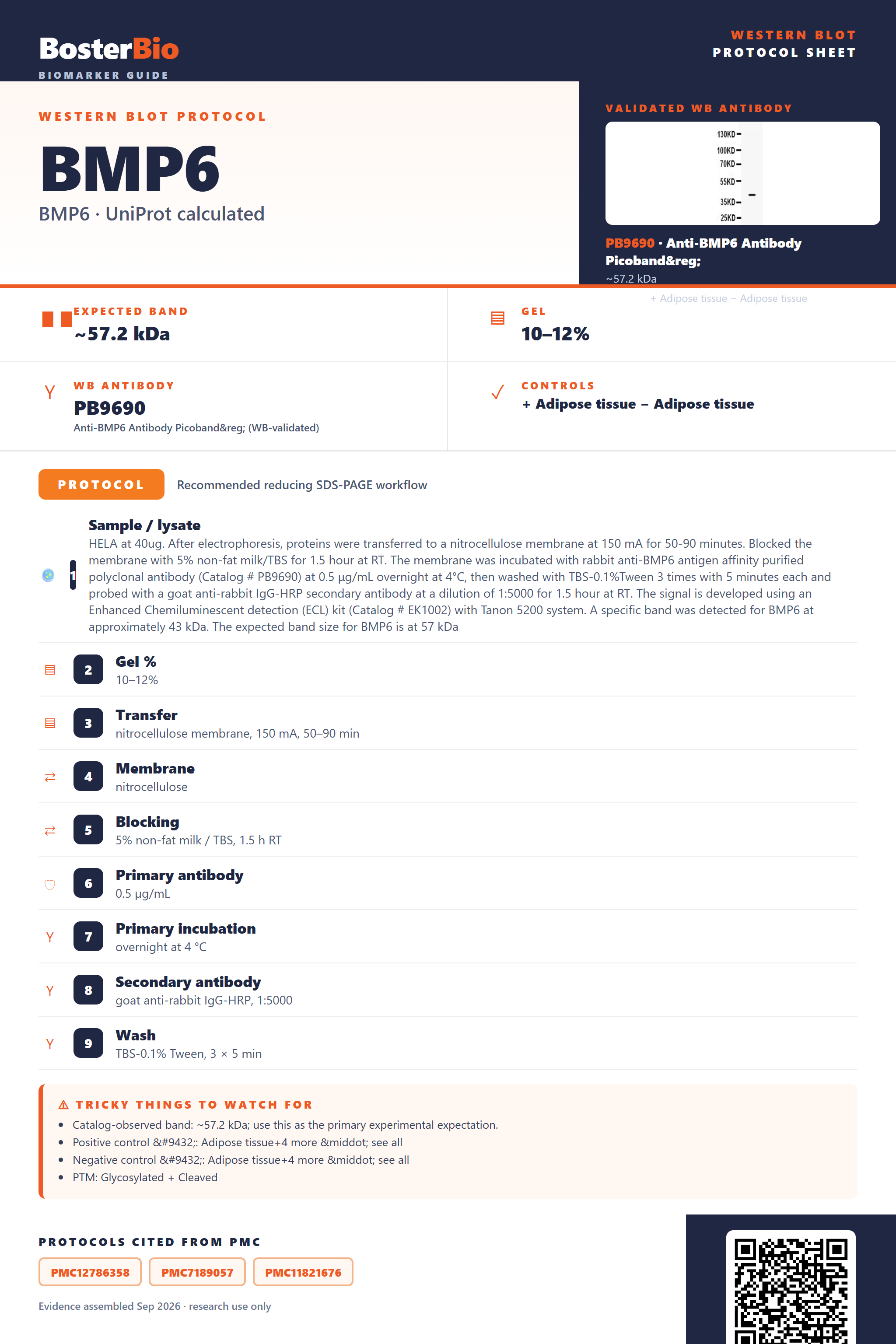
Expected bands, validated protocols, controls and antibodies — the at-a-glance facts below, then the full design guide.
| Expected band | ~57.2 kDa | |
| Observed band | 43 kDa | |
| Gel | 10–12% | |
| Positive control | Adipose tissue+4 more · see all | |
| Negative control | Adipose tissue+4 more · see all |
| PTM | Glycosylated + Cleaved | |
| Caveat | Partial propeptide cleavage | |
| Regulation | Bile acid metabolism | |
| Isoform | 1 isoform(s) |
Literature-validated Western blot parameters for BMP6 — gel percentage, transfer, blocking, antibody incubation and detection, extracted from published methods.
| Sample / lysate | HELA at 40ug. After electrophoresis, proteins were transferred to a nitrocellulose membrane at 150 mA for 50-90 minutes. Blocked the membrane with 5% non-fat milk/TBS for 1.5 hour at RT. The membrane was incubated with rabbit anti-BMP6 antigen affinity purified polyclonal antibody (Catalog # PB9690) at 0.5 μg/mL overnight at 4°C, then washed with TBS-0.1%Tween 3 times with 5 minutes each and probed with a goat anti-rabbit IgG-HRP secondary antibody at a dilution of 1:5000 for 1.5 hour at RT. The signal is developed using an Enhanced Chemiluminescent detection (ECL) kit (Catalog # EK1002) with Tanon 5200 system. A specific band was detected for BMP6 at approximately 43 kDa. The expected band size for BMP6 is at 57 kDa |
| Gel % | 10–12% |
| Transfer | nitrocellulose membrane, 150 mA, 50–90 min |
| Membrane | nitrocellulose |
| Blocking | 5% non-fat milk / TBS, 1.5 h RT |
| Primary antibody | 0.5 µg/mL |
| Primary incubation | overnight at 4 °C |
| Secondary antibody | goat anti-rabbit IgG-HRP, 1:5000 |
| Wash | TBS-0.1% Tween, 3 × 5 min |
| Detection | ECL |
| Exposure / imaging | Tanon 5200 |
| Observed band | 43 kDa |
BMP6 has a 57.2 kDa predicted backbone but is commonly observed near 43 kDa due to signal-peptide and propeptide processing, with glycosylation and disulfide-linked dimerization further shifting apparent size.
| single band near 43 kDa in whole-cell lysate | this is the empirically observed BMP6 species, running below the 57.2 kDa unmodified full-length prediction, consistent with signal-peptide removal and partial propeptide processing |
| diffuse or smeared band around the 43 kDa region rather than a razor-sharp line | heterogeneous occupancy across the 5 N-glycosylation sites (Asn241, 269, 386, 404, 454) adds variable carbohydrate mass to the backbone |
| a band roughly double the 43 kDa monomer size (around 80-90 kDa) under non-reducing conditions | the interchain disulfide bond (Cys477-Cys477) covalently links two BMP6 monomers into a homodimer typical of the TGF-beta/BMP family |
| an additional smaller band well below 43 kDa | full cleavage of the large propeptide (residues 21-374) from the ~139-residue mature C-terminal growth-factor domain yields a much smaller processed fragment |
| little or no BMP6 signal in unconcentrated whole-cell lysate | BMP6 is a secreted protein, so intracellular retention is low and most protein is exported before lysis |
| predicted mass from UniProt (57.2 kDa) | baseline unmodified full-length mass before any cleavage or modification is applied |
| signal peptide cleavage (residues 1-20) | removes a small N-terminal segment, contributing to migration below the raw predicted mass |
| propeptide region (residues 21-374) | its presence or removal determines whether the detected species is closer to the ~43 kDa proprotein or a much smaller mature cystine-knot fragment |
| N-glycosylation at Asn241, 269, 386, 404, 454 | added carbohydrate at up to 5 sites increases apparent mass relative to the bare polypeptide and can broaden the band |
| interchain disulfide bond (Cys477-Cys477) homodimer | covalent dimerization roughly doubles the apparent band size when reduction is incomplete |
| bacterial/non-glycosylated recombinant standard | recombinant BMP6 lacking eukaryotic glycosylation machinery runs lower than the native ~43 kDa species |
| Situation | Likely cause | Next action |
|---|---|---|
| No band in lysate | BMP6 is secreted, so whole-cell lysate retains little of the protein compared to conditioned medium | probe concentrated conditioned media or supernatant alongside or instead of whole-cell lysate |
| Band higher than expected | incomplete reduction leaves the Cys477-Cys477 interchain disulfide intact, preserving the covalent homodimer near 80-90 kDa | add fresh reducing agent (DTT or beta-mercaptoethanol) and boil the sample longer before loading |
| Band lower than expected | complete proteolytic processing removes the propeptide, leaving only the smaller mature cystine-knot domain | confirm whether the antibody epitope lies in the propeptide or mature region and adjust expected size accordingly |
| Broad smear instead of sharp band | heterogeneous occupancy across the 5 N-glycosylation sites produces a range of glycoforms around 43 kDa | treat lysate with PNGase F to collapse the smear into a single deglycosylated band for confirmation |
| Multiple bands | co-migration of the uncleaved proprotein, the ~43 kDa partially processed form, and the fully cleaved mature fragment | compare the pattern against expected precursor and mature sizes and use domain-specific antibodies to assign each band |
| Fragments below expected size | separation of the mature growth-factor domain from the propeptide, or proteolytic degradation during sample handling | add protease inhibitors during lysis and use fresh lysate to distinguish true processing from degradation |

Comprehensive Human Protein Atlas IHC scoring per tissue. Rows are taken directly from the HPA tissue chart — click any row's HPA link to view the source.
| Tissue | Cell type | Level | Evidence | Source |
|---|---|---|---|---|
| Adipose tissue | adipocytes | Not detected | Protein (IHC) | HPA → |
| Adrenal gland | glandular cells | Not detected | Protein (IHC) | HPA → |
| Appendix | glandular cells | Not detected | Protein (IHC) | HPA → |
| Bone marrow | hematopoietic cells | Not detected | Protein (IHC) | HPA → |
| Breast | adipocytes | Not detected | Protein (IHC) | HPA → |
| Tissue | Cell type | Level | Evidence | Source |
|---|---|---|---|---|
| Adipose tissue | adipocytes | Not detected | Protein (IHC) | HPA → |
| Adrenal gland | glandular cells | Not detected | Protein (IHC) | HPA → |
| Appendix | glandular cells | Not detected | Protein (IHC) | HPA → |
| Bone marrow | hematopoietic cells | Not detected | Protein (IHC) | HPA → |
| Breast | adipocytes | Not detected | Protein (IHC) | HPA → |
Deeper troubleshooting and optimisation questions for BMP6, answered from its protein features.
BosterBio's BMP6 antibodies are among the best-performing WB antibodies on the market — well cited, thoroughly validated, and orthogonally cross-validated against negative tissues and complementary methods.
One BMP6 antibody is available in this catalog, PB9690, supported by a documented Western blot image on HeLa whole-cell lysate. No additional citation, orthogonal-validation, or negative-tissue data were supplied for this SKU.
Which to pick: Only PB9690 is listed for BMP6, so it's the default choice. It has a real WB validation image (HeLa lysate), which is useful for confirming expected band size before your experiment, though no other antibodies are available for comparison here.