This website uses cookies to ensure you get the best experience on our website.


- Table of Contents
Plan chromogenic BMX IHC around the cytoplasmic pattern in epididymal glandular cells (HPA tissue IHC) and an IHC-validated antibody diluted 1:50–1:200 (datasheet: A02537-1). Include a no-primary control and interpret staining with the reported presumed off-target binding in mind (HPA tissue IHC).

Expected localisation, validated protocols, controls and antibodies — the at-a-glance facts below, then the full design guide.
| Expected localisation | Cytoplasmic staining in epididymal glandular cells (HPA tissue IHC) | |
| Staining pattern | Distinct cytoplasmic signal in epididymal glandular cells (HPA tissue IHC) | |
| Antigen retrieval | Citrate pH 6.0 HIER, 95–98 °C, 20 min (rule: cytoplasmic / membrane antigen) | |
| Positive control | Epididymis+1 more · see all | |
| Negative control | Adipose tissue+4 more · see all |
| Fixation | Keep fixation consistent across sections (standard IHC practice; not target-specific). Selected-image fixative and duration unreported (datasheet A02537-1); verify before use. | |
| Caveat | Presumed off-target binding may confound staining (HPA tissue IHC) | |
| Regulation | High in cells with migratory potential (UniProt) | |
| Isoform / epitope | No isoforms; one 1–675 chain; epitope site unknown (UniProt) |
The catalog antibody’s IHC-P protocol is paired with two published BMX IHC protocols (PMC5564764; PMC10178277).
| Sample | Tissue sections; selected-image fixative not specified (standard IHC workflow) |
| Fixation | Image fixative and duration unreported (datasheet A02537-1); verify before use. |
| Sectioning | 4–5 µm sections on charged slides (standard) |
| Deparaffinisation | Xylene, graded ethanol series to water (standard) |
| Antigen retrieval | Heat-induced epitope retrieval in citrate buffer, pH 6.0, 20 min at 95–98 °C (standard rule: cytoplasmic / membrane antigen) |
| Peroxidase block | 3% H2O2, 10 min, room temperature (standard) |
| Blocking | 10% normal serum of the secondary host, 30 min, room temperature (standard) |
| Primary antibody | Rabbit anti-BMX, 1:50-1:200 (datasheet A02537-1) |
| Primary incubation | Overnight at 4 °C (standard) |
| Detection | HRP-polymer secondary, DAB chromogen 5–10 min (standard) |
| Counterstain | Hematoxylin, blue, dehydrate and mount (standard) |
| Expected result | BMX-positive staining in glandular cells of epididymis (HPA tissue IHC: High). HPA tissue profile: Distinct cytoplasmic expression in epididymis. No signal in the no-primary control. |
In paraffin-section IHC, the clearest expected BMX signal is distinct cytoplasmic staining in epididymal glandular cells, with medium staining reported in testicular Sertoli cells (HPA: tissue IHC). BMX is a cytoplasmic kinase without a transmembrane segment (UniProt P51813: location and topology). HPA rates its tissue IHC evidence Enhanced, while reporting medium staining–RNA consistency, presumed off-target binding that was disregarded, and pending external verification (HPA: reliability).
| Distinct cytoplasmic staining in epididymal glandular cells, with comparatively weaker staining in Sertoli cells. | This follows the reported high epididymal and medium Sertoli-cell patterns (HPA: tissue IHC). Read intensity relative to controls on the same run; absolute darkness alone does not establish specificity (general IHC practice). |
| Predominantly membranous or nuclear staining replaces the expected cytoplasmic pattern in tissue sections. | Review before calling it BMX: tissue IHC reports cytoplasm (HPA: tissue IHC). Membrane localization is supported and nucleoplasmic localization is approved in ICC-IF (HPA: subcellular), so those findings alone do not prove artefact; the assay context matters. |
| Strong staining appears in cells recorded as not detected, such as adipocytes or bone-marrow hematopoietic cells. | This conflicts with the sampled tissue IHC pattern (HPA: tissue IHC). Consider nonspecific antibody staining or endogenous detection activity (general IHC practice); compare with a detection-only control before assigning a cause. |
| Broad, diffuse color covers tissue and spaces between cells rather than outlining individual cytoplasms. | That distribution cannot be read confidently as the distinct epididymal cytoplasmic signal (HPA: tissue IHC). Excess detection background, inadequate blocking, or incomplete washing are possible general IHC causes (general IHC practice). |
| The epididymal glandular-cell positive control has no convincing cytoplasmic staining. | A negative study slide is uninterpretable if the reported high-staining control also fails (HPA: High in epididymal glandular cells; general IHC practice). Check control tissue, antibody and detection performance before concluding that the study sample lacks BMX. |
| Tissue and cell choice | Epididymal glandular cells provide the strongest reported tissue IHC reference; Sertoli cells are medium, while several listed cell populations are not detected (HPA: tissue IHC). Score the named cells, not the whole section. |
| Assay-dependent localization | Tissue IHC describes distinct cytoplasm, whereas ICC-IF supports plasma membrane localization and also reports nucleoplasm (HPA: tissue IHC and subcellular). Use the tissue IHC pattern for this paraffin-section guide. |
| Validation limits | Two listed antibodies have Enhanced IHC status (HPA: antibody validation). HPA also notes presumed off-target binding and pending external verification, so agreement with the reference pattern still needs controls (HPA: reliability). |
| Protein organization | BMX has no transmembrane segment and is annotated as cytoplasmic; localization at spreading-cell edges is reported when complexed with BCAR1 (UniProt P51813: topology and location). This does not specify a paraffin-section edge-staining pattern. |
| Protocol and epitope uncertainty | The supplied UniProt and HPA records give no antibody epitope or BMX-specific antigen-retrieval result (UniProt P51813; HPA: supplied records). Treat retrieval conditions as an assay optimization choice, without predicting a BMX-specific fixation effect. |
| Situation | Likely cause | Next action |
|---|---|---|
| No signal in both the study section and epididymal positive control. | The reported high-staining glandular-cell reference has failed (HPA: High in epididymis). Antibody, detection, tissue preservation, or retrieval workflow problems remain possible (general IHC practice). | Confirm that glandular cells are present; check reagent application and detection controls, then follow the antibody's validated IHC-P conditions where available (general IHC practice). Do not score the study section as BMX-negative yet. |
| Epididymis stains, but the study tissue is negative. | Expression may differ by sampled cell population; HPA reports several populations as not detected (HPA: tissue IHC). A working positive control does not establish expression in every tissue (general IHC practice). | Identify the cell population being scored and report its result against the stained control. Avoid extrapolating UniProt's endothelial or migratory-cell expression statement to a particular IHC section (UniProt P51813: tissue specificity). |
| Strong signal appears in an HPA negative reference population. | That disagrees with the reported not-detected pattern (HPA: tissue IHC); nonspecific binding or endogenous chromogen-generating activity is possible (general IHC practice). | Compare a detection-only section, review blocking of endogenous activity, and assess whether the color is cell-associated (general IHC practice). HPA's Enhanced rating does not remove its off-target caveat (HPA: reliability). |
| Color is diffuse across the section or obscures cell boundaries. | The slide cannot resolve the distinct cytoplasmic pattern reported for epididymal glandular cells (HPA: tissue IHC). Concentrated reagent, incomplete washing, or nonspecific detection can contribute (general IHC practice). | Compare with the negative detection control; review blocking, washes, antibody concentration, and detection development using validated IHC-P guidance (general IHC practice). Reassess compartment only after background is controlled. |
| Membrane or nuclear signal dominates the paraffin-section result. | It differs from the tissue IHC cytoplasmic profile (HPA: tissue IHC). ICC-IF separately reports supported plasma membrane and approved nucleoplasmic localization, so the compartment alone cannot identify the cause (HPA: subcellular). | Compare the same run's epididymal control and detection-only control, then describe compartment and cell type explicitly (general IHC practice). Do not use the ICC-IF localization record as an IHC-P protocol or as proof of tissue-section specificity. |
| Only faint, scattered cells stain in the epididymal control. | The result falls short of the reported high glandular-cell staining, although section composition and assay performance can affect interpretation (HPA: tissue IHC; general IHC practice). No BMX-specific fixation sensitivity is supplied (UniProt P51813; HPA: supplied records). | Check that glandular epithelium is represented, inspect control and detection performance, and compare retrieval and dilution with the antibody's validated IHC-P instructions if available (general IHC practice). Do not infer a BMX-specific fixation cause. |
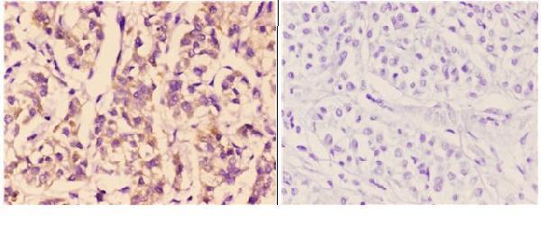
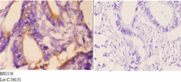
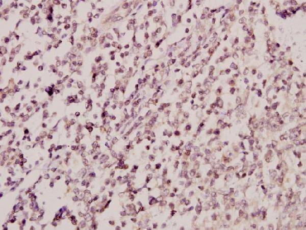


Comprehensive Human Protein Atlas IHC scoring per tissue (reliability: Enhanced — Medium consistency between antibody staining and RNA expression data. Presumed off target binding observed and disregarded. Pending external verification.). Rows are taken directly from the HPA tissue chart — click any row's HPA link to view the source.
| Tissue | Cell type | Level | Evidence | Source |
|---|---|---|---|---|
| Epididymis | Glandular cells | High | Protein (IHC) | HPA → |
| Testis | Sertoli cells | Medium | Protein (IHC) | HPA → |
| Tissue | Cell type | Level | Evidence | Source |
|---|---|---|---|---|
| Adipose tissue | Adipocytes | Not detected | Protein (IHC) | HPA → |
| Adrenal gland | Glandular cells | Not detected | Protein (IHC) | HPA → |
| Appendix | Glandular cells | Not detected | Protein (IHC) | HPA → |
| Bone marrow | Hematopoietic cells | Not detected | Protein (IHC) | HPA → |
| Breast | Adipocytes | Not detected | Protein (IHC) | HPA → |
Use the paraffin-section image and tissue-expression data as controls while evaluating BMX staining by chromogenic IHC.
Both anti-BMX antibodies have paraffin-section IHC images and are listed for IF, with Human, Mouse and Rat reactivity (IHC image captions; catalog applications/reactivity).
A02537-1 has paraffin-section IHC images from human liver and rectum carcinoma, and is listed for IF (A02537-1 image captions; catalog applications). A02537Y43 has a paraffin-section IHC image from human tonsil carcinoma and is listed for IF (A02537Y43 image caption; catalog applications).
Which to pick: For tissue IHC, choose A02537-1 when its documented cytoplasmic and cell-membrane staining and PBS primary-omission control are useful; choose A02537Y43 when a tonsil carcinoma IHC example is more relevant (A02537-1 image captions; A02537Y43 image caption). For IF, either rabbit polyclonal antibody is catalog-listed; the payload provides no IF image or ICC validation (catalog host, dilution_raw, applications and image alts). Both list Human, Mouse and Rat reactivity, while their IHC captions specify paraffin sections without reporting a fixative (catalog reactivity; IHC image captions).