BNIP3 / BCL2/adenovirus E1B 19 kDa protein-interacting protein 3 · IHC design guide

Design Immunohistochemistry for BNIP3

Plan chromogenic BNIP3 IHC in paraffin sections using cytoplasmic staining as the tissue benchmark (HPA tissue IHC). Kidney collecting ducts and pancreatic exocrine glandular cells show high staining and can guide positive-control selection (HPA tissue IHC).

Evidence assembled Oct 2026 · For research use; verify linked source records and product datasheet before use
Immunohistochemistry protocol sheet for BNIP3 (IHC for BNIP3): expected localisation Cytoplasmic in tissue (HPA tissue IHC); mitochondrial outer membrane (UniProt), antibody M01469, validated IHC image, and IHC protocol steps
Printable BNIP3 IHC protocol sheet — expected localisation Cytoplasmic in tissue (HPA tissue IHC); mitochondrial outer membrane (UniProt), antibody M01469, controls and protocol steps. Open the full BNIP3 IHC guide →

BNIP3 Immunohistochemistry Experimental Design Guide

Expected localisation, validated protocols, controls and antibodies — the at-a-glance facts below, then the full design guide.

Must know before staining
Expected localisation Cytoplasmic in tissue (HPA tissue IHC); mitochondrial outer membrane (UniProt)
Staining pattern Cytoplasmic in most tissues; high in kidney collecting ducts (HPA tissue IHC)
Antigen retrieval Citrate pH 6.0 HIER, 95–98 °C, 20 min (rule: cytoplasmic / membrane antigen)
Positive control ⓘ Cerebellum+4 more · see all
Negative control ⓘ None in HPA (detected in all 45 tissues); use no-primary + isotype controls
Important caveats
Reasons your staining may differ from the expected pattern.
Fixation Matched tissue-IHC evidence does not establish the fixation claim. Validate the specimen-specific method before use. (selected-SKU IHC image M01469)
Caveat Staining and RNA show medium consistency (HPA tissue IHC)
Regulation PPTC7 promotes BNIP3 degradation (UniProt)
Isoform / epitope No annotated isoforms or processed chains (UniProt)
Section 1

Recommended BNIP3 IHC & IF Protocols

The catalog antibody’s IHC-P protocol is accompanied by published BNIP3 methods for human tissue sections (PMC5463433), tissue microarrays (PMC5602480), and kidney sections (PMC3654981).

Recommended immunohistochemistry (IHC-P) protocol parameters
SampleParaffin-embedded Human pancreatic cancer tissue; fixative not specified (datasheet M01469)
FixationImage fixative and duration unreported (datasheet M01469); verify before use.
Sectioning4–5 µm sections on charged slides (standard)
DeparaffinisationXylene, graded ethanol series to water (standard)
Antigen retrievalHeat-induced epitope retrieval in citrate buffer, pH 6.0, 20 min at 95–98 °C (standard rule: cytoplasmic / membrane antigen)
Peroxidase block3% H2O2, 10 min, room temperature (standard)
Blocking10% normal serum of the secondary host, 30 min, room temperature (standard)
Primary antibodyRabbit monoclonal (clone AAEI-2) anti-BNIP3, 1:50-1:200 (datasheet M01469)
Primary incubationOvernight at 4 °C (standard)
DetectionHRP-polymer secondary, DAB chromogen 5–10 min (standard)
CounterstainHematoxylin, blue, dehydrate and mount (standard)
Expected resultBNIP3-positive staining in cells in granular layer of cerebellum (HPA tissue IHC: High). HPA tissue profile: Cytoplasmic expression in most tissues. No signal in the no-primary control.
💡Decision noteStart with citrate at pH 6.0, 95–98 °C for 20 min (page antigen-retrieval setting); a kidney-section study used heated acetic acid (PMC3654981).
Section 2

What Is the Expected BNIP3 Staining Pattern?

BNIP3 is an outer mitochondrial membrane protein with a transmembrane segment at residues 164–184 (UniProt Q12983 topology). In tissue IHC, expect cytoplasmic staining, including strong staining in pancreatic exocrine cells and kidney collecting ducts (HPA tissue IHC). HPA rates the tissue pattern Supported, with medium consistency between staining and RNA data (HPA tissue IHC).

What am I looking at on my slide?
Strong cytoplasmic staining in pancreatic exocrine cells (HPA tissue IHC).Matches a reported high-staining cell population (HPA tissue IHC).
Diffuse nuclear staining across cells (HPA tissue IHC; UniProt Q12983).Unexpected for the usual pattern; assess artefact (HPA tissue IHC; UniProt Q12983).
Staining confined to germinal center cells (HPA tissue IHC).These cells stain low; check cross-reactivity or endogenous activity (HPA tissue IHC; general IHC practice).
Uniform color over cells and surrounding tissue (general IHC practice).Suggests background from detection or inadequate blocking (general IHC practice).
No signal in pancreatic exocrine cells (HPA tissue IHC).Unexpected in a reported high-staining population; check the IHC workflow (HPA tissue IHC).
💡Expected BNIP3 appearanceCall positive when pancreatic exocrine cells show strong cytoplasmic staining; diffuse nuclear color is suspect (HPA tissue IHC; UniProt Q12983).
How each factor affects the staining
Membrane topology (UniProt Q12983 topology).BNIP3 has one transmembrane segment at 164–184 (UniProt Q12983 topology).
Tissue choice (HPA tissue IHC).Pancreatic exocrine cells stain high; thyroid glandular cells stain low (HPA tissue IHC).
IHC evidence (HPA tissue IHC; HPA antibodies).Tissue pattern is Supported; HPA003015 and CAB011676 are IHC Supported (HPA tissue IHC; HPA antibodies).
IF/ICC expectation? (HPA ICC-IF).HPA summarizes membrane staining but gives no main location or cell-line images (HPA ICC-IF).
Nuclear envelope exception (UniProt Q12983).E1B 19-kDa protein coexpression can shift BNIP3 to the nuclear envelope (UniProt Q12983).
Why is my staining missing, weak or wrong?
SituationLikely causeNext action
No color in pancreatic exocrine cells (HPA tissue IHC).Detection or retrieval may have failed (general IHC practice).Check reagent controls and retrieval conditions (general IHC practice).
Diffuse nuclear color (HPA tissue IHC; UniProt Q12983).Possible nonspecific staining (general IHC practice).Review controls; consider the E1B-dependent envelope exception (general IHC practice; UniProt Q12983).
Uniform tissue-wide haze (general IHC practice).Blocking or detection background (general IHC practice).Check no-primary control and optimize blocking and washes (general IHC practice).
Color in low-staining germinal center cells (HPA tissue IHC).Possible cross-reactivity or endogenous activity (general IHC practice).Check no-primary control and compare with known high-staining cells (general IHC practice; HPA tissue IHC).
Signal obscured by counterstain (general IHC practice).Counterstain may be too strong (general IHC practice).Reduce counterstain and reassess cellular localization (general IHC practice).

Sample controls for BNIP3 IHC & IF

🧪Run cerebellum first and score staining in granular-layer cells, where BNIP3 is High (HPA: cerebellum, cells in granular layer, High). HPA detects BNIP3 in all 45 scored tissues, so there is no supported negative tissue; use no-primary and isotype controls, and treat any unstained cells on the positive slide as background comparators rather than confirmed BNIP3-negative cells (HPA: no negative rows; detected in all 45 scored tissues).
Positive control tissue: Cerebellum (Cells in granular layer, HPA High)
Negative control tissue: None in HPA: BNIP3 is detected in all 45 scored tissues. Use a no-primary (secondary-only) and an isotype control instead.
ICC-IF cell lines (HPA subcellular resource): HPA carries no ICC-IF cell line for BNIP3; derive a cell-line control from the positive tissue's cell type (Cells in granular layer) and confirm it by RNA or western blot first.
Technical controls: Include a no-primary, secondary-only slide and an isotype control matched to the primary antibody’s host species and immunoglobulin class; a BNIP3 knockout specimen can provide a biological specificity control (standard IHC practice). Block endogenous peroxidase for chromogenic IHC, and assess tissue autofluorescence if using cerebellum for IF (standard IHC/IF practice).
⚠️Feasibility: No target-specific fixation window, fixation effect, or retrieval dependency is reported in the supplied evidence; test antigen retrieval on the paraffin sections (supplied target/application evidence; standard IHC practice). The M01469 pancreatic-cancer paraffin-section caption reports 1:100 dilution but leaves the fixative unreported, so it does not establish fixation (selected tissue-IHC caption: M01469). The supplied evidence does not establish whether frozen sections or IF are easier; for cerebellar IF, check autofluorescent background before scoring signal (HPA: no cell lines with ICC-IF images; standard IF practice). The selected M01469 tissue-IHC caption documents paraffin sections, but does not specify the fixative (selected-SKU IHC image M01469).

HPA tissue IHC evidence for BNIP3

Comprehensive Human Protein Atlas IHC scoring per tissue (reliability: Supported — Medium consistency between antibody staining and RNA expression data.). Rows are taken directly from the HPA tissue chart — click any row's HPA link to view the source.

Positive expression · recommended positive controls

TissueCell typeLevelEvidenceSource
Cerebellum Cells in granular layer High Protein (IHC) HPA →
Cerebral cortex Neuronal cells High Protein (IHC) HPA →
Hippocampus Neuronal cells High Protein (IHC) HPA →
Kidney Collecting ducts High Protein (IHC) HPA →
Pancreas Exocrine glandular cells High Protein (IHC) HPA →

Undetected expression · recommended negative controls

TissueCell typeLevelEvidenceSource
None in HPA: BNIP3 is detected in all 45 scored tissues. Use a no-primary (secondary-only) and an isotype control instead.
Section 3

Advanced BNIP3 IHC Tips

Troubleshoot BNIP3 staining in paraffin section chromogenic IHC using the catalog antibody’s tissue image, protein annotations, and tissue expression data.

What retrieval conditions should I try first when BNIP3 staining is weak?
Start with citrate buffer at pH 6.0, heated to 95–98 °C for 20 min (page retrieval rule). The catalog antibody has a paraffin section image at 1:100, but its caption does not report retrieval conditions, so use an adjacent section to assess whether this starting condition exposes the epitope (catalog antibody IHC caption). Include tissue with expected staining, such as pancreatic exocrine glandular cells, alongside the specimen (HPA: High in pancreatic exocrine glandular cells). If staining remains weak, compare a modified retrieval condition on adjacent sections while holding antibody concentration, detection, and counterstaining constant (standard IHC practice).
Could fixation explain weak or uneven BNIP3 staining?
BNIP3 sensitivity to any particular fixative or fixation duration is unknown from the supplied evidence; the catalog image says paraffin embedded but does not state its fixative (catalog antibody IHC caption). Record the specimen’s fixative and processing history, then compare sections with different known histories using the same citrate pH 6.0, 95–98 °C, 20 min retrieval (page retrieval rule; standard IHC practice). Examine morphology and staining in a positive reference section before changing antibody conditions (standard IHC practice). Treat differences between specimens as possible processing effects until controls show reproducible BNIP3 staining under matched conditions (standard IHC practice).
How should I evaluate diffuse cytoplasmic versus punctate BNIP3 staining?
BNIP3 is annotated at mitochondria and the mitochondrial outer membrane, with a transmembrane segment at residues 164–184 (UniProt Q12983 localisation and topology). HPA reports cytoplasmic staining across most tissues, so a cytoplasmic chromogenic signal can be compatible with its tissue IHC profile even when individual mitochondria cannot be resolved (HPA: tissue IHC profile; standard IHC practice). Compare the signal with cell boundaries and morphology at higher magnification, and check whether a known positive cell population stains in the same run (standard IHC practice). Predominantly nuclear staining warrants investigation; UniProt describes a nuclear envelope shift specifically with E1B 19-kDa protein coexpression (UniProt Q12983 localisation).
How do epitope location and BNIP3 modifications affect staining decisions?
The supplied record lists 0 annotated isoforms and one 1–194 amino acid chain, so an isoform-specific staining interpretation is unsupported here (UniProt Q12983 processing and isoforms). Its membrane-spanning segment lies at residues 164–184, but the catalog image does not identify the antibody epitope (UniProt Q12983 topology; catalog antibody IHC caption). Annotated phosphoserines include residues 54, 66, 86, 92, and 95; their effect on this antibody’s IHC signal is unknown (UniProt Q12983 modified residues; supplied antibody evidence). Check the antibody’s epitope documentation before assigning a staining difference to topology or phosphorylation, and keep retrieval conditions matched during comparisons (standard IHC practice).
How can I adapt BNIP3 assessment to a multiplex IF experiment?
For an IF experiment, pair BNIP3 with a marker identifying the expected cell population, such as pancreatic exocrine glandular cells when examining pancreas, and inspect each channel separately before interpreting overlap (HPA: High in pancreatic exocrine glandular cells; standard IF practice). Choose fluorophores after measuring the tissue’s autofluorescence, favoring a spectrally distinct channel for a weak BNIP3 signal (standard IF practice). BNIP3 spans the mitochondrial outer membrane at residues 164–184, but the antibody epitope and its membrane-facing side are unspecified (UniProt Q12983 topology; supplied antibody evidence). Set permeabilisation according to the documented epitope side, and verify accessibility with matched controls rather than transferring the paraffin IHC conditions to IF (standard IF practice).
What controls help identify nonspecific BNIP3 chromogenic signal?
Run a no-primary control and inspect tissue edges, damaged regions, pigment, and precipitate before assigning chromogen to BNIP3 (standard IHC practice). Apply a peroxidase block when using enzyme-based chromogenic detection, and assess whether residual endogenous activity contributes to signal in the control (standard IHC practice). The catalog antibody’s paraffin section image uses 1:100; use that reported dilution as a reference while checking whether background rises with stronger antibody conditions (catalog antibody IHC caption; standard IHC practice). Compare the specimen with pancreatic exocrine glandular cells as a positive reference and thyroid glandular cells as a low-expression reference, without treating either as absolute proof of specificity (HPA: High in pancreas; Low in thyroid gland).
How should I score BNIP3 IHC across specimens? ⚠ ANSWER MARKED FOR VERIFICATION
Define the cell population and region before scoring, then use an H-score or report the percentage of positive cells with an intensity category (standard IHC practice). If measuring stained area, report signal density per mm² of viable tissue and normalise to the eligible cell count or tissue area used for each specimen (standard IHC practice). Score comparable compartments consistently, because HPA describes broadly cytoplasmic tissue staining while UniProt places BNIP3 at mitochondria and their outer membrane (HPA: tissue IHC profile; UniProt Q12983 localisation). Keep retrieval at citrate pH 6.0, 95–98 °C for 20 min and hold imaging and threshold settings constant across the comparison (page retrieval rule; standard IHC practice).
When is a BNIP3-positive stain biologically credible rather than artefactual?
A credible result has cellular staining consistent with BNIP3’s mitochondrial outer membrane annotation and the cytoplasmic pattern reported in tissue IHC (UniProt Q12983 localisation; HPA: tissue IHC profile). Check whether expected cells stain, such as pancreatic exocrine glandular cells, while recognizing that HPA rates its tissue evidence Supported with medium RNA–staining consistency (HPA: High in pancreatic exocrine glandular cells; HPA: reliability). Treat signal confined to section edges, necrotic areas, or no-primary controls as suspect, and check endogenous enzyme activity in chromogenic detection (standard IHC practice). An isolated nuclear pattern needs independent validation; the annotated nuclear envelope shift occurs with E1B 19-kDa protein coexpression (UniProt Q12983 localisation).
Boster reagents

Best BNIP3 / BCL2/adenovirus E1B 19 kDa protein-interacting protein 3 IHC Antibodies

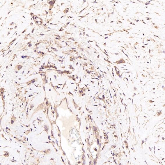
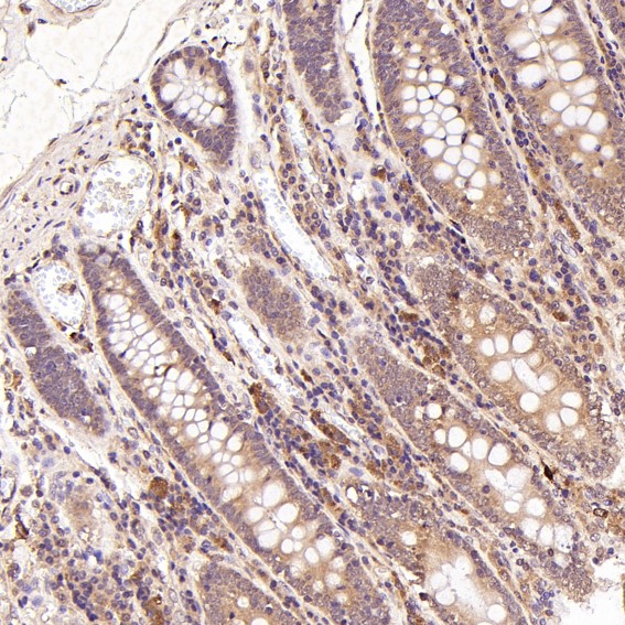
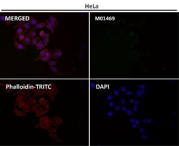
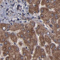
M01469 has IHC images from human pancreatic cancer, placenta, thyroid cancer and kidney (catalog image captions), plus an IF image with no sample identified (catalog IF caption).

Real IHC data Immunohistochemical analysis of paraffin-embedded Human pancreatic cancer, using the Antibody at 1:100 dilution.
Anti-BNIP3 Rabbit Monoclonal Antibody
Cat # M01469

M01469 is the SKU that will render; its IHC captions identify paraffin-embedded human pancreatic cancer, placenta and thyroid cancer, plus human kidney with processing unreported (catalog IHC captions). Its application list includes IHC and IF/ICC, and its listed reactivity is Human, Mouse and Rat (catalog applications and reactivity).

Which to pick: Choose M01469 for tissue IHC: its images include paraffin-embedded human samples, but the fixative is unreported (catalog IHC captions). For IF/ICC, M01469 has a listed IF/ICC application and an IF image whose specimen is unspecified (catalog applications and IF caption). Both M01469 and M01469-2 are rabbit monoclonals with listed Human, Mouse and Rat reactivity; cross-species IHC performance is not established by the human-only image captions (catalog host, clone and reactivity; catalog IHC captions).

Each figure is that product's own IHC / IF validation image from its datasheet.

References

  1. UniProt Consortium. UniProt entry Q12983 (BNIP3_HUMAN, BCL2/adenovirus E1B 19 kDa protein-interacting protein 3).
  2. Human Protein Atlas. BNIP3 tissue IHC expression (reliability: Supported).
  3. Human Protein Atlas. BNIP3 subcellular location (ICC-IF): Membrane.
  4. Human Protein Atlas. BNIP3 antibody validation summary (2 antibodies).
  5. Chemoresistance of colorectal cancer to 5-fluorouracil is associated with silencing of the BNIP3 gene through aberrant methylation. Journal of Cancer 2017 — PMC5463433.
  6. Expression and Functional Characterization of the BNIP3 Protein in Renal Cell Carcinomas. Translational oncology 2017 — PMC5602480.
  7. Upregulation of BCL2 19 kD Protein-Interacting Protein 3 (BNIP3) is Predictive of Unfavorable Prognosis in Uveal Melanoma. Medical science monitor : international medical journal of experimental and clinical research 2018 — PMC6070000.
  8. Peroxisome proliferator-activated receptor δ agonist, HPP593, prevents renal necrosis under chronic ischemia. PloS one 2013 — PMC3654981.
  9. PubMed PMID:7954800 — UniProt-cited evidence.
  10. PubMed PMID:9396766 — UniProt-cited evidence.
  11. PubMed PMID:15164054 — UniProt-cited evidence.