This website uses cookies to ensure you get the best experience on our website.


- Table of Contents
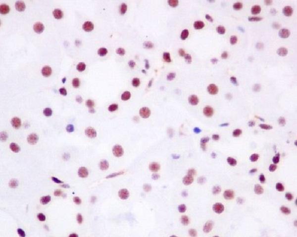
Plan paraffin BRD4 IHC around its ubiquitous nuclear tissue pattern (HPA tissue IHC). Use the documented human kidney IHC example and compare nuclear staining across consistently handled sections (datasheet: M00123; standard IHC practice).

Expected localisation, validated protocols, controls and antibodies — the at-a-glance facts below, then the full design guide.
| Expected localisation | Ubiquitous nuclear staining (HPA tissue IHC) | |
| Staining pattern | Nuclear staining across many cell types (HPA tissue IHC) | |
| Antigen retrieval | Tris-EDTA pH 9.0 HIER, 95–98 °C, 20 min (rule: nuclear antigen) | |
| Positive control | Adipose tissue+4 more · see all | |
| Negative control | None in HPA (detected in all 45 tissues); use no-primary + isotype controls |
| Fixation | Keep fixation consistent across sections. (standard IHC practice; not target-specific) | |
| Caveat | Histone deacetylation can release BRD4 from chromatin (UniProt) | |
| Regulation | Deacetylation reduces chromatin binding (UniProt) | |
| Isoform / epitope | 3 isoforms (A, B, C); verify epitope coverage (UniProt) |
The catalog antibody protocol is accompanied by published BRD4 IHC methods for heart, breast, peripheral nerve, and gastric tissue (PMC12203807; PMC6053709; PMC9278623; PMC5458191).
| Sample | Paraffin-embedded human kidney tissue; fixative not specified (datasheet M00123) |
| Fixation | Image fixative and duration unreported (datasheet M00123); verify before use. |
| Sectioning | 4–5 µm sections on charged slides (standard) |
| Deparaffinisation | Xylene, graded ethanol series to water (standard) |
| Antigen retrieval | Heat-induced epitope retrieval in Tris-EDTA buffer, pH 9.0, 20 min at 95–98 °C (standard rule: nuclear antigen) |
| Peroxidase block | 3% H2O2, 10 min, room temperature (standard) |
| Blocking | 10% normal serum of the secondary host, 30 min, room temperature (standard) |
| Primary antibody | Rabbit monoclonal (clone DBH-2) anti-BRD4, 1:50 (datasheet M00123) |
| Primary incubation | Overnight at 4 °C (standard) |
| Detection | HRP-polymer secondary, DAB chromogen 5–10 min (standard) |
| Counterstain | Hematoxylin, blue, dehydrate and mount (standard) |
| Expected result | BRD4-positive staining in adipocytes of adipose tissue (HPA tissue IHC: High). HPA tissue profile: Ubiquitous nuclear expression. No signal in the no-primary control. |
BRD4 is nuclear and chromatin associated (UniProt O60885). Expect broad nuclear staining, including in adipocytes, glandular cells and hematopoietic cells (HPA: High in each). HPA rates its tissue IHC pattern Supported; BRD4 has no transmembrane segment (UniProt O60885).
| Strong nuclear staining in adipocytes | Expected pattern (HPA: High in adipocytes; UniProt: nucleus). |
| Cytoplasmic-only or membrane-only staining | Discordant with BRD4 localization; assess artefact (UniProt: nucleus; no transmembrane segment). |
| Strong signal restricted to an unexpected cell type | Consider cross-reactivity or endogenous detection activity (general IHC practice). |
| Diffuse staining across the section | May reflect nonspecific background; check controls (general IHC practice). |
| No nuclear signal in adipocytes | Unexpected in this positive tissue; check the assay (HPA: High in adipocytes). |
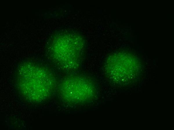
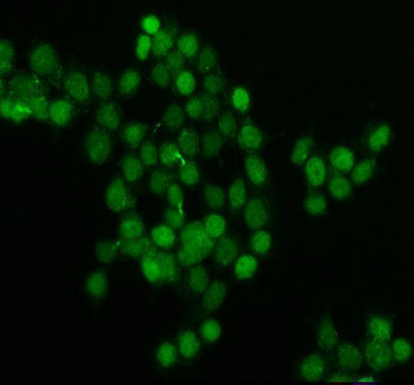
| What should IF/ICC show? | Mainly nucleoplasm, with mitotic chromosomes also stained (HPA: ICC-IF). |
| Cell-cycle localization | BRD4 associates with acetylated chromatin and mitotic chromosomes (UniProt O60885). |
| Isoforms | BRD4 has three isoforms; check which regions the antibody recognizes (UniProt: A, C, B). |
| HPA antibody validation | IHC is Supported for HPA015055, HPA061646, CAB068177 and CAB068178 (HPA antibodies). |
| Situation | Likely cause | Next action |
|---|---|---|
| No signal in adipocyte nuclei | Assay failure is possible (HPA: High in adipocytes). | Check retrieval, detection and a positive control (general IHC practice). |
| Cytoplasmic-only signal | Localization conflicts with nuclear BRD4 (UniProt O60885). | Review cell morphology and antibody specificity (general IHC practice). |
| Signal in an unexpected cell type | Cross-reactivity or endogenous activity is possible (general IHC practice). | Run a no-primary control and compare the HPA cell pattern (general IHC practice; HPA tissue). |
| Diffuse background | Nonspecific binding or inadequate blocking is possible (general IHC practice). | Optimize blocking, washes and antibody dilution (general IHC practice). |
| Uneven nuclear staining | Uneven retrieval or detection is possible (general IHC practice). | Check section coverage and retrieval consistency (general IHC practice). |



Comprehensive Human Protein Atlas IHC scoring per tissue (reliability: Supported — High consistency between antibody staining and RNA expression data.). Rows are taken directly from the HPA tissue chart — click any row's HPA link to view the source.
| Tissue | Cell type | Level | Evidence | Source |
|---|---|---|---|---|
| Adipose tissue | Adipocytes | High | Protein (IHC) | HPA → |
| Adrenal gland | Glandular cells | High | Protein (IHC) | HPA → |
| Appendix | Glandular cells | High | Protein (IHC) | HPA → |
| Bone marrow | Hematopoietic cells | High | Protein (IHC) | HPA → |
| Breast | Adipocytes | High | Protein (IHC) | HPA → |
| Tissue | Cell type | Level | Evidence | Source |
|---|---|---|---|---|
| None in HPA: BRD4 is detected in all 45 scored tissues. Use a no-primary (secondary-only) and an isotype control instead. | ||||
Troubleshoot BRD4 staining by checking retrieval, nuclear localisation, antibody epitope coverage and matched controls before comparing signal across paraffin sections.
The catalog includes anti-BRD4 antibodies with IHC data from paraffin-embedded human kidney and IF/ICC data from human cell lines; all three list Human, Mouse, and Rat reactivity (catalog applications, image captions, and reactivity).
M00123 lists IHC and IF/ICC applications and shows IHC staining in paraffin-embedded human kidney (M00123 catalog applications and IHC image caption). A00123-2 shows IF/ICC in PC-3 cells, and A00123-3 shows IF/ICC in MCF-7 cells (respective IF image captions and catalog applications).
Which to pick: Choose M00123 for tissue IHC: it is a rabbit monoclonal antibody with an IHC application and an image from paraffin-embedded human kidney; the caption does not report the fixative (M00123 catalog clone, applications, and IHC image caption). For IF/ICC, A00123-2 has a PC-3 image and A00123-3 has an MCF-7 image; M00123 also lists IF/ICC (respective catalog applications and image captions). All three list Human, Mouse, and Rat reactivity, but the supplied IHC image documents human kidney only (catalog reactivity; M00123 IHC image caption).