This website uses cookies to ensure you get the best experience on our website.


- Table of Contents
Plan BRF1 paraffin-section IHC around variable nuclear staining in most tissues (HPA tissue IHC). Compare kidney tubular cells with high staining against adipocytes with no detected staining (HPA tissue IHC), and titrate the catalog antibody within 1:100–1:300 (datasheet).

Expected localisation, validated protocols, controls and antibodies — the at-a-glance facts below, then the full design guide.
| Expected localisation | Nuclear staining (HPA tissue IHC) | |
| Staining pattern | Variable nuclear staining in most tissues (HPA tissue IHC) | |
| Antigen retrieval | Tris-EDTA pH 9.0 HIER, 95–98 °C, 20 min (rule: nuclear antigen) | |
| Positive control | Adrenal gland+4 more · see all | |
| Negative control | Adipose tissue+4 more · see all |
| Fixation | Keep fixation consistent across sections (standard IHC practice; not target-specific) | |
| Caveat | Presumed off-target staining was observed (HPA tissue IHC) | |
| Regulation | Expression regulation unreported (UniProt) | |
| Isoform / epitope | 9 isoforms; check epitope coverage (UniProt) |
The catalog antibody protocol is paired with four published BRF1 IHC examples (PMC8081602; PMC4868753; PMC7336232; PMC11369479).
| Sample | Paraffin-embedded human brain tissue; fixative not specified (datasheet A03761-1) |
| Fixation | Image fixative and duration unreported (datasheet A03761-1); verify before use. |
| Sectioning | 4–5 µm sections on charged slides (standard) |
| Deparaffinisation | Xylene, graded ethanol series to water (standard) |
| Antigen retrieval | Heat-induced epitope retrieval in Tris-EDTA buffer, pH 9.0, 20 min at 95–98 °C (standard rule: nuclear antigen) |
| Peroxidase block | 3% H2O2, 10 min, room temperature (standard) |
| Blocking | 10% normal serum of the secondary host, 30 min, room temperature (standard) |
| Primary antibody | Rabbit monoclonal (clone 2F1) anti-BRF1, 1:100-1:300 (datasheet A03761-1) |
| Primary incubation | Overnight at 4 °C (standard) |
| Detection | HRP-polymer secondary, DAB chromogen 5–10 min (standard) |
| Counterstain | Hematoxylin, blue, dehydrate and mount (standard) |
| Expected result | BRF1-positive staining in glandular cells of adrenal gland (HPA tissue IHC: High). HPA tissue profile: Nuclear expression of varying levels in most tissues. No signal in the no-primary control. |
BRF1 should show predominantly nuclear staining in paraffin tissue sections (UniProt Q92994: nucleus; HPA: nuclear expression at varying levels in most tissues). High staining is reported in kidney tubular cells, cerebral cortex neurons, and testis spermatogonia (HPA: High). A membrane pattern is unexpected (UniProt Q92994: no transmembrane segment). HPA rates its tissue IHC evidence Approved, while noting presumed off-target binding was disregarded and external verification is pending.
| Distinct nuclear staining in kidney tubular cells, with nuclei in the same cell population staining more strongly than surrounding cytoplasm. | This fits BRF1 localisation and a reported high-staining cell population (UniProt Q92994: nucleus; HPA: High in kidney tubular cells). Judge the nuclear pattern within identified tubules; staining elsewhere in the section needs its own cell-type assessment. |
| Strong cytoplasmic or membrane staining dominates while nuclei remain pale. | This conflicts with the expected compartment (UniProt Q92994: nucleus; HPA: nuclear expression). Treat the pattern as suspect and check controls and detection conditions before scoring it as BRF1; HPA notes that presumed off-target binding was disregarded in its tissue assessment (HPA: Approved reliability description). |
| Prominent staining appears in adipocytes or prostate glandular cells, especially without convincing nuclear staining in an expected positive population. | Those named cell populations were reported as not detected (HPA: adipocytes, Not detected; prostate glandular cells, Not detected). Investigate nonspecific antibody binding or endogenous detection activity (standard IHC practice). The HPA observations do not establish that every cell in either whole tissue must be negative. |
| A uniform haze covers nuclei, cytoplasm, and extracellular spaces, obscuring cell boundaries. | Diffuse background prevents a reliable compartment call (standard IHC practice). Assess blocking, washing, antibody concentration, and detection controls (standard IHC practice); the supplied UniProt and HPA records do not identify a BRF1-specific cause of background. |
| No nuclear signal is visible in an included high-staining cell population. | A blank kidney tubular, neuronal, or spermatogonial population is discordant with the corresponding HPA High observations (HPA: kidney tubules, cerebral cortex neurons, testis spermatogonia). Check that the expected cells are present, then review assay controls and workflow (standard IHC practice) before interpreting the specimen as negative. |
| Cell type and baseline abundance | Compare like cells with like cells: HPA reports high staining in thyroid glandular cells and medium staining in appendix glandular cells, alongside low staining in stomach glandular cells (HPA: tissue IHC). A weak result in a low-staining population carries less weight than a blank high-staining population. |
| IHC antibody evidence | The listed tissue-IHC antibody has Approved status (HPA: HPA074990 IHC Approved). HPA also reports presumed off-target binding was disregarded and external verification is pending (HPA: reliability description). Interpret unexpected staining against cell type and compartment rather than treating Approved as proof of specificity in every specimen. |
| Antigen retrieval | Retrieval conditions can be evaluated with an expected positive tissue and detection controls when nuclear staining is absent (standard IHC practice). No BRF1-specific retrieval condition or fixation sensitivity is established by the supplied UniProt and HPA evidence; a failed signal alone cannot identify epitope masking as its cause. |
| Isoforms and epitope coverage | UniProt lists nine BRF1 isoforms and distinct transcriptional roles for isoforms 1 and 2 (UniProt Q92994). The payload gives no antibody epitope or isoform-coverage data, so staining cannot be assigned to one isoform, and isoform differences cannot explain a particular negative slide from these sources alone. |
| IF/ICC Q&A: Where should BRF1 fluorescence appear? | HPA places BRF1 in the nucleoplasm and nuclear bodies, both Approved (HPA: subcellular ICC-IF). That supports a nuclear IF/ICC interpretation; the separately listed ICC antibody is HPA051918, ICC Approved (HPA: antibodies). These observations do not supply an IF/ICC protocol for this IHC section. |
| Situation | Likely cause | Next action |
|---|---|---|
| Expected high-staining nuclei are blank. | The assay may have failed, the expected cell population may be absent, or detection may be inadequate (standard IHC practice); HPA alone cannot distinguish these causes. | Confirm the named cells on the counterstained section, inspect a known positive control, and review retrieval and detection steps (standard IHC practice; HPA: High in kidney tubular cells). |
| Signal is mainly cytoplasmic or membranous. | The pattern disagrees with the reported nuclear location (UniProt Q92994: nucleus; HPA: nuclear tissue expression). Its precise cause is unproven. | Check a positive tissue and a detection control, then reassess whether any reproducible nuclear signal remains (standard IHC practice). Do not score dominant membrane staining as the expected BRF1 pattern (UniProt Q92994: no transmembrane segment). |
| Reported not-detected cells stain strongly. | Nonspecific binding or endogenous detection activity is possible (standard IHC practice); HPA has also noted presumed off-target binding in its tissue assessment (HPA: reliability description). | Identify the stained cell type, compare it with HPA's cell-level result, and inspect antibody-omission and detection controls (standard IHC practice; HPA: adipocytes and prostate glandular cells, Not detected). |
| Brown haze is widespread and nuclei cannot be distinguished. | High background can obscure a real nuclear pattern (standard IHC practice); the supplied sources do not establish a BRF1-specific mechanism. | Review blocking, washes, antibody concentration, and chromogenic detection controls (standard IHC practice). Reassess only when nuclear staining can be separated from the haze (UniProt Q92994: nucleus). |
| Staining is weak in a low-staining population. | Low signal may match the reported baseline; HPA lists stomach and small-intestine glandular cells as Low (HPA: tissue IHC). | Compare the same cell type and use a high-staining control population to judge assay performance (standard IHC practice; HPA: kidney tubular cells, High). Avoid calling the run failed solely from a weak low-staining population. |
| A positive tissue stains, but the study tissue is negative. | BRF1 staining varies among tissues and cell types (HPA: nuclear expression at varying levels in most tissues); the supplied evidence gives no diagnosis-specific expectation for the study specimen. | Score the identified cell population and nuclear signal with the positive control in view (standard IHC practice; UniProt Q92994: nucleus). Report the study result as observed, without inferring an isoform or fixation effect from absence alone. |
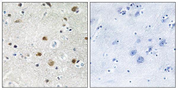
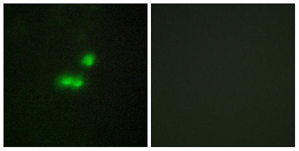
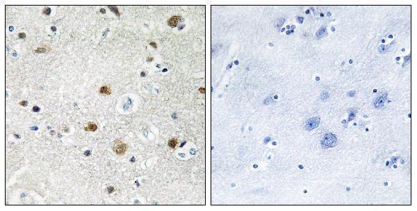
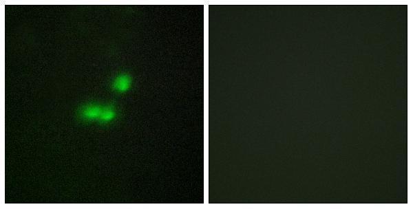


Comprehensive Human Protein Atlas IHC scoring per tissue (reliability: Approved — Presumed off target binding observed and disregarded. Pending external verification.). Rows are taken directly from the HPA tissue chart — click any row's HPA link to view the source.
| Tissue | Cell type | Level | Evidence | Source |
|---|---|---|---|---|
| Adrenal gland | Glandular cells | High | Protein (IHC) | HPA → |
| Cerebral cortex | Neuronal cells | High | Protein (IHC) | HPA → |
| Endometrium | Cells in endometrial stroma | High | Protein (IHC) | HPA → |
| Kidney | Cells in tubules | High | Protein (IHC) | HPA → |
| Testis | Spermatogonia cells | High | Protein (IHC) | HPA → |
| Tissue | Cell type | Level | Evidence | Source |
|---|---|---|---|---|
| Adipose tissue | Adipocytes | Not detected | Protein (IHC) | HPA → |
| Liver | Cholangiocytes | Not detected | Protein (IHC) | HPA → |
| Ovary | Ovarian stroma cells | Not detected | Protein (IHC) | HPA → |
| Parathyroid gland | Glandular cells | Not detected | Protein (IHC) | HPA → |
| Prostate | Glandular cells | Not detected | Protein (IHC) | HPA → |
Troubleshoot BRF1 staining in paraffin sections by checking nuclear localisation, tissue controls and the limits of antibody specific evidence (UniProt Q92994; HPA tissue IHC).
Two anti-BRF1 antibodies have IHC images of paraffin-embedded human brain and IF images of MCF7 cells (catalog image captions); both list human and mouse reactivity (catalog).
A03761-1 lists IHC and IF, with its own images showing paraffin-embedded human brain IHC and MCF7 IF (A03761-1 catalog/image captions). A30544 lists IHC, IF and ICC, with its own images showing paraffin-embedded human brain IHC and MCF7 IF (A30544 catalog/image captions).
Which to pick: For tissue IHC, choose A03761-1 if a defined rabbit clone is useful: clone 2F1 has a listed IHC dilution of 1:100–1:300, and its own image shows a paraffin-embedded human brain section; the fixative is unreported (A03761-1 catalog/IHC caption). For IF/ICC, choose polyclonal A30544 because both applications are listed and its own IF image shows MCF7 cells; its own IHC image also shows paraffin-embedded human brain, with fixative unreported (A30544 catalog/IF and IHC captions). For cross-species planning, either SKU lists human and mouse reactivity, although both supplied IHC images show human tissue (A03761-1 and A30544 catalogs/IHC captions).