BTN3A1 / Butyrophilin subfamily 3 member A1 · IHC design guide

Design Immunohistochemistry for BTN3A1

Plan BTN3A1 paraffin IHC around cytoplasmic tissue staining (HPA tissue IHC), with membrane localization as a molecular expectation (UniProt). Compare positive kidney tubules with adipocytes lacking detectable staining (HPA tissue IHC), and account for the catalog antibody’s BTN3A1/2/3 reactivity (datasheet).

Evidence assembled Oct 2026 · For research use; verify linked source records and product datasheet before use
Immunohistochemistry protocol sheet for BTN3A1 (IHC for BTN3A1): expected localisation Cytoplasmic in tissue (HPA tissue IHC); membrane (UniProt), antibody A04257, validated IHC image, and IHC protocol steps
Printable BTN3A1 IHC protocol sheet — expected localisation Cytoplasmic in tissue (HPA tissue IHC); membrane (UniProt), antibody A04257, controls and protocol steps. Open the full BTN3A1 IHC guide →

BTN3A1 Immunohistochemistry Experimental Design Guide

Expected localisation, validated protocols, controls and antibodies — the at-a-glance facts below, then the full design guide.

Must know before staining
Expected localisation Cytoplasmic in tissue (HPA tissue IHC); membrane (UniProt)
Staining pattern Cytoplasmic staining in glandular cells across several tissues (HPA tissue IHC)
Antigen retrieval Citrate pH 6.0 HIER, 95–98 °C, 20 min (rule: cytoplasmic / membrane antigen)
Positive control ⓘ Adrenal gland+4 more · see all
Negative control ⓘ Adipose tissue+4 more · see all
Important caveats
Reasons your staining may differ from the expected pattern.
Fixation Keep fixation consistent across sections. (standard IHC practice; not target-specific)
Caveat Catalog antibody targets BTN3A1/2/3 (datasheet)
Regulation No specific expression regulator reported (UniProt)
Isoform / epitope 4 isoforms; check extracellular vs cytoplasmic epitope (UniProt)
Section 1

Recommended BTN3A1 IHC & IF Protocols

The catalog antibody’s IHC-P protocol is followed by 4 published BTN3A1 IHC protocols (PMC11165028; PMC9772689; PMC8409294; PMC13458587).

Recommended immunohistochemistry (IHC-P) protocol parameters
SampleParaffin-embedded human kidney tissue; fixative not specified (datasheet A04257)
FixationImage fixative and duration unreported (datasheet A04257); verify before use.
Sectioning4–5 µm sections on charged slides (standard)
DeparaffinisationXylene, graded ethanol series to water (standard)
Antigen retrievalHeat-induced epitope retrieval in citrate buffer, pH 6.0, 20 min at 95–98 °C (standard rule: cytoplasmic / membrane antigen)
Peroxidase block3% H2O2, 10 min, room temperature (standard)
Blocking10% normal serum of the secondary host, 30 min, room temperature (standard)
Primary antibodyRabbit anti-BTN3A1, 1:50 recommended; image 1:200 (datasheet A04257)
Primary incubationOvernight at 4 °C (standard)
DetectionHRP-polymer secondary, DAB chromogen 5–10 min (standard)
CounterstainHematoxylin, blue, dehydrate and mount (standard)
Expected resultBTN3A1-positive staining in glandular cells of adrenal gland (HPA tissue IHC: High). HPA tissue profile: Cytoplasmic expression in several tissues. No signal in the no-primary control.
💡Decision noteStart with citrate pH 6.0 retrieval at 95–98 °C for 20 min (page antigen retrieval); use a paper’s stated retrieval when reproducing its protocol.
Section 2

What Is the Expected BTN3A1 Staining Pattern?

BTN3A1 is a cell membrane protein with an extracellular region and a cytoplasmic region (UniProt O00481 topology). In paraffin section IHC, expect predominantly cytoplasmic staining in selected glandular cells, kidney tubule cells and lymph node non-germinal center cells (HPA: tissue IHC). HPA rates its tissue pattern Approved, with medium consistency between staining and RNA data (HPA: tissue IHC).

What am I looking at on my slide?
Cytoplasmic staining in colon glandular cells or kidney tubule cells.This matches cell types scored High by HPA (HPA: tissue IHC). Score the named cells, since staining of an entire tissue section need not be uniform (HPA: tissue IHC).
Clear cell borders accompany cytoplasmic staining in an expected cell population.A membrane component is compatible with BTN3A1 topology (UniProt O00481 topology); HPA describes the tissue IHC pattern as cytoplasmic (HPA: tissue IHC). Do not require a continuous membrane rim.
Predominantly nuclear staining, with little staining in the expected cytoplasm.This conflicts with HPA tissue IHC and UniProt membrane localization (HPA: tissue IHC; UniProt O00481). Treat it as a possible artefact and check controls before scoring it as BTN3A1.
Strong staining in adipocytes or cardiomyocytes.HPA reports BTN3A1 as not detected in these cells (HPA: tissue IHC). Investigate cross-reactivity or endogenous detection activity; an unexpected cell pattern alone does not identify the cause.
A uniform haze covers cells and tissue spaces, or an expected positive cell population is blank.Haze that obscures cell boundaries limits scoring (general IHC practice). A blank colon glandular or kidney tubule population conflicts with HPA's High observations and calls for a positive-control and workflow check (HPA: tissue IHC; general IHC practice).
💡Expected BTN3A1 appearanceCall a result positive when interpretable cytoplasmic staining occurs in expected cells, such as High colon glandular or kidney tubule cells (HPA: tissue IHC); nuclear-only staining or uniform haze is inconsistent with that pattern (HPA: tissue IHC; general IHC practice).
How each factor affects the staining
Compartment and epitopeBTN3A1 spans the membrane at residues 255–271, with extracellular residues 30–254 and cytoplasmic residues 272–513 (UniProt O00481 topology). The supplied sources do not locate the IHC antibody epitope, so they cannot establish which region its staining reports.
Cell-specific reference patternHPA scores several glandular populations, kidney tubule cells and lymph node non-germinal center cells High, but adipocytes and cardiomyocytes not detected (HPA: tissue IHC). Compare like cell types within sections.
Antibody evidenceHPA lists one antibody, HPA012565, as IHC Approved and ICC Uncertain (HPA: antibody validation). Approved tissue staining, with medium RNA consistency, supports a reference pattern but does not prove every stained cell is specific (HPA: tissue IHC).
Processing and variantsUniProt lists a signal peptide at residues 1–29, one glycosylation site at residue 115, and four isoforms (UniProt O00481). With no supplied antibody epitope, their effect on this IHC result cannot be predicted.
IF/ICC Q: What pattern should be expected?A: HPA reports mainly vesicular localization, marked uncertain, from ICC-IF images in U-251MG and U2OS; HPA012565 is ICC Uncertain (HPA: subcellular; HPA: antibody validation). Interpret IF/ICC on its own guide page.
Why is my staining missing, weak or wrong?
SituationLikely causeNext action
Expected colon glandular or kidney tubule cells have no visible signal.The result conflicts with HPA's High cell-level observations; the cause is undetermined (HPA: tissue IHC).Confirm that the expected cells are present, then review the catalog antibody's IHC-P procedure, retrieval, antibody dilution and detection controls (general IHC practice).
The section shows broad, diffuse brown staining without clear cell boundaries.Background from nonspecific binding or detection chemistry may obscure a cellular pattern (general IHC practice).Compare a no-primary control, review blocking and washes, and score only distinguishable cellular staining (general IHC practice).
Nuclei dominate the signal while expected cytoplasmic staining is absent.Nuclear dominance is inconsistent with HPA's cytoplasmic tissue pattern and UniProt membrane localization (HPA: tissue IHC; UniProt O00481).Review the counterstain and no-primary control, then reassess staining in expected cells before assigning positivity (general IHC practice).
Adipocytes or cardiomyocytes stain strongly.These cells are listed as not detected by HPA; cross-reactivity or endogenous detection activity is possible, not established (HPA: tissue IHC; general IHC practice).Compare expected positive cells and a no-primary control; if using peroxidase detection, check the endogenous peroxidase block (general IHC practice).
Signal varies markedly among cells in one tissue.HPA assigns levels to specific cell types, and its overall tissue profile has medium staining–RNA consistency (HPA: tissue IHC).Identify and score the relevant cell population, then compare a reference positive section processed in the same run (HPA: tissue IHC; general IHC practice).
A membrane rim is weak, but cytoplasmic staining is clear.HPA reports cytoplasmic tissue staining, although UniProt places BTN3A1 at the cell membrane (HPA: tissue IHC; UniProt O00481).Judge the IHC result against HPA's cell-specific cytoplasmic pattern; do not reject it solely for lacking a continuous rim (HPA: tissue IHC).

Sample controls for BTN3A1 IHC & IF

🧪Run colon first and score its glandular cells for staining (HPA: High in colon glandular cells); use cervix glandular cells as the negative comparison (HPA: Not detected in cervix glandular cells). No internal-negative cell population is identified for the colon slide in the supplied HPA rows; independently verified negative cells should show only background, and immune cells should not be assumed negative (UniProt O00481: BTN3A1 detected on T cells, natural killer cells, dendritic cells and macrophages).
Positive control tissue: Adrenal gland (Glandular cells, HPA High)
Negative control tissue: Adipose tissue (HPA Not detected)
ICC-IF cell lines (HPA subcellular resource): HPA ICC-IF images show BTN3A1 in U-251MG, U2OS, with annotated localisation: Vesicles (uncertain) (HPA subcellular).
Technical controls: Include a no-primary, secondary-only control and an irrelevant IgG control matched to the primary antibody’s host species and, for a monoclonal antibody, its isotype; use BTN3A1 knockout material or an immunizing-peptide block, if available, to assess target specificity (standard IHC practice). For colon sections, block endogenous peroxidase for chromogenic detection, block endogenous biotin if using avidin–biotin detection, and assess autofluorescence if using IF (standard IHC/IF practice).
⚠️Feasibility: No supplied evidence reports a BTN3A1-specific fixation window or retrieval effect; the A04257 paraffin-kidney caption leaves the fixative unreported (selected SKU caption: paraffin-embedded human kidney, 1:200; fixative not stated), so optimize retrieval empirically. The evidence does not establish whether frozen sections or IF/ICC are easier; ICC-IF images exist for U-251MG and U2OS, with mainly vesicular localization marked uncertain (HPA subcellular), while cell-membrane localization is reported separately (UniProt O00481). In colon, mucin and inflammatory cells can complicate chromogenic scoring (standard IHC practice); score the glandular compartment separately (HPA: High in colon glandular cells).

HPA tissue IHC evidence for BTN3A1

Comprehensive Human Protein Atlas IHC scoring per tissue (reliability: Approved — Medium consistency between antibody staining and RNA expression data.). Rows are taken directly from the HPA tissue chart — click any row's HPA link to view the source.

Positive expression · recommended positive controls

TissueCell typeLevelEvidenceSource
Adrenal gland Glandular cells High Protein (IHC) HPA →
Appendix Glandular cells High Protein (IHC) HPA →
Colon Glandular cells High Protein (IHC) HPA →
Endometrium Glandular cells High Protein (IHC) HPA →
Gallbladder Glandular cells High Protein (IHC) HPA →

Undetected expression · recommended negative controls

TissueCell typeLevelEvidenceSource
Adipose tissue Adipocytes Not detected Protein (IHC) HPA →
Breast Adipocytes Not detected Protein (IHC) HPA →
Caudate Glial cells Not detected Protein (IHC) HPA →
Cervix Glandular cells Not detected Protein (IHC) HPA →
Epididymis Glandular cells Not detected Protein (IHC) HPA →
Section 3

Advanced BTN3A1 IHC Tips

Troubleshoot BTN3A1 staining by checking retrieval, antibody epitope, cellular compartment, and cell type before comparing signal intensity across paraffin sections.

What retrieval should I try when BTN3A1 staining is weak in paraffin sections?
Start with citrate buffer at pH 6.0 for heat induced retrieval at 95–98 °C for 20 min (page retrieval setting). Run a known positive control alongside the test section; kidney tubule cells are a reasonable comparison because high staining is reported there (HPA: kidney). If signal remains weak, test a different retrieval condition on serial sections while keeping antibody dilution and detection constant (standard IHC practice). Check tissue preservation and staining in the control before treating stronger background as improved detection (standard IHC practice). The selected paraffin kidney image reports antibody dilution of 1:200, but does not state a retrieval method (catalog caption: A04257).
Could fixation explain inconsistent BTN3A1 staining between paraffin blocks?
BTN3A1 specific sensitivity to fixation is unknown because no target specific fixation comparison is supplied (provided evidence). The selected kidney image establishes staining of a paraffin section at 1:200, while its fixative is unreported (catalog caption: A04257). Compare sections with documented processing histories using the same citrate pH 6.0 retrieval and detection run (page retrieval setting; standard IHC practice). If one block fails, inspect morphology and stain a second antigen control to assess overall antigen preservation (standard IHC practice). Record fixation conditions prospectively before attributing differences to BTN3A1 biology (standard IHC practice).
Should BTN3A1 appear at the membrane or in the cytoplasm?
BTN3A1 is annotated as a cell membrane protein with an extracellular region spanning residues 30–254 and a transmembrane segment at 255–271 (UniProt O00481 topology). Tissue IHC also reports cytoplasmic expression across several tissues, while subcellular imaging suggests vesicles with uncertain confidence (HPA: tissue profile; HPA: subcellular). Score membrane and cytoplasmic staining separately, using the same compartment definitions across all sections (standard IHC practice). In kidney, compare staining within tubule cells because that compartment has reported high signal (HPA: kidney). Diffuse staining outside intact cells requires review against morphology and negative controls before assignment to BTN3A1 (standard IHC practice).
How can an unknown antibody epitope complicate BTN3A1 interpretation?
The record lists 4 BTN3A1 isoforms, but does not identify which sequences the catalog antibody recognizes (UniProt O00481 isoforms; provided evidence). Its extracellular region spans residues 30–254, whereas the cytoplasmic region spans 272–513 (UniProt O00481 topology). Request the immunogen or mapped epitope and check whether it is shared by the isoforms relevant to the experiment (standard antibody validation practice). Compare an independently validated antibody targeting another region if compartment patterns conflict, and keep retrieval and detection comparable (standard IHC practice). Do not infer isoform abundance from staining intensity without isoform specific validation (standard antibody validation practice).
How should I investigate BTN3A1 staining in a multiplex IF follow up?
Treat IF as a separate assay: the paraffin kidney IHC image at 1:200 does not establish IF performance or fixation conditions (catalog caption: A04257). Pair BTN3A1 with a validated marker for the cell population being assessed; macrophages and T cells are among the reported protein expressing populations (UniProt O00481 tissue specificity). Inspect unstained tissue and choose fluorophores in channels with less autofluorescence, using single stain controls to assess spectral bleed through (standard IF practice). If the antibody recognizes the extracellular region at residues 30–254, start without permeabilisation; an intracellular epitope may require carefully titrated permeabilisation (UniProt O00481 topology; standard IF practice). Confirm the antibody’s epitope before using either approach to interpret absent signal (standard antibody validation practice).
How can I reduce diffuse brown background without losing BTN3A1 signal?
First inspect the no primary control and the tissue morphology to distinguish detection background from plausible cellular staining (standard chromogenic IHC practice). Block endogenous peroxidase before horseradish peroxidase detection, and check whether a shorter DAB development time reduces diffuse colour (standard chromogenic IHC practice). Titrate the primary around the image’s 1:200 dilution while keeping citrate pH 6.0 retrieval constant (catalog caption: A04257; page retrieval setting; standard IHC practice). Use an appropriate protein block and thorough washes, then compare signal in kidney tubule cells against adjacent tissue (standard IHC practice; HPA: kidney). Persistent edge staining should be recorded separately from intracellular signal (standard IHC practice).
What is a defensible way to score BTN3A1 across tissue sections? ⚠ ANSWER MARKED FOR VERIFICATION
Define the cell population and compartment before scoring; kidney tubule cells are a reported high staining population, while cardiomyocytes are reported as undetected (HPA: tissue IHC). For a chromogenic readout, record the percentage of positive cells and intensity, then calculate an H-score from their product across intensity classes (standard IHC scoring practice). Alternatively, count positive cells per mm² when the question concerns cell density (standard histology practice). Normalise to the number of eligible cells or measured viable tissue area, using the same threshold and illumination settings across images (standard image analysis practice). Exclude folds, edges, and necrotic regions by predefined criteria (standard IHC scoring practice).
Which staining patterns would make a BTN3A1 result questionable?
Interpret staining in light of BTN3A1 membrane topology and the reported cytoplasmic tissue pattern; neither alone proves that every brown deposit is specific (UniProt O00481 topology; HPA: tissue profile). Kidney tubule staining is supported by the tissue IHC profile, whereas a strong cardiomyocyte result conflicts with its reported undetected category and warrants validation (HPA: kidney; HPA: heart muscle). Review unexpected nuclear only signal, staining at section edges, and deposits over necrotic tissue against morphology and controls (standard IHC practice). Use a no primary control to assess endogenous enzyme or detection signal, especially when colour appears outside intact cells (standard chromogenic IHC practice). Treat HPA’s approved rating as qualified by medium RNA and staining consistency (HPA: reliability).
Boster reagents

Best BTN3A1 / Butyrophilin subfamily 3 member A1 IHC Antibodies

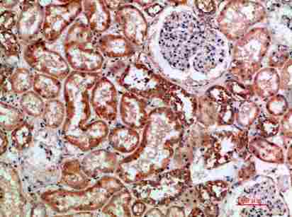
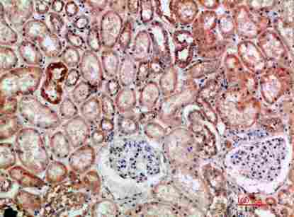
A04257 is a human-reactive anti-BTN3A1/2/3 antibody listed for IHC and IF (catalog: A04257 applications/reactivity). Its IHC image documents paraffin-embedded human kidney (A04257 image caption).

Real IHC data Immunohistochemical analysis of paraffin-embedded human-kidney, antibody was diluted at 1:200.
Anti-BTN3A1/2/3 Antibody
Cat # A04257

A04257 is listed for human IHC and IF (catalog: A04257 applications/reactivity). Its IHC image shows paraffin-embedded human kidney at 1:200; no IF image is supplied (A04257 image caption; catalog: A04257 IF image alts).

Which to pick: Choose A04257 for paraffin-section IHC based on its human-kidney image; the fixative is unreported (A04257 image caption). For IF, A04257 is listed at 1:50, but no IF image or ICC validation is supplied (catalog: A04257 applications/IF dilution/IF image alts). No cross-species choice is supported: A04257 is a rabbit polyclonal listed for human reactivity only (catalog: A04257 host/clonality/reactivity).

Each figure is that product's own IHC / IF validation image from its datasheet.

References

  1. UniProt Consortium. UniProt entry O00481 (BT3A1_HUMAN, Butyrophilin subfamily 3 member A1).
  2. Human Protein Atlas. BTN3A1 tissue IHC expression (reliability: Approved).
  3. Human Protein Atlas. BTN3A1 subcellular location (ICC-IF): Mainly localized to vesicles..
  4. Human Protein Atlas. BTN3A1 antibody validation summary (1 antibodies).
  5. High expression of BTN3A1 is associated with clinical and immunological characteristics and predicts a poor prognosis in advanced human gliomas. Frontiers in immunology 2024 — PMC11165028.
  6. Targeting of colorectal cancer organoids with zoledronic acid conjugated to the anti-EGFR antibody cetuximab. Journal for immunotherapy of cancer 2022 — PMC9772689.
  7. Comprehensive analysis of BTN3A1 in cancers: mining of omics data and validation in patient samples and cellular models. FEBS open bio 2021 — PMC8409294.
  8. Metformin sensitizes esophageal squamous cell carcinoma to Vγ9Vδ2 T cell-mediated cytotoxicity by upregulating BTN3A1 and BTN2A1. Cell death & disease 2026 — PMC13458587.
  9. PubMed PMID:9382921 — UniProt-cited evidence.
  10. PubMed PMID:9149941 — UniProt-cited evidence.
  11. PubMed PMID:14702039 — UniProt-cited evidence.