This website uses cookies to ensure you get the best experience on our website.


- Table of Contents
Plan chromogenic C1QBP IHC around granular cytoplasmic tissue staining (HPA tissue IHC). This guide covers fixation, controls and scoring while accounting for reported secretion (UniProt).

Expected localisation, validated protocols, controls and antibodies — the at-a-glance facts below, then the full design guide.
| Expected localisation | Granular cytoplasm in tissue (HPA tissue IHC) | |
| Staining pattern | Cells broadly show granular cytoplasmic staining (HPA tissue IHC) | |
| Antigen retrieval | EDTA pH 8.0 HIER, heat-mediated (datasheet M01439) | |
| Positive control | Appendix+4 more · see all | |
| Negative control | Liver+1 more · see all |
| Fixation | Keep fixation consistent across sections (standard IHC practice; not target-specific) | |
| Caveat | Secretion may complicate cell-associated staining (UniProt) | |
| Regulation | Activated lymphocytes secrete C1QBP (UniProt) | |
| Isoform / epitope | No annotated isoforms; mature chain spans residues 74–282 (UniProt) |
The catalog antibody’s IHC protocol uses EDTA pH 8.0 retrieval (datasheet M01439). One published paraffin section protocol is included below (PMC13069790).
| Sample | Paraffin-embedded human breast cancer tissue; fixative not specified (datasheet M01439) |
| Fixation | Image fixative and duration unreported (datasheet M01439); verify before use. |
| Sectioning | 4–5 µm sections on charged slides (standard) |
| Deparaffinisation | Xylene, graded ethanol series to water (standard) |
| Antigen retrieval | Heat retrieval: EDTA pH 8.0 (datasheet M01439); 20 min, 95–100 °C (standard) |
| Peroxidase block | 3% H2O2, 10 min, room temperature (standard) |
| Blocking | 10% goat serum (datasheet M01439) |
| Primary antibody | Rabbit monoclonal (clone 17C69) anti-C1QBP, 1:50-1:200 (datasheet M01439) |
| Primary incubation | Overnight at 4 °C (datasheet M01439) |
| Detection | HRP-conjugated secondary, DAB chromogen (datasheet M01439) |
| Counterstain | Hematoxylin, blue, dehydrate and mount (standard) |
| Expected result | C1QBP-positive staining in germinal center cells of appendix (HPA tissue IHC: High). HPA tissue profile: Ubiquitous cytoplasmic expression with a granular pattern. No signal in the no-primary control. |
C1QBP is predominantly mitochondrial (UniProt Q07021), while tissue IHC shows ubiquitous, granular cytoplasmic staining (HPA: Supported; HPA: low tissue specificity). Strong staining is reported in germinal center cells, glandular cells, endocrine cells, ovarian follicle cells and splenic white pulp cells (HPA: High in each listed cell type). Cell surface expression is also reported (UniProt Q07021); C1QBP has no transmembrane segment (UniProt Q07021 topology).
| Granular cytoplasmic staining in the expected cells, with stronger signal in germinal centers or endocrine cells. | This fits the tissue IHC profile (HPA: ubiquitous granular cytoplasm; High in germinal center and endocrine cells). Compare cells within the same section; the profile does not require equal intensity everywhere (HPA: low tissue specificity; variable reported staining levels). |
| Predominantly nuclear, nucleolar or smooth diffuse staining, with little granular cytoplasm. | This departs from the usual tissue IHC pattern (HPA: granular cytoplasm). Nuclear and nucleolar localization are reported (UniProt Q07021), so compartment alone cannot prove artefact. Check whether the pattern is restricted to identifiable cells and survives appropriate controls (general IHC practice). |
| Strong staining in cholangiocytes or skeletal myocytes while nearby expected cells stain weakly. | Those cell types were not detected in the supplied tissue profile (HPA: cholangiocytes and skeletal myocytes, Not detected). Cross-reactivity or endogenous chromogen activity is possible; compare a control without primary antibody and an expected positive section (general IHC practice). |
| A broad haze or uniform color over cells, stroma and blank areas. | That distribution does not resemble the reported cellular granules (HPA: granular cytoplasm). Background from nonspecific binding, endogenous detection activity or incomplete washing can obscure interpretation; inspect the control without primary antibody (general IHC practice). |
| No staining in an expected positive section, including its germinal center or endocrine cells. | This conflicts with the supplied positive profile (HPA: High in germinal center and endocrine cells). First verify that the expected cells are present, then check the antibody, detection reagents and run controls (general IHC practice); a blank slide alone cannot establish absent C1QBP. |
| Cell population and tissue choice | The RNA profile has low tissue specificity, but reported protein intensity differs by cell type (HPA: low tissue specificity; High, Low and Not detected groups). Score the named cell population rather than treating a whole section as uniformly positive (general IHC practice). |
| Compartment and assay | Tissue IHC is described as granular cytoplasmic (HPA: tissue IHC). ICC-IF instead lists plasma membrane as the main location and mitochondria as an additional location (HPA: ICC-IF, both supported). Interpret each assay against its own evidence. |
| Processing and cell surface signal | The annotated mature chain spans residues 74–282 and no transmembrane segment is annotated (UniProt Q07021 processing and topology). Cell surface expression is reported for macrophages and monocyte-derived dendritic cells (UniProt Q07021). The supplied record gives no antibody epitope, so it cannot predict which forms this antibody detects. |
| Extracellular distribution | A secreted form is reported, including secretion by activated lymphocytes (UniProt Q07021). HPA notes that secreted variants can complicate RNA–protein correspondence (HPA: reliability description). Extracellular color alone therefore does not identify its producing cell; assess cellular morphology and controls (general IHC practice). |
| Validation and retrieval limits | The tissue pattern has Supported reliability (HPA: tissue IHC), and HPA026483 is listed as IHC Supported (HPA: antibody validation). Neither entry supplies an antigen retrieval condition or target-specific fixation effect; use the IHC-validated antibody’s documented conditions if available (general IHC practice). |
| Situation | Likely cause | Next action |
|---|---|---|
| Expected positive tissue is blank. | The section may lack the reported positive cells (HPA: cell-specific High groups), or an IHC reagent or run step may have failed (general IHC practice). | Confirm cell identity on the section and inspect a concurrently processed positive control; review antibody dilution, detection and chromogen steps against the antibody instructions (general IHC practice). |
| Only uniformly dark tissue or diffuse haze is visible. | The pattern differs from granular cytoplasm (HPA: tissue IHC). Nonspecific binding or endogenous detection activity can contribute (general IHC practice). | Compare the control without primary antibody; review blocking, washing and chromogen development, then score only discrete cellular signal (general IHC practice). |
| Cholangiocytes or skeletal myocytes appear strongly positive. | These cells are reported as Not detected (HPA: tissue IHC); cross-reactivity or endogenous detection activity is possible (general IHC practice). | Check a control without primary antibody and an expected positive cell population in the same run; withhold a C1QBP call if control staining persists (general IHC practice). |
| Signal appears nuclear or nucleolar with little cytoplasmic granularity. | This is atypical for the tissue IHC profile (HPA: granular cytoplasm), although nuclear and nucleolar localization is reported (UniProt Q07021). | Record the cell type and distribution, compare with a positive tissue and the control without primary antibody, and avoid calling artefact from location alone (general IHC practice). |
| Some cells in an otherwise positive section look weak or negative. | Reported levels vary across cell populations, from High to Low or Not detected (HPA: tissue IHC); whole-section uniformity is not expected from those observations. | Score identified cell populations separately and compare their staining with the supplied HPA cell-type profile (HPA: tissue IHC; general IHC practice). |
| IF/ICC shows a membrane pattern: does that contradict the IHC result? | HPA assigns plasma membrane as the main ICC-IF location and mitochondria as an additional one (HPA: ICC-IF, both supported); its tissue IHC summary describes granular cytoplasm (HPA: tissue IHC). | Use the assay-specific pattern to interpret each result; for IF/ICC design and controls, consult its separate guide (HPA: ICC-IF; general IHC/IF practice). |
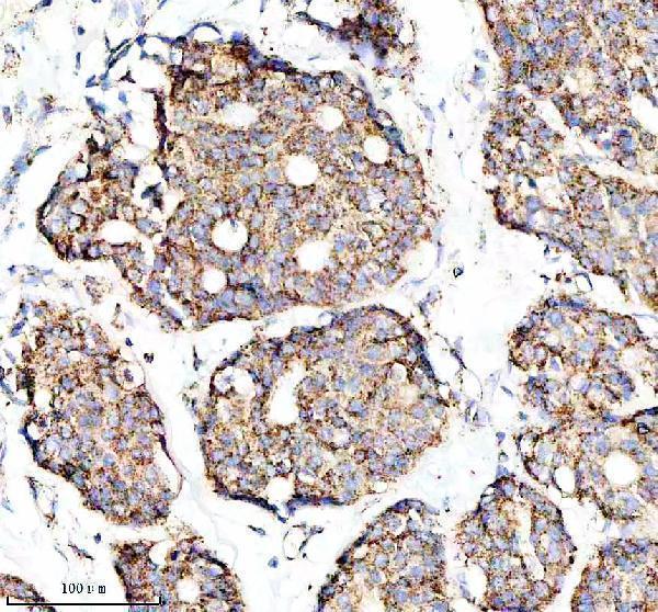
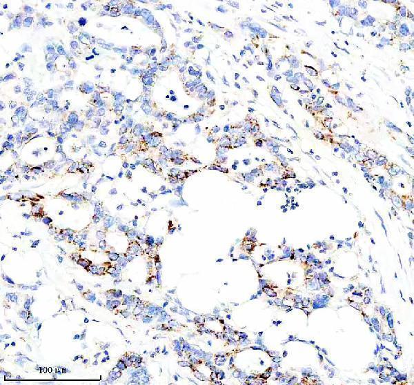


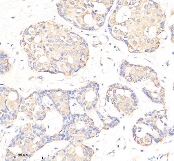
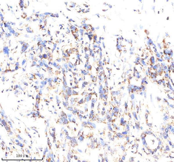


Comprehensive Human Protein Atlas IHC scoring per tissue (reliability: Supported — High consistency between antibody staining and RNA expression data. At least one protein variant secreted, tissue location of RNA and protein might differ and correlation is complex.). Rows are taken directly from the HPA tissue chart — click any row's HPA link to view the source.
| Tissue | Cell type | Level | Evidence | Source |
|---|---|---|---|---|
| Appendix | Germinal center cells | High | Protein (IHC) | HPA → |
| Breast | Glandular cells | High | Protein (IHC) | HPA → |
| Colon | Endocrine cells | High | Protein (IHC) | HPA → |
| Lymph node | Germinal center cells | High | Protein (IHC) | HPA → |
| Ovary | Follicle cells | High | Protein (IHC) | HPA → |
Troubleshoot C1QBP staining in paraffin sections by checking retrieval, compartment patterns, controls and scoring before interpreting chromogenic signal.
Anti-C1QBP antibodies have IHC images from human breast cancer and tonsil sections, plus mouse and rat tissues; one also has an IF image from U20S cells (catalog image captions).
M01439 has paraffin-section IHC images from human breast and pancreas cancers and mouse and rat midbrain; M01439-1 has images from human breast and pancreas cancers and rat cerebellum (catalog IHC captions). PB9921 has paraffin-section IHC images from human tonsil and mouse and rat spleen, plus an IF image from U20S cells (catalog image captions).
Which to pick: For paraffin-section IHC, choose rabbit monoclonal M01439 or M01439-1 when their imaged tissues match your study; each caption uses EDTA retrieval at pH 8.0 and a 1:50 primary dilution (catalog product details and IHC captions). For IF/ICC, choose PB9921: IF and ICC are listed applications, and its U20S cell IF caption uses 2 μg/mL primary antibody (catalog applications and IF caption). All three list human, mouse and rat reactivity, but their IHC captions document different tissues and retrieval conditions; the fixative is unreported in those captions (catalog reactivity and IHC captions).