Click image to see more details
-

-
-
-
-
+4
Product Info Summary
| SKU: | M01439 |
|---|---|
| Size: | 100 μl/vial |
| Reactive Species: | Human, Mouse, Rat |
| Host: | Rabbit |
| Application: | IHC, WB |
Customers Who Bought This Also Bought

Product info
Product Name
Anti-GC1q R Rabbit Monoclonal Antibody
SKU/Catalog Number
M01439
BM5284 is an alternative SKU for this antibody, used in previous lots.
Size
100 μl/vial
Form
Liquid
Description
Boster Bio Anti-GC1q R Rabbit Monoclonal Antibody catalog # M01439. Tested in WB, IHC applications. This antibody reacts with Human, Mouse, Rat.
Storage & Handling
Store at -20°C for one year. For short term storage and frequent use, store at 4°C for up to one month. Avoid repeated freeze-thaw cycles.
Cite This Product
Anti-GC1q R Rabbit Monoclonal Antibody (Boster Biological Technology, Pleasanton CA, USA, Catalog # M01439)
Host
Rabbit
Contents
Rabbit IgG in stabilizing components, phosphate buffered saline, pH 7.4, 150mM NaCl, 0.02% sodium azide and 50% glycerol.
*This antibody is supplied in a stabilized formulation.
Compatibility with conjugation reactions depends on the chemistry of the conjugation method used.
For conjugation methods that are not compatible with the stabilizing components present in this formulation, a carrier-free antibody format is required.
Clonality
Monoclonal
Clone Number
17C69
Isotype
IgG
Immunogen
A synthesized peptide derived from human GC1q R
Reactive Species
M01439 is reactive to C1QBP in Human, Mouse, Rat
Observed Molecular Weight
35 kDa
Calculated molecular weight
31.4 kDa
Antibody Validation
Boster validates all antibodies on WB, IHC, ICC, Immunofluorescence, and ELISA with known positive control and negative samples to ensure specificity and high affinity, including thorough antibody incubations.
Application & Images
Applications
M01439 is guaranteed for IHC, WB Boster Guarantee
Recommend Dilution
WB 1:500-2000
IHC 1:50-200
Tested application
Suggested blocking solution with 5% non-fat milk or BSA; (*)Recommended protein loading: 20-40 µg per lane
Validation Images & Assay Conditions
Click image to see more details
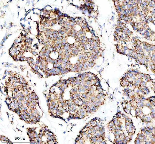
IHC analysis of C1QBP using anti-C1QBP antibody (M01439).
C1QBP was detected in a paraffin-embedded section of human breast cancer tissue. Heat mediated antigen retrieval was performed in EDTA buffer (pH 8.0, epitope retrieval solution). The tissue section was blocked with 10% goat serum. The tissue section was then incubated with 1:50 rabbit anti-C1QBP Antibody (M01439) overnight at 4°C. Peroxidase Conjugated Goat Anti-rabbit IgG was used as secondary antibody and incubated for 30 minutes at 37°C. The tissue section was developed using HRP Conjugated Rabbit IgG Super Vision Assay Kit (Catalog # SV0002) with DAB as the chromogen.

Click image to see more details
Western blot analysis of C1QBP using anti-C1QBP antibody (M01439).
Electrophoresis was performed on a 5-20% SDS-PAGE gel at 70V (Stacking gel) / 90V (Resolving gel) for 2-3 hours. The sample well of each lane was loaded with 30 ug of sample under reducing conditions.
Lane 1: human Hela whole cell lysates,
Lane 2: human 293T whole cell lysates,
Lane 3: human A549 whole cell lysates,
Lane 4: human HepG2 whole cell lysates.
After electrophoresis, proteins were transferred to a nitrocellulose membrane at 150 mA for 50-90 minutes. Blocked the membrane with 5% non-fat milk/TBS for 1.5 hour at RT. The membrane was incubated with rabbit anti-C1QBP antigen affinity purified monoclonal antibody (M01439) at 1:500 overnight at 4°C, then washed with TBS-0.1%Tween 3 times with 5 minutes each and probed with a goat anti-rabbit IgG-HRP secondary antibody at a dilution of 1:5000 for 1.5 hour at RT. The signal is developed using an Enhanced Chemiluminescent detection (ECL) kit (Catalog # EK1002) with Tanon 5200 system. A specific band was detected for C1QBP at approximately 35 kDa. The expected band size for C1QBP is at 31 kDa.

Click image to see more details
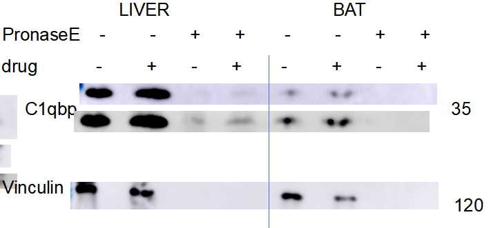
Western blot analysis of C1QBP using anti-C1QBP antibody (M01439).
Electrophoresis was performed on a 5-20% SDS-PAGE gel at 70V (Stacking gel) / 90V (Resolving gel) for 2-3 hours. The sample well of each lane was loaded with 30 ug of sample under reducing conditions.
Lane 1: mouse liver tissue lysates,
Lane 2: drug treatment-mouse liver tissue lysates,
Lane 3: pronaseE treatment-mouse liver tissue lysates,
Lane 4: pronaseE and drug treatment-mouse liver tissue lysates,
Lane 5: mouse brown adipose tissue lysates,
Lane 6: drug treatment-mouse brown adipose tissue lysates,
Lane 7: pronaseE treatment-mouse brown adipose tissue lysates,
Lane 8: pronaseE and drug treatment-mouse brown adipose tissue lysates.
After electrophoresis, proteins were transferred to a nitrocellulose membrane at 150 mA for 50-90 minutes. Blocked the membrane with 5% non-fat milk/TBS for 1.5 hour at RT. The membrane was incubated with rabbit anti-C1QBP antigen affinity purified monoclonal antibody (M01439) at 1:1500 overnight at 4°C, then washed with TBS-0.1%Tween 3 times with 5 minutes each and probed with a goat anti-rabbit IgG-HRP secondary antibody for 1 hour at RT. The signal is developed using an Enhanced Chemiluminescent detection (ECL) kit (Catalog # EK1002) with AI680 system. A specific band was detected for C1QBP at approximately 35 kDa. The expected band size for C1QBP is at 31 kDa.

Click image to see more details
IHC analysis of C1QBP using anti-C1QBP antibody (M01439).
C1QBP was detected in a paraffin-embedded section of human pancreas cancer tissue. Heat mediated antigen retrieval was performed in EDTA buffer (pH 8.0, epitope retrieval solution). The tissue section was blocked with 10% goat serum. The tissue section was then incubated with 1:50 rabbit anti-C1QBP Antibody (M01439) overnight at 4°C. Peroxidase Conjugated Goat Anti-rabbit IgG was used as secondary antibody and incubated for 30 minutes at 37°C. The tissue section was developed using HRP Conjugated Rabbit IgG Super Vision Assay Kit (Catalog # SV0002) with DAB as the chromogen.

Click image to see more details
IHC analysis of C1QBP using anti-C1QBP antibody (M01439).
C1QBP was detected in a paraffin-embedded section of mouse midbrain tissue. Heat mediated antigen retrieval was performed in EDTA buffer (pH 8.0, epitope retrieval solution). The tissue section was blocked with 10% goat serum. The tissue section was then incubated with 1:50 rabbit anti-C1QBP Antibody (M01439) overnight at 4°C. Peroxidase Conjugated Goat Anti-rabbit IgG was used as secondary antibody and incubated for 30 minutes at 37°C. The tissue section was developed using HRP Conjugated Rabbit IgG Super Vision Assay Kit (Catalog # SV0002) with DAB as the chromogen.

Click image to see more details
IHC analysis of C1QBP using anti-C1QBP antibody (M01439).
C1QBP was detected in a paraffin-embedded section of rat midbrain tissue. Heat mediated antigen retrieval was performed in EDTA buffer (pH 8.0, epitope retrieval solution). The tissue section was blocked with 10% goat serum. The tissue section was then incubated with 1:50 rabbit anti-C1QBP Antibody (M01439) overnight at 4°C. Peroxidase Conjugated Goat Anti-rabbit IgG was used as secondary antibody and incubated for 30 minutes at 37°C. The tissue section was developed using HRP Conjugated Rabbit IgG Super Vision Assay Kit (Catalog # SV0002) with DAB as the chromogen.

Click image to see more details
IHC analysis of C1QBP using anti-C1QBP antibody (M01439).
C1QBP was detected in a paraffin-embedded section of mouse cerebellum tissue. Heat mediated antigen retrieval was performed in EDTA buffer (pH 8.0, epitope retrieval solution). The tissue section was blocked with 10% goat serum. The tissue section was then incubated with 1:50 rabbit anti-C1QBP Antibody (M01439) overnight at 4°C. Peroxidase Conjugated Goat Anti-rabbit IgG was used as secondary antibody and incubated for 30 minutes at 37°C. The tissue section was developed using HRP Conjugated Rabbit IgG Super Vision Assay Kit (Catalog # SV0002) with DAB as the chromogen.

Click image to see more details
IHC analysis of C1QBP using anti-C1QBP antibody (M01439).
C1QBP was detected in a paraffin-embedded section of rat cerebellum tissue. Heat mediated antigen retrieval was performed in EDTA buffer (pH 8.0, epitope retrieval solution). The tissue section was blocked with 10% goat serum. The tissue section was then incubated with 1:50 rabbit anti-C1QBP Antibody (M01439) overnight at 4°C. Peroxidase Conjugated Goat Anti-rabbit IgG was used as secondary antibody and incubated for 30 minutes at 37°C. The tissue section was developed using HRP Conjugated Rabbit IgG Super Vision Assay Kit (Catalog # SV0002) with DAB as the chromogen.
Specific Publications For Anti-GC1q R Rabbit Monoclonal Antibody (M01439)
Loading publications
Recommended Resources
Here are featured tools and databases that you might find useful.
- Boster's Pathways Library
- Protein Databases
- Bioscience Research Protocol Resources
- Data Processing & Analysis Software
- Photo Editing Software
- Scientific Literature Resources
- Research Paper Management Tools
- Molecular Biology Software
- Primer Design Tools
- Bioinformatics Tools
- Phylogenetic Tree Analysis
Customer Reviews
Have you used Anti-GC1q R Rabbit Monoclonal Antibody?
Share your experimental results or join a short interview to earn up to $1,000 in product credits or other rewards.
1 Reviews For Anti-GC1q R Rabbit Monoclonal Antibody
This antibody is highly specific and efficient, suitable for Western blot detection of C1QBP protein in mouse liver and brown adipose tissue, with no nonspecific bands observed. However, due to experimental conditions and the particular characteristics of
Excellent
| SKU | M01439 |
|---|---|
| Application | Western Blot |
| Sample | rat colon tissue |
| Sample Processing Description | RIPA lysis buffer with protease inhibitor PMSF (100:1) was used to lyse the sample for 10 minutes, followed by centrifugation at 12,000 rpm for 15 minutes. The supernatant was mixed with 5× loading buffer, denatured at 100°C for 10 minutes, and then loaded onto SDS-PAGE. |
| Other Reagents | Blocking buffer |
| Primary Antibody | Anti-GC1q R Rabbit Monoclonal Antibody |
| Primary Incubation | 1:1000, overnight at 4 ℃ |
| Secondary Antibody | HRP Conjugated AffiniPure Goat Anti-Rabbit IgG (H+L) |
| Secondary Incubation | 1:5000, 1 hour in room temperature |
| Detection | Substrate: ECL, Imaging system:ChemiDoc MP |
| Results Summary | The figure shows Western blot results of the target protein and the loading control in mouse liver and brown adipose tissue from normal and treated groups, with or without enzymatic digestion. The target bands in the liver are clear and well defined, and the experimental results are satisfactory. |
Jiaqi Xin, Capital Medical University
Verified customer
Submitted 2026-01-08
Customer Q&As
Have a question?
Find answers in Q&As, reviews.
Can't find your answer?
Submit your question
