This website uses cookies to ensure you get the best experience on our website.


- Table of Contents
Plan chromogenic IHC-P for C1QTNF1 with the IHC-validated antibody starting at 10 μg/mL (datasheet: A09550-1). Compare cytoplasmic staining in kidney tubules and colon glands, while accounting for uncertain tissue localisation of this secreted protein (HPA tissue IHC; UniProt).

Expected localisation, validated protocols, controls and antibodies — the at-a-glance facts below, then the full design guide.
| Expected localisation | Cytoplasmic staining in most tissues (HPA tissue IHC) | |
| Staining pattern | Cytoplasmic staining in kidney tubules and colon glands (HPA tissue IHC) | |
| Antigen retrieval | Citrate pH 6.0 HIER, 95–98 °C, 20 min (rule: cytoplasmic / membrane antigen) | |
| Positive control | Colon+4 more · see all | |
| Negative control | Caudate+1 more · see all |
| Fixation | Matched tissue-IHC evidence does not establish the fixation claim. Validate the specimen-specific method before use. (selected-SKU IHC image A09550-1) | |
| Caveat | Secreted variants may separate RNA and protein locations (HPA tissue IHC) | |
| Regulation | Regulation unreported (UniProt) | |
| Isoform / epitope | 2 isoforms; mature chain spans residues 26–281; epitope unmapped (UniProt) |
Compare the catalog antibody’s IHC-P protocol (page contract) with a published C1QTNF1 tissue IHC method (PMC10387556).
| Sample | Tissue sections; selected-image fixative not specified (standard IHC workflow) |
| Fixation | Image fixative and duration unreported (datasheet A09550-1); verify before use. |
| Sectioning | 4–5 µm sections on charged slides (standard) |
| Deparaffinisation | Xylene, graded ethanol series to water (standard) |
| Antigen retrieval | Heat-induced epitope retrieval in citrate buffer, pH 6.0, 20 min at 95–98 °C (standard rule: cytoplasmic / membrane antigen) |
| Peroxidase block | 3% H2O2, 10 min, room temperature (standard) |
| Blocking | 10% normal serum of the secondary host, 30 min, room temperature (standard) |
| Primary antibody | Rabbit anti-C1QTNF1, 10 μg/mL (datasheet A09550-1) |
| Primary incubation | Overnight at 4 °C (standard) |
| Detection | HRP-polymer secondary, DAB chromogen 5–10 min (standard) |
| Counterstain | Hematoxylin, blue, dehydrate and mount (standard) |
| Expected result | C1QTNF1-positive staining in glandular cells of colon (HPA tissue IHC: High). HPA tissue profile: Cytoplasmic expression in most tissues. No signal in the no-primary control. |
C1QTNF1 is a secreted protein with a cleaved signal peptide and no transmembrane segment (UniProt Q9BXJ1 topology). In paraffin-section IHC, HPA reports cytoplasmic staining in most tissues, including high staining in several epithelial and other cell populations (HPA tissue IHC). Treat this as a provisional pattern: HPA rates the tissue IHC evidence Uncertain because secretion can separate protein staining from the cells expressing its RNA (HPA tissue IHC reliability).
| Clear cytoplasmic staining in colon or duodenal glandular cells, or kidney tubular cells (HPA tissue IHC: High). | This matches reported high-staining cell populations (HPA tissue IHC). Compare the named cells with nearby structures and a negative reagent control before calling the section positive (standard IHC practice). |
| Predominantly nuclear or sharply membrane-restricted chromogenic staining, with little cytoplasmic signal. | This departs from the reported tissue IHC profile (HPA tissue IHC: cytoplasmic). Check morphology, detection background and antibody specificity (standard IHC practice). Nuclear staining in ICC-IF is a separate HPA observation and does not validate nuclear IHC staining (HPA subcellular ICC-IF). |
| Strong staining mainly in cells outside the reported positive population of the selected tissue. | Treat the cell assignment as unresolved; cross-reactivity or endogenous detection activity are possible explanations (standard IHC practice). HPA's uncertain tissue reliability and C1QTNF1 secretion prevent a firm cell-of-origin claim from staining alone (HPA tissue IHC; UniProt Q9BXJ1 subcellular location). |
| Diffuse color across cells, stroma and blank areas, without a recognizable cell pattern. | This is background until controls establish otherwise (standard IHC practice). It cannot be scored against HPA's reported cytoplasmic cell pattern (HPA tissue IHC); inspect reagent controls and detection conditions (standard IHC practice). |
| No detectable staining in colon glandular cells or kidney tubular cells (HPA tissue IHC: High). | Investigate assay performance using a control section and the antibody's IHC-P instructions (standard IHC practice). A negative run does not overturn HPA's reported staining, and its Uncertain reliability means that report is not an absolute positive-control guarantee (HPA tissue IHC). |
| Secretion and topology (UniProt Q9BXJ1). | C1QTNF1 has a signal peptide, is secreted and has no transmembrane segment (UniProt Q9BXJ1). Staining need not identify the cell that made the protein; tissue RNA and protein locations may differ (HPA tissue IHC reliability). |
| Processing and epitope position (UniProt Q9BXJ1). | The annotated signal peptide spans residues 1–25 and the mature chain spans 26–281 (UniProt Q9BXJ1). Interpret an antibody result in light of its stated immunogen or epitope, if available (standard IHC practice); no epitope is supplied here. |
| Variants and molecular features (UniProt Q9BXJ1). | UniProt lists 2 isoforms, one glycosylation site at residue 93, a collagen-like region and a C1q domain (UniProt Q9BXJ1). These annotations alone do not establish which isoform or feature an IHC antibody detects. |
| Antibody and tissue-evidence limits (HPA antibodies; HPA tissue IHC). | The listed IHC antibody CAB025429 is rated Uncertain, while HPA051101 is Approved for ICC only (HPA antibodies). HPA also rates the tissue IHC pattern Uncertain (HPA tissue IHC); do not treat ICC approval as IHC validation. |
| IF/ICC Q&A: should nuclear signal be expected? | HPA reports approved nucleoplasm and cytosol locations in ICC-IF images from A-549, PC-3 and U2OS (HPA subcellular ICC-IF). That observation answers the IF/ICC question; it does not change the cytoplasmic tissue IHC reference pattern (HPA tissue IHC). |
| Choice of tissue control (HPA tissue IHC). | HPA reports high staining in colon glandular cells and kidney tubular cells, but no detected staining in caudate or hippocampal glial cells (HPA tissue IHC). Use these as comparison patterns with HPA's Uncertain reliability in mind (HPA tissue IHC reliability). |
| Situation | Likely cause | Next action |
|---|---|---|
| No signal in a reported high-staining cell population (HPA tissue IHC: High). | The IHC run may have failed, or this specimen may differ from the reported HPA pattern (standard IHC practice; HPA tissue IHC reliability: Uncertain). | Check a control section, reagent omission control and the catalog antibody's IHC-P instructions; assess retrieval and detection conditions as general IHC variables (standard IHC practice). |
| Color appears throughout the section, including spaces without cells. | Nonspecific reagent binding or detection background may obscure the reported cytoplasmic pattern (standard IHC practice; HPA tissue IHC). | Compare with a primary-antibody omission control, then review blocking, washing and chromogen development (standard IHC practice). |
| Brown deposit persists when the primary antibody is omitted. | Endogenous enzyme activity or another detection-system source is possible in chromogenic IHC (standard IHC practice). | Review the detection-system controls and apply the appropriate endogenous-activity block for that system (standard IHC practice). |
| A bright nuclear pattern dominates the paraffin-section result. | It conflicts with HPA's cytoplasmic tissue IHC profile, although HPA reports nucleoplasm staining in ICC-IF (HPA tissue IHC; HPA subcellular ICC-IF). | Score the IHC result separately from ICC-IF; verify cell boundaries, controls and staining with an independently validated IHC reagent if available (standard IHC practice). |
| Strong staining occurs in an unexpected cell population. | Cross-reactivity or detection background is possible; secreted C1QTNF1 also complicates assigning protein to its producing cell (standard IHC practice; UniProt Q9BXJ1 subcellular location). | Inspect the cell type and controls, then seek independent specificity evidence before assigning cellular expression (standard IHC practice; HPA tissue IHC reliability: Uncertain). |
| Glial cells stain strongly where HPA reports no detection (HPA tissue IHC: caudate and hippocampus). | The result differs from those HPA observations; nonspecific staining or specimen variation remains possible (HPA tissue IHC; standard IHC practice). | Check morphology and omission controls, then compare with a reported high-staining tissue in the same run; avoid treating HPA's Uncertain pattern as a definitive exclusion rule (standard IHC practice; HPA tissue IHC reliability). |


Comprehensive Human Protein Atlas IHC scoring per tissue (reliability: Uncertain — At least one protein variant secreted, tissue location of RNA and protein might differ and correlation is complex. Pending external verification.). Rows are taken directly from the HPA tissue chart — click any row's HPA link to view the source.
| Tissue | Cell type | Level | Evidence | Source |
|---|---|---|---|---|
| Colon | Glandular cells | High | Protein (IHC) | HPA → |
| Duodenum | Glandular cells | High | Protein (IHC) | HPA → |
| Esophagus | Squamous epithelial cells | High | Protein (IHC) | HPA → |
| Kidney | Cells in tubules | High | Protein (IHC) | HPA → |
| Lymph node | Germinal center cells | High | Protein (IHC) | HPA → |
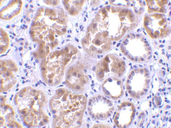
Use the kidney IHC example as a reference point while checking retrieval, tissue morphology, cell identity, and the limits of the available validation (selected A09550-1 tissue-IHC caption).
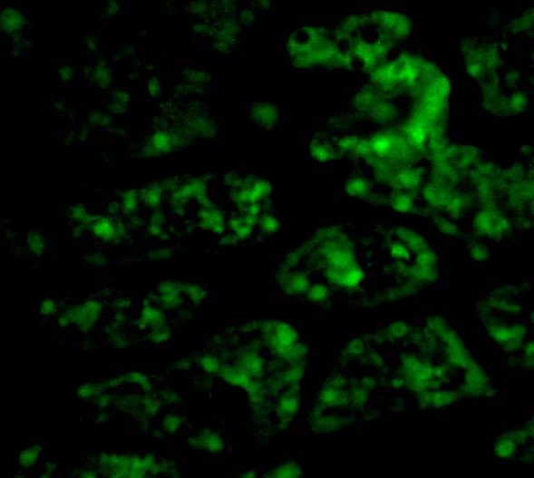
A09550-1 has human kidney tissue IHC and human kidney cell IF images (catalog image captions), with listed Human, Mouse and Rat reactivity (catalog reactivity).
A09550-1 is listed for IHC-P and IF (catalog applications). Its images show IHC in human kidney tissue at 10 μg/mL and IF in human kidney cells at 20 μg/mL (A09550-1 image captions).
Which to pick: For paraffin-section IHC, choose A09550-1: IHC-P is listed, and its own IHC caption shows human kidney tissue at 10 μg/mL; the fixative is unreported (catalog applications; A09550-1 IHC caption). For cell IF, choose A09550-1 based on its IF listing and human kidney cell image; ICC-specific validation is unreported (catalog applications; A09550-1 IF caption). For mouse or rat samples, A09550-1 has listed reactivity, though its displayed examples are human only and no clone is specified (catalog reactivity; image captions; catalog clone field).