This website uses cookies to ensure you get the best experience on our website.


- Table of Contents
Plan chromogenic CACNA1H IHC-P with the catalog antibody starting at 5 μg/mL (datasheet: IHC-P). Evaluate Leydig-cell staining and cytoplasmic/nuclear signal cautiously because presumed off-target binding was observed (HPA tissue IHC).

Expected localisation, validated protocols, controls and antibodies — the at-a-glance facts below, then the full design guide.
| Expected localisation | Tissue: cytoplasm/nuclei (HPA tissue IHC); expected: cell membrane (UniProt) | |
| Staining pattern | Highest in Leydig cells; cytoplasmic/nuclear across tissues (HPA tissue IHC) | |
| Antigen retrieval | Citrate pH 6.0 HIER, 95–98 °C, 20 min (rule: cytoplasmic / membrane antigen) | |
| Positive control | Testis+4 more · see all | |
| Negative control | Adipose tissue+4 more · see all |
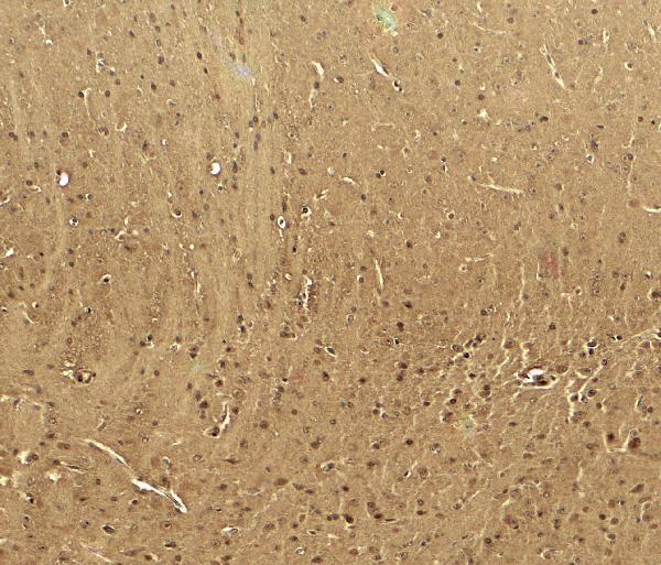
| Fixation | Keep fixation consistent (standard IHC practice; not target-specific); Matched tissue-IHC evidence does not establish the fixation claim. Validate the specimen-specific method before use. (selected-SKU IHC image A01406-1) | |
| Caveat | Presumed off-target staining and low RNA concordance (HPA tissue IHC) | |
| Regulation | STAC increases cell-membrane expression (UniProt) | |
| Isoform / epitope | 2 isoforms; check if the epitope is cytoplasmic or extracellular (UniProt) |
The catalog antibody’s IHC-P protocol is accompanied by published chromogenic IHC protocols for PPGL and breast tumor specimens (PMC11301417; PMC13271060).
| Sample | Tissue sections; selected-image fixative not specified (standard IHC workflow) |
| Fixation | Image fixative and duration unreported (datasheet A01406-1); verify before use. |
| Sectioning | 4–5 µm sections on charged slides (standard) |
| Deparaffinisation | Xylene, graded ethanol series to water (standard) |
| Antigen retrieval | Heat-induced epitope retrieval in citrate buffer, pH 6.0, 20 min at 95–98 °C (standard rule: cytoplasmic / membrane antigen) |
| Peroxidase block | 3% H2O2, 10 min, room temperature (standard) |
| Blocking | 10% normal serum of the secondary host, 30 min, room temperature (standard) |
| Primary antibody | Rabbit anti-CACNA1H, 5 μg/mL (datasheet A01406-1) |
| Primary incubation | Overnight at 4 °C (standard) |
| Detection | HRP-polymer secondary, DAB chromogen 5–10 min (standard) |
| Counterstain | Hematoxylin, blue, dehydrate and mount (standard) |
| Expected result | CACNA1H-positive staining in leydig cells of testis (HPA tissue IHC: High). HPA tissue profile: Cytoplasmic and nuclear expression in several tissues, Most abundant in Leydig cells of testis, stromal cells in ovary and glandular cells. No signal in the no-primary control. |
CACNA1H is a 24-pass cell-membrane channel (UniProt O95180 topology). In paraffin IHC, HPA reports the strongest staining in testicular Leydig cells, with moderate staining in several other cell types (HPA tissue IHC). HPA also describes cytoplasmic and nuclear staining, but rates tissue IHC Approved with low consistency against RNA data and presumed off-target binding (HPA tissue IHC). Interpret compartment and cell identity together.
| Strong staining in testicular Leydig cells, with cell-associated signal. | This matches the strongest listed tissue result: High in Leydig cells (HPA tissue IHC). Compare adjacent cell types and tissue structure before scoring a slide; intensity alone cannot resolve the off-target concern in the HPA reliability note (HPA tissue IHC). |
| Moderate staining in adrenal glandular cells or ciliary rootlets of bronchial or fallopian-tube ciliated cells. | These are listed at Medium intensity (HPA tissue IHC). The ciliary-rootlet pattern is a specific reported observation; score the stained cell and structure, rather than treating every stained area in those tissues as equivalent (HPA tissue IHC). |
| Predominantly nuclear staining without a convincing cell-associated pattern. | Treat this as uncertain: a membrane location is annotated (UniProt O95180 subcellular location), while nuclear tissue staining is also reported (HPA tissue IHC). HPA notes presumed off-target binding, so nuclear signal alone does not establish specific CACNA1H detection (HPA tissue IHC). |
| Strong staining in adipocytes or pancreatic exocrine glandular cells. | Both are listed as Not detected (HPA tissue IHC). Recheck cell identification, slide background and detection controls; unexpected staining may reflect cross-reactivity or endogenous detection activity (general IHC practice). It cannot be assigned to CACNA1H from intensity alone. |
| Uniform haze across cells and extracellular areas, or no staining in Leydig cells. | Haze obscures the reported cell-selective pattern; consider background from staining or detection steps (general IHC practice). A blank Leydig-cell section conflicts with HPA’s High observation, but may reflect assay performance or sample variation (HPA tissue IHC; general IHC practice). |
| Membrane topology and epitope access | CACNA1H has 24 transmembrane segments and intracellular and extracellular regions (UniProt O95180 topology). Epitope location is not supplied; assess the antibody’s documented epitope before inferring whether staining should outline a membrane or require permeabilisation (general IHC practice). |
| Reported intracellular patterns | HPA tissue IHC describes cytoplasmic and nuclear staining, while UniProt annotates the cell membrane (HPA tissue IHC; UniProt O95180 subcellular location). This disagreement limits compartment-only calls; HPA also reports low RNA–staining consistency and presumed off-target binding (HPA tissue IHC). |
| Isoforms and processing | UniProt lists 2 isoforms and one chain spanning residues 1–2353, with no signal peptide or propeptide annotated (UniProt O95180 isoforms and processing). The supplied record does not map an antibody epitope to either isoform, so isoform-specific IHC predictions are unavailable. |
| Retrieval and detection controls | Antigen retrieval, blocking and detection controls are general paraffin-IHC workflow choices (general IHC practice). The supplied HPA and UniProt records give no CACNA1H-specific retrieval condition or fixation sensitivity; do not infer one from topology or staining intensity (HPA tissue IHC; UniProt O95180 topology). |
| Situation | Likely cause | Next action |
|---|---|---|
| Leydig cells lack signal in a section expected to be positive. | The result differs from HPA’s High Leydig-cell observation; tissue preservation, retrieval, antibody or detection performance may need checking (HPA tissue IHC; general IHC practice). | Confirm Leydig-cell identity and tissue integrity, then review the validated IHC procedure and a suitable positive control (general IHC practice). Do not infer a CACNA1H-specific fixation effect from this failure. |
| Strong nuclear signal dominates the section. | HPA reports nuclear staining but also presumed off-target binding; UniProt locates CACNA1H at the cell membrane (HPA tissue IHC; UniProt O95180 subcellular location). | Record nuclear and nonnuclear patterns separately. Compare cell types with HPA’s tissue observations and inspect controls before calling the nuclear signal specific (HPA tissue IHC; general IHC practice). |
| Adipocytes, pancreatic exocrine cells or other listed negative cells stain strongly. | These examples conflict with HPA’s Not detected entries (HPA tissue IHC). Cross-reactivity or endogenous detection activity is possible (general IHC practice). | Check cell identification and a no-primary control; review blocking and detection steps for the assay used (general IHC practice). Keep the unexpected staining separate from a positive CACNA1H score. |
| Color is diffuse, including extracellular spaces. | Field-wide haze can arise from background staining or detection steps (general IHC practice); it does not reproduce HPA’s cell-specific observations (HPA tissue IHC). | Compare the section with a no-primary control, inspect wash and blocking steps, and score only resolvable cells after background is addressed (general IHC practice). |
| A tissue shows moderate staining, but the stained cell type is unclear. | HPA assigns levels to particular cells, including adrenal glandular cells and bronchial ciliary rootlets, rather than to every cell in a tissue (HPA tissue IHC). | Use morphology and counterstain to identify the stained cells; document compartment and intensity with the cell name (general IHC practice). Mark uncertain identities as unresolved. |
| Can HPA’s IF/ICC images define the paraffin-IHC positive pattern? | HPA IF/ICC lists approved nucleoplasm and vesicles in A-549 and MCF-7 images; tissue IHC has a separate staining profile and reliability note (HPA subcellular ICC-IF; HPA tissue IHC). | Use those IF/ICC locations as a modality-specific comparison only. Base paraffin-IHC cell selection and scoring on tissue-IHC observations, with their low-consistency caveat (HPA tissue IHC). |


Comprehensive Human Protein Atlas IHC scoring per tissue (reliability: Approved — Low consistency between antibody staining and RNA expression data. Presumed off target binding observed and disregarded.). Rows are taken directly from the HPA tissue chart — click any row's HPA link to view the source.
| Tissue | Cell type | Level | Evidence | Source |
|---|---|---|---|---|
| Testis | Leydig cells | High | Protein (IHC) | HPA → |
| Adrenal gland | Glandular cells | Medium | Protein (IHC) | HPA → |
| Bronchus | Ciliated cells (ciliary rootlets) | Medium | Protein (IHC) | HPA → |
| Esophagus | Squamous epithelial cells | Medium | Protein (IHC) | HPA → |
| Fallopian tube | Ciliated cells (ciliary rootlets) | Medium | Protein (IHC) | HPA → |
| Tissue | Cell type | Level | Evidence | Source |
|---|---|---|---|---|
| Adipose tissue | Adipocytes | Not detected | Protein (IHC) | HPA → |
| Pancreas | Exocrine glandular cells | Not detected | Protein (IHC) | HPA → |
| Seminal vesicle | Glandular cells | Not detected | Protein (IHC) | HPA → |
| Smooth muscle | Smooth muscle cells | Not detected | Protein (IHC) | HPA → |
| Spleen | Cells in red pulp | Not detected | Protein (IHC) | HPA → |
Troubleshoot CACNA1H chromogenic IHC by testing retrieval and detection conditions, then judging staining against membrane topology and cell-level controls.
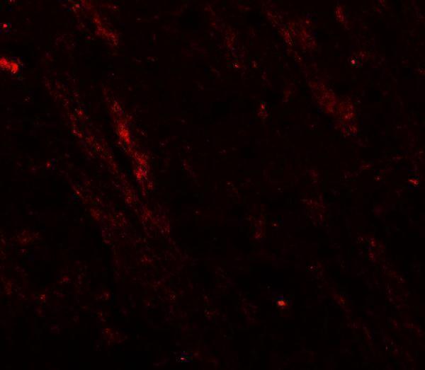
A01406-1 has mouse brain IHC and IF images (catalog: image captions) and listed reactivity with human, mouse, and rat (catalog: reactivity).
A01406-1 is listed for IHC-P and IF (catalog: applications), with mouse brain images at 5 μg/mL for IHC and 20 μg/mL for IF (catalog: image captions). The supplied images document mouse brain only (catalog: image captions); human and rat are listed as reactive species (catalog: reactivity).
Which to pick: For paraffin-section IHC, choose A01406-1 (catalog: IHC-P); its IHC image shows mouse brain at 5 μg/mL (catalog: IHC image caption), but the fixative is unreported (catalog: IHC image caption). For IF, choose A01406-1 based on its mouse brain image at 20 μg/mL (catalog: IF image caption); ICC is not separately listed (catalog: applications). For work across species, A01406-1 lists human, mouse, and rat reactivity (catalog: reactivity), although its supplied IHC and IF images show mouse brain only (catalog: image captions); the host is rabbit and clonality is unspecified (catalog: host; clone).