CACYBP / Calcyclin-binding protein · IHC design guide

Design Immunohistochemistry for CACYBP

Plan chromogenic IHC on paraffin sections with the IHC-validated RP1104 antibody at 0.5–1 µg/mL (datasheet: IHC-P). This guide covers cytoplasmic scoring, using strongly stained endocrine cells and undetected myocytes as tissue references (HPA tissue IHC).

Evidence assembled Oct 2026 · For research use; verify linked source records and product datasheet before use
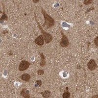
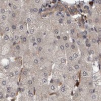
Immunohistochemistry protocol sheet for CACYBP (IHC for CACYBP): expected localisation Cytoplasmic staining in most tissues (HPA tissue IHC), antibody RP1104, validated IHC image, and IHC protocol steps
Printable CACYBP IHC protocol sheet — expected localisation Cytoplasmic staining in most tissues (HPA tissue IHC), antibody RP1104, controls and protocol steps. Open the full CACYBP IHC guide →

CACYBP Immunohistochemistry Experimental Design Guide

Expected localisation, validated protocols, controls and antibodies — the at-a-glance facts below, then the full design guide.

Must know before staining
Expected localisation Cytoplasmic staining in most tissues (HPA tissue IHC)
Staining pattern Cytoplasmic in most tissues; endocrine cells can stain strongly (HPA tissue IHC)
Antigen retrieval EDTA pH 8.0 HIER, heat-mediated (datasheet RP1104)
Positive control ⓘ Appendix+4 more · see all
Negative control ⓘ Heart muscle+3 more · see all
Important caveats
Reasons your staining may differ from the expected pattern.
Fixation Keep fixation consistent across sections (standard IHC practice; not target-specific)
Caveat Cell type matters: myocytes are undetected (HPA tissue IHC)
Regulation RA/calcium can shift compartment signal (UniProt)
Isoform / epitope 3 isoforms; epitope coverage is unspecified (UniProt)
Section 1

Recommended CACYBP IHC & IF Protocols

The catalog antibody has a datasheet IHC-P protocol (datasheet RP1104); four published CACYBP IHC protocols provide tissue-specific examples (PMC10779320; PMC7051089; PMC3996359; PMC10299707).

Recommended immunohistochemistry (IHC-P) protocol parameters
SampleParaffin-embedded human intestinal cancer tissue; fixative not specified (datasheet RP1104)
FixationImage fixative and duration unreported (datasheet RP1104); verify before use.
Sectioning4–5 µm sections on charged slides (standard)
DeparaffinisationXylene, graded ethanol series to water (standard)
Antigen retrievalHeat retrieval: EDTA pH 8.0 (datasheet RP1104); 20 min, 95–100 °C (standard)
Peroxidase block3% H2O2, 10 min, room temperature (standard)
Blocking10% goat serum (datasheet RP1104)
Primary antibodyRabbit anti-CACYBP, 0.5-1μg/ml (datasheet RP1104)
Primary incubationOvernight at 4 °C (datasheet RP1104)
DetectionStreptavidin-biotin complex (SABC), DAB chromogen (datasheet RP1104)
CounterstainHematoxylin, blue, dehydrate and mount (standard)
Expected resultCACYBP-positive staining in endocrine cells of appendix (HPA tissue IHC: High). HPA tissue profile: Cytoplasmic expression in most tissues. No signal in the no-primary control.
💡Decision noteStart with heat-mediated EDTA pH 8.0 retrieval for the catalog antibody (datasheet RP1104); the published protocols used citrate pH 6.0 with their antibodies (PMC10779320; PMC7051089; PMC3996359; PMC10299707).
Section 2

What Is the Expected CACYBP Staining Pattern?

CACYBP is mainly cytoplasmic in tissue IHC (HPA: tissue IHC) and mainly cytosolic with additional nucleoplasmic signal in ICC-IF (HPA: subcellular). Expect staining in endocrine cells of the appendix and colon, neurons of the cerebral cortex, and germinal center cells of lymph nodes (HPA: High in each). HPA rates tissue IHC reliability Enhanced, while noting medium agreement with RNA expression (HPA: reliability). CACYBP has no transmembrane segment (UniProt Q9HB71 topology).

What am I looking at on my slide?
Clear cytoplasmic staining in appendix or colon endocrine cells, with identifiable cell outlines (HPA: High in each).This matches the reported cell types and tissue IHC compartment (HPA: tissue IHC). Compare staining with adjacent cells and morphology before scoring; intensity alone cannot identify an endocrine cell (general IHC practice).
Staining appears exclusively nuclear, with little or no cytoplasmic signal in otherwise positive tissue (HPA: tissue IHC).A nuclear-only pattern is discordant with HPA's predominantly cytoplasmic tissue profile, so investigate staining or interpretation artefact (HPA: tissue IHC; general IHC practice). Some nuclear localization remains biologically plausible (UniProt Q9HB71 subcellular; HPA: subcellular).
Strong signal appears in cardiomyocytes or skeletal myocytes while expected positive cells remain weak (HPA: Not detected in those muscle cells).Treat the mismatch as possible cross-reactivity or endogenous chromogenic activity; inspect morphology and detection controls before calling CACYBP positive (HPA: tissue IHC; general IHC practice).
Color spreads across extracellular spaces or obscures cell boundaries throughout the section (general IHC practice).This is diffuse background rather than an interpretable cell-localized pattern. Check primary-antibody concentration, blocking and detection controls before assigning a cell type (general IHC practice).
No signal is seen in appendix endocrine cells or cerebral cortex neurons expected to stain strongly (HPA: High in each).A negative result in these cells makes the run inconclusive for other sections. Check that the positive cells are present, then review retrieval, antibody and detection performance (HPA: tissue IHC; general IHC practice).
💡Expected CACYBP appearanceCall a result positive when identifiable expected cells show clear, predominantly cytoplasmic chromogenic staining (HPA: tissue IHC); nuclear-only staining or color in HPA-undetected muscle cells warrants investigation rather than a positive call (HPA: tissue IHC; general IHC practice).
How each factor affects the staining
Compartment and calcium state (UniProt Q9HB71 subcellular).UniProt describes cytoplasmic localization at low calcium and both nuclear and cytoplasmic localization after retinoic acid induction with increased calcium in neuroblastoma cells; that cell-model observation does not predict a shift in every paraffin tissue (UniProt Q9HB71 subcellular).
Tissue and cell selection (HPA: tissue IHC).HPA reports High staining in several specific cell populations but low tissue RNA specificity overall. Score the identified cell population, not every cell in a nominally positive tissue; HPA also reports Not detected in cardiomyocytes and myocytes (HPA: tissue IHC).
Antibody evidence (HPA: antibodies; HPA: reliability).HPA lists IHC Enhanced for two rabbit polyclonal antibodies, HPA025753 and HPA057038, while the tissue profile notes medium staining-to-RNA consistency. This supports comparison with the reported pattern without making every unexpected signal target-specific (HPA: antibodies; HPA: reliability).
Isoforms and epitope information (UniProt Q9HB71 isoforms).Three isoforms are listed, but the supplied record does not map the catalog antibody's epitope or establish isoform-specific IHC staining. Avoid assigning a staining difference to one isoform from these data alone (UniProt Q9HB71 isoforms).
Chromogenic detection activity (general IHC practice).Endogenous activity or nonspecific detection can produce color unrelated to the primary antibody. Use an appropriate detection control when unexpected cells stain; this is a general IHC concern, not a reported CACYBP-specific effect (general IHC practice).
Why is my staining missing, weak or wrong?
SituationLikely causeNext action
Expected appendix or colon endocrine cells show no chromogenic signal (HPA: High in each).The selected cells may be absent from the examined field, or the IHC workflow may have failed; the supplied sources do not identify a CACYBP-specific fixation sensitivity (HPA: tissue IHC; general IHC practice).Confirm cell identity and section quality, then compare a known-positive section processed in the same run. Review the validated antibody dilution, retrieval conditions and detection steps without assuming a target-specific fixation effect (general IHC practice).
Signal is predominantly nuclear in tissue IHC (HPA: tissue IHC).The pattern differs from HPA's cytoplasmic tissue profile, although nuclear localization is reported by UniProt and ICC-IF (HPA: tissue IHC; UniProt Q9HB71 subcellular; HPA: subcellular).Check counterstain, cell boundaries and controls; report the compartment explicitly. Seek independent confirmation before interpreting a nuclear-only tissue pattern as CACYBP (general IHC practice).
Cardiomyocytes, skeletal myocytes or soft-tissue fibroblasts stain strongly (HPA: Not detected in each).The result conflicts with the listed HPA cell patterns and may reflect cross-reactivity or endogenous detection activity (HPA: tissue IHC; general IHC practice).Compare with an appropriate control that omits the primary antibody and inspect whether color follows tissue structures rather than cells; repeat with adjusted detection conditions if needed (general IHC practice).
Diffuse staining makes cell-level scoring impossible (general IHC practice).Excess antibody or detection signal, inadequate blocking, or insufficient washing can raise nonspecific background (general IHC practice).Review the catalog antibody's validated IHC-P instructions, titrate within its stated range if supplied, and check blocking and wash steps. Do not infer a CACYBP-specific epitope or fixation effect from background alone (general IHC practice).
A nominally positive tissue looks weak overall despite a technically adequate run (HPA: tissue IHC).HPA's High calls apply to named cell populations, and its reliability note reports only medium agreement with RNA expression; whole-section intensity can obscure a localized positive population (HPA: tissue IHC; HPA: reliability).Find and score the specified cells, such as lymph-node germinal center cells or cerebral cortex neurons, using morphology and a comparable positive control (HPA: High in each; general IHC practice).
Q: Should IF/ICC show the same compartment pattern as tissue IHC (HPA: tissue IHC; HPA: subcellular)?A: HPA reports mainly cytosolic signal with additional nucleoplasmic signal in ICC-IF, while tissue IHC is described as cytoplasmic in most tissues (HPA: subcellular; HPA: tissue IHC).Interpret IF/ICC compartment calls against the HPA subcellular record and use the separate IF/ICC guide for that application; do not treat an IF image as a substitute for the IHC tissue and cell controls (HPA: subcellular; general IHC practice).

Sample controls for CACYBP IHC & IF

🧪Run appendix first: endocrine cells should stain strongly (HPA: High in appendix endocrine cells); use heart muscle as the negative tissue, where cardiomyocytes are not detected (HPA: Not detected in heart muscle cardiomyocytes). On the appendix slide, adjacent cells without visible signal should show only background chromogen, but their CACYBP-negative status is unverified (HPA: appendix result identifies endocrine cells).
Positive control tissue: Appendix (Endocrine cells, HPA High)
Negative control tissue: Heart muscle (HPA Not detected)
ICC-IF cell lines (HPA subcellular resource): HPA ICC-IF images show CACYBP in A-431, U-251MG, U2OS, with annotated localisation: Cytosol (enhanced) (HPA subcellular).
Technical controls: Include a no-primary, secondary-only control and a host- and clonality-matched rabbit isotype control alongside a CACYBP-knockout biological negative (selected caption: rabbit primary antibody). For appendix sections, quench endogenous peroxidase and check endogenous biotin before interpreting the biotin-based DAB signal (selected caption: biotinylated secondary, streptavidin-biotin complex and DAB).
⚠️Feasibility: A target-specific fixation window and fixation effect are unreported, and the selected RP1104 tissue-IHC caption does not state the fixative. The caption reports heat retrieval in EDTA at pH 8.0 for a paraffin section, but does not establish that retrieval is required; the supplied evidence also does not establish whether frozen sections or IF would be easier (selected RP1104 tissue-IHC caption; HPA: ICC-IF images available). In appendix, endogenous peroxidase or biotin can complicate interpretation of the caption’s biotin-based DAB detection (selected RP1104 tissue-IHC caption).

HPA tissue IHC evidence for CACYBP

Comprehensive Human Protein Atlas IHC scoring per tissue (reliability: Enhanced — Medium consistency between antibody staining and RNA expression data.). Rows are taken directly from the HPA tissue chart — click any row's HPA link to view the source.

Positive expression · recommended positive controls

TissueCell typeLevelEvidenceSource
Appendix Endocrine cells High Protein (IHC) HPA →
Cerebellum Cells in molecular layer High Protein (IHC) HPA →
Cerebral cortex Neuronal cells High Protein (IHC) HPA →
Colon Endocrine cells High Protein (IHC) HPA →
Duodenum Endocrine cells High Protein (IHC) HPA →

Undetected expression · recommended negative controls

TissueCell typeLevelEvidenceSource
Heart muscle Cardiomyocytes Not detected Protein (IHC) HPA →
Skeletal muscle Myocytes Not detected Protein (IHC) HPA →
Smooth muscle Smooth muscle cells Not detected Protein (IHC) HPA →
Soft tissue Fibroblasts Not detected Protein (IHC) HPA →
Section 3

Advanced CACYBP IHC Tips

Troubleshoot CACYBP staining in paraffin sections by checking retrieval, compartment, cell identity and controls before comparing signal intensity.

Which retrieval conditions should I start with for CACYBP IHC?
Start with heat-mediated antigen retrieval in EDTA at pH 8.0 (datasheet RP1104). The selected paraffin-section example used this condition before incubating with 1 μg/ml catalog antibody overnight at 4°C (datasheet RP1104). If staining is weak, compare a shorter and longer heating interval on adjacent sections while keeping antibody concentration and detection constant (standard IHC practice). Check section integrity and background at each interval; excessive heating can damage morphology or increase nonspecific staining (standard IHC practice). Record the heating method and cooling time so that a useful condition can be repeated (standard IHC practice).
How should I troubleshoot weak staining when fixation history is uncertain?
CACYBP-specific fixation sensitivity is unknown from the supplied evidence; the selected tissue caption identifies a paraffin section but does not state its fixative (datasheet RP1104). Record the fixative, fixation duration and processing history for each specimen before comparing staining across cases (standard IHC practice). Run a known positive section in the same batch, using the selected example’s EDTA pH 8.0 retrieval and 1 μg/ml primary concentration as starting conditions (datasheet RP1104). If both test and control sections are weak, check retrieval and detection before attributing the result to fixation (standard IHC practice). If only one specimen is weak, report its fixation history as a possible limitation, without claiming a CACYBP-specific fixation effect (standard IHC practice).
Should I score nuclear CACYBP staining as positive?
Score cytoplasmic staining first: tissue IHC shows cytoplasmic expression in most tissues, and cell imaging places CACYBP mainly in the cytosol (HPA: tissue IHC; HPA: subcellular). Nuclear signal can be plausible because nucleoplasmic localization is also reported (HPA: subcellular; UniProt Q9HB71: localization). Record nuclear and cytoplasmic staining separately, with the percentage of positive cells and intensity for each compartment (standard IHC practice). Compare the pattern with an expected positive cell population on the same slide, such as colon endocrine cells (HPA: High in colon endocrine cells). Treat isolated nuclear staining cautiously if adjacent controls lack convincing cytoplasmic signal (standard IHC practice).
Could isoforms or epitope accessibility explain discordant IHC results?
CACYBP has 3 annotated isoforms, while the supplied catalog caption does not identify the antibody epitope (UniProt Q9HB71: isoforms; datasheet RP1104). Its CS region spans residues 73–167 and its SGS region spans 168–228; neither location identifies this antibody’s binding site (UniProt Q9HB71: domains; datasheet RP1104). Check the antibody’s documented immunogen before interpreting differences between antibodies as isoform-specific (standard IHC practice). Processing and retrieval can alter epitope accessibility in paraffin sections, so compare antibodies on adjacent sections under documented conditions (standard IHC practice). Report discordant patterns as antibody-dependent until epitope coverage and staining specificity are established (standard IHC practice).
How can IF help resolve an ambiguous chromogenic CACYBP pattern?
Use IF as a separate localization check, multiplexing CACYBP with a validated marker for the cell population being assessed, such as endocrine cells in colon (HPA: High in colon endocrine cells; standard IF practice). Choose a far-red CACYBP fluorophore when the tissue has substantial shorter-wavelength autofluorescence, and include unstained and single-label controls (standard IF practice). Permeabilize to reach cytosolic and nucleoplasmic epitopes: CACYBP has no transmembrane segment and is reported in both compartments (UniProt Q9HB71: topology and localization; HPA: subcellular). Titrate permeabilization and antibody concentration on matched sections to preserve morphology and limit diffuse signal (standard IF practice). Interpret IF alongside the chromogenic section, since staining conditions and detection methods differ (standard IHC/IF practice).
What should I check when DAB signal appears throughout the section?
First compare a no-primary control with the stained section to identify signal from detection reagents or tissue rather than primary-antibody binding (standard IHC practice). The selected example used 10% goat serum, a biotinylated secondary antibody, an avidin–biotin detection complex and DAB (datasheet RP1104). Include a peroxidase block and assess endogenous biotin when using this detection scheme, especially if background persists in the no-primary control (standard IHC practice). If background depends on the primary antibody, titrate from the reported 1 μg/ml concentration and review blocking and wash conditions (datasheet RP1104; standard IHC practice). Preserve the same retrieval and development conditions across comparison sections (standard IHC practice).
How should I quantify CACYBP staining across specimens? ⚠ ANSWER MARKED FOR VERIFICATION
Define the cell population and compartment before scoring because CACYBP is mainly cytosolic, with additional nucleoplasmic localization reported (HPA: subcellular). For chromogenic IHC, record % positive cells and intensity, then calculate an H-score if the study requires a combined measure (standard IHC practice). For sparse populations, count positive cells per mm² of viable tissue and retain the denominator for each specimen (standard IHC practice). Normalize comparisons to the same cell population, viable area, retrieval batch and image-analysis thresholds (standard IHC practice). Keep nuclear and cytoplasmic scores separate, and document whether endocrine or neuronal cells were included, since both show high staining in listed tissues (HPA: tissue IHC).
How can I distinguish true CACYBP staining from artefact?
A credible pattern includes cytoplasmic staining in an expected population, such as colon endocrine cells or cerebral cortex neuronal cells (HPA: tissue IHC). Additional nuclear staining can be credible, but a pattern confined to an unexpected compartment needs control review (UniProt Q9HB71: localization; HPA: subcellular; standard IHC practice). Inspect section edges, folds and necrotic regions for staining that tracks tissue damage rather than cell boundaries (standard IHC practice). Compare no-primary and peroxidase-block controls before assigning diffuse DAB signal to CACYBP (standard IHC practice). Weigh any negative result against the assay controls and the HPA report of only medium consistency between antibody staining and RNA data (HPA: reliability description).
Boster reagents

Best CACYBP / Calcyclin-binding protein IHC Antibodies

CACYBP antibody RP1104 has paraffin-section IHC images from human intestinal cancer and mouse and rat testis (catalog image captions); PA1759 has IF data from HeLa cells (catalog image caption).

Real IHC data IHC analysis of CACYBP using anti-CACYBP antibody (RP1104). CACYBP was detected in a paraffin-embedded section of human intestinal cancer tissue. Heat mediated antigen retrieval was performed in EDTA buffer (pH 8.0, epitope retrieval solution). The tissue section was blocked with 10% goat serum. The tissue section was then incubated with 1 μg/ml rabbit anti-CACYBP Antibody (RP1104) overnight at 4°C. Biotinylated goat anti-rabbit IgG was used as secondary antibody and incubated for 30 minutes at 37°C. The tissue section was developed using Strepavidin-Biotin-Complex (SABC) (Catalog # SA1022) with DAB as the chromogen.
Anti-SIAH Interacting Protein/CACYBP Antibody ®
Cat # RP1104
Real IF data IF analysis of CACYBP using anti-CACYBP antibody (PA1759). CACYBP was detected in an immunocytochemical section of Hela cells. Enzyme antigen retrieval was performed using IHC enzyme antigen retrieval reagent (AR0022) for 15 mins. The cells were blocked with 10% goat serum. And then incubated with 5 μg/mL rabbit anti-CACYBP Antibody (PA1759) overnight at 4°C. DyLight®488 Conjugated Goat Anti-Rabbit IgG (BA1127) was used as secondary antibody at 1:500 dilution and incubated for 30 minutes at 37°C. The section was counterstained with DAPI. Visualize using a fluorescence microscope and filter sets appropriate for the label used.
Anti-SIAH Interacting Protein/CACYBP Antibody ®
Cat # PA1759

RP1104 will render with paraffin-section IHC data from human intestinal cancer tissue; its catalog also shows mouse and rat testis IHC images (catalog image captions). PA1759 will render with IF data from HeLa cells and is listed for IF/ICC in human, mouse and rat (catalog image caption; catalog applications and reactivity).

Which to pick: Choose RP1104 for tissue IHC: it is listed for IHC in human, mouse and rat, and its own images document paraffin-section staining in all three species (catalog applications, reactivity and image captions). Choose PA1759 for IF/ICC when the HeLa IF example is relevant; both antibodies list human, mouse and rat reactivity, but PA1759 has no listed IHC application (catalog image caption; catalog applications and reactivity). RP1104’s IHC captions specify paraffin embedding and EDTA retrieval at pH 8.0; the fixative is unreported (catalog image captions).

Each figure is that product's own IHC / IF validation image from its datasheet.

References

  1. UniProt Consortium. UniProt entry Q9HB71 (CYBP_HUMAN, Calcyclin-binding protein).
  2. Human Protein Atlas. CACYBP tissue IHC expression (reliability: Enhanced).
  3. Human Protein Atlas. CACYBP subcellular location (ICC-IF): Mainly localized to the cytosol. In addition localized to the nucleoplasm..
  4. Human Protein Atlas. CACYBP antibody validation summary (2 antibodies).
  5. Evaluation of the Expression and Localization of the Multifunctional Protein CacyBP/SIP and Elements of the MAPK Signaling Pathway in the Adrenal Glands of Rats with Primary and Secondary Hypertension. International journal of molecular sciences 2023 — PMC10779320.
  6. Ageing-related changes in the levels of β-catenin, CacyBP/SIP, galectin-3 and immunoproteasome subunit LMP7 in the heart of men. PloS one 2020 — PMC7051089.
  7. Up-regulation of CacyBP/SIP during rat breast cancer development. Breast cancer (Tokyo, Japan) 2014 — PMC3996359.
  8. The Effect of CacyBP/SIP on the Phosphorylation of ERK1/2 and p38 Kinases in Clear Cell Renal Cell Carcinoma. International journal of molecular sciences 2023 — PMC10299707.
  9. PubMed PMID:12006993 — UniProt-cited evidence.
  10. PubMed PMID:14702039 — UniProt-cited evidence.
  11. PubMed PMID:16710414 — UniProt-cited evidence.