This website uses cookies to ensure you get the best experience on our website.


- Table of Contents
Plan CALB2 chromogenic IHC on paraffin sections with tissue controls and consistent fixation. Interpret cytoplasmic staining by cell type, including brain cells and testicular Leydig cells (HPA tissue IHC).

Expected localisation, validated protocols, controls and antibodies — the at-a-glance facts below, then the full design guide.
| Expected localisation | Cytoplasmic staining (HPA tissue IHC); dendrites and synapses (UniProt) | |
| Staining pattern | Cytoplasmic signal in brain cells and Leydig cells (HPA tissue IHC) | |
| Antigen retrieval | EDTA pH 8.0 HIER, heat-mediated (datasheet A04255) | |
| Positive control | Adrenal gland+4 more · see all | |
| Negative control | Appendix+4 more · see all |
| Fixation | Keep fixation consistent (standard IHC practice; not target-specific); Matched tissue-IHC evidence does not establish the fixation claim. Validate the specimen-specific method before use. (selected-SKU IHC image A04255) | |
| Caveat | Staining varies by cell type and tissue (HPA tissue IHC) | |
| Regulation | RNA enhanced in adipose and brain (HPA tissue RNA) | |
| Isoform / epitope | No isoforms; one 1–271 chain, with no extracellular domain (UniProt) |
The catalog antibody protocol (datasheet A04255) is followed by three published chromogenic IHC protocols for CALB2 in gonadal tissue and medulla (PMC7552167; PMC7729007; PMC12567713).
| Sample | Paraffin-embedded human rectal cancer tissue; fixative not specified (datasheet A04255) |
| Fixation | Image fixative and duration unreported (datasheet A04255); verify before use. |
| Sectioning | 4–5 µm sections on charged slides (standard) |
| Deparaffinisation | Xylene, graded ethanol series to water (standard) |
| Antigen retrieval | Heat retrieval: EDTA pH 8.0 (datasheet A04255); 20 min, 95–100 °C (standard) |
| Peroxidase block | 3% H2O2, 10 min, room temperature (standard) |
| Blocking | 10% goat serum (datasheet A04255) |
| Primary antibody | Rabbit anti-CALB2, 0.5-1μg/ml (datasheet A04255) |
| Primary incubation | Overnight at 4 °C (datasheet A04255) |
| Detection | Streptavidin-biotin complex (SABC), DAB chromogen (datasheet A04255) |
| Counterstain | Hematoxylin, blue, dehydrate and mount (standard) |
| Expected result | CALB2-positive staining in glandular cells of adrenal gland (HPA tissue IHC: High). HPA tissue profile: Cytoplasmic expression in several tissues and cells, including brain, adipose tissue, fallopian tube, retina, endocrine pancreas and Leydig cells. No signal in the no-primary control. |
CALB2 staining should be predominantly cytoplasmic in selected cells, including neuronal cells and Leydig cells (HPA tissue IHC: High in caudate and cortical neuronal cells; High in Leydig cells). Signal may extend into neuronal projections (HPA tissue IHC: High in hypothalamic neuronal projections; UniProt P22676: dendrite and synapse). CALB2 has no transmembrane segment (UniProt P22676 topology). The tissue IHC profile has Enhanced reliability, reflecting high agreement between staining and RNA expression (HPA tissue IHC: Enhanced).
| Defined cytoplasmic staining in caudate or cortical neuronal cells, with stained processes where visible (HPA tissue IHC: High in both neuronal populations; High in hypothalamic projections). | This fits the expected cellular distribution (HPA tissue IHC: cytoplasmic profile; UniProt P22676: dendrite and synapse). Assess recognizable cells and processes rather than judging an entire brain section uniformly positive (HPA tissue IHC: cell-specific observations). |
| Predominantly nuclear staining, especially without accompanying cytoplasmic signal. | An isolated nuclear pattern lacks support from the supplied localization records (HPA subcellular: supported cytosol; UniProt P22676: dendrite and synapse). Treat it as suspect and compare it with a known-positive section and detection controls before interpreting it as CALB2 (general IHC practice). |
| Strong staining in appendix glandular cells or bone-marrow hematopoietic cells. | Those particular cell populations are recorded as Not detected (HPA tissue IHC: appendix glandular cells; bone-marrow hematopoietic cells). Investigate antibody cross-reactivity or detection background (general IHC practice). Do not generalize this finding to every glandular cell: adrenal glandular cells are High (HPA tissue IHC). |
| Diffuse color across cells, stroma, and section edges, obscuring cell boundaries. | A broad haze cannot establish the cell-restricted CALB2 pattern (HPA tissue IHC: cell-specific staining). It can arise from nonspecific binding, incomplete blocking, or detection background (general IHC practice); interpret only after the background is reduced and positive cells remain distinct. |
| No signal in morphologically identifiable Leydig cells or caudate neuronal cells. | These are reported High populations (HPA tissue IHC: testis Leydig cells; caudate neuronal cells). A blank result there raises a workflow or reagent question (general IHC practice), but one negative section alone cannot establish absent CALB2 expression. |
| Compartment and topology (HPA subcellular: supported cytosol; UniProt P22676 topology). | CALB2 has no transmembrane segment, and its supported cellular location is cytosolic (UniProt P22676 topology; HPA subcellular). Expect intracellular staining; a continuous cell-surface rim alone is insufficient, although cerebellar GLUC cells are annotated as cytoplasm/membrane (HPA tissue IHC). |
| Choice of comparison cells (HPA tissue IHC). | Use a reported High cell population as the positive reference and a specified Not detected population as a comparison (HPA tissue IHC: caudate neuronal cells High; appendix glandular cells Not detected). Low staining in lung macrophages is recorded, so faint macrophage signal is not automatically abnormal (HPA tissue IHC: Low). |
| Antibody validation (HPA antibodies: IHC Enhanced). | HPA007305, HPA007306, and CAB000106 each have Enhanced IHC status (HPA antibodies). That status supports the reported IHC pattern through agreement between independent antibodies or orthogonal data; it does not validate an unexpected pattern in every specimen (HPA antibody-validation definition; general IHC interpretation). |
| Antigen retrieval and fixation (general IHC practice). | The supplied UniProt and HPA records provide no CALB2-specific retrieval condition or fixation-sensitivity result. Compare routine paraffin-section retrieval conditions only if the known-positive control fails, and judge them by specific signal versus background (general IHC practice). |
| Protein form and epitope limits (UniProt P22676). | The record lists one chain spanning residues 1–271, no signal peptide or propeptide, and no annotated isoforms (UniProt P22676 processing). It also lists EF-hand domains and phosphotyrosine at residue 214; without an antibody epitope map, these details do not predict retrieval needs or a staining difference (UniProt P22676; general IHC interpretation). |
| Situation | Likely cause | Next action |
|---|---|---|
| A known-positive section is blank (HPA tissue IHC: caudate neuronal cells High). | The target cells may be absent from the examined field, or a staining step may have failed (general IHC practice). | Verify the cell population on the section, then check antibody application, retrieval, detection reagents, and counterstain against a working positive control (general IHC practice). |
| Nuclei stain more strongly than cytoplasm. | Predominantly nuclear localization conflicts with the supported cytosolic location (HPA subcellular: supported cytosol); nonspecific binding or detection background is possible (general IHC practice). | Compare the same run with a known-positive section and a primary-antibody omission control; adjust blocking or antibody concentration if background persists (general IHC practice). |
| Appendix glandular cells show strong signal (HPA tissue IHC: Not detected). | The pattern may reflect cross-reactivity or endogenous detection activity rather than CALB2 (general IHC practice; HPA tissue IHC: appendix glandular cells Not detected). | Check a primary-antibody omission control and the detection-system control, then confirm that an expected High cell population still stains specifically (general IHC practice; HPA tissue IHC). |
| Color is diffuse throughout the section. | Nonspecific binding or excessive chromogen development can obscure cell boundaries (general IHC practice). | Review blocking, washes, antibody concentration, and development time; retain conditions that resolve individual positive cells with a clear background (general IHC practice; HPA tissue IHC: cell-specific profile). |
| Only faint signal appears in lung macrophages. | Low staining is reported for this population (HPA tissue IHC: lung macrophages Low), so intensity alone cannot establish a failed assay. | Judge the run using a reported High population and a specified Not detected population; avoid treating the whole lung as a negative control (HPA tissue IHC; general IHC practice). |
| IF/ICC Q: Where should CALB2 signal appear? | A: Cytosol is the supported ICC-IF location (HPA subcellular: supported cytosol). | Interpret IF/ICC on its own guide page; compare localization with the supported cytosolic record, while using this page's cell-specific tissue observations only for IHC interpretation (HPA subcellular; HPA tissue IHC). |

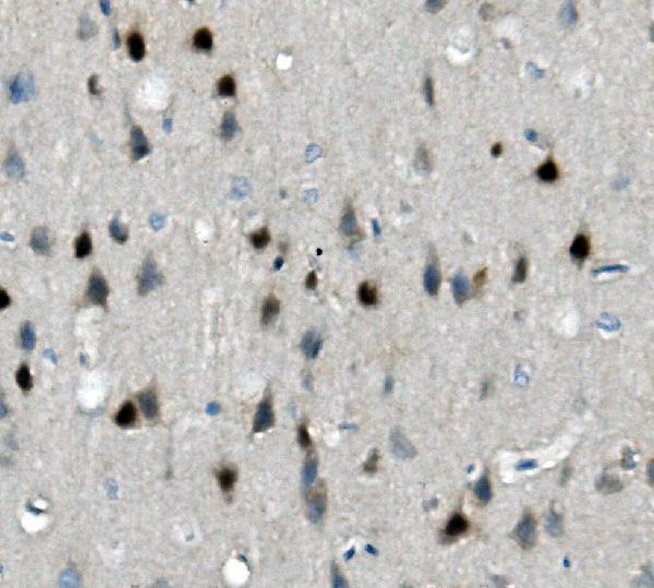
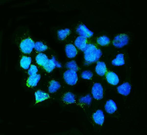


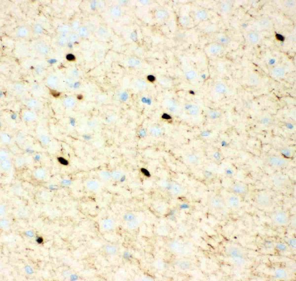
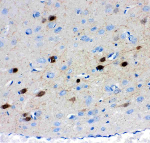

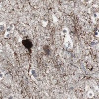
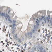

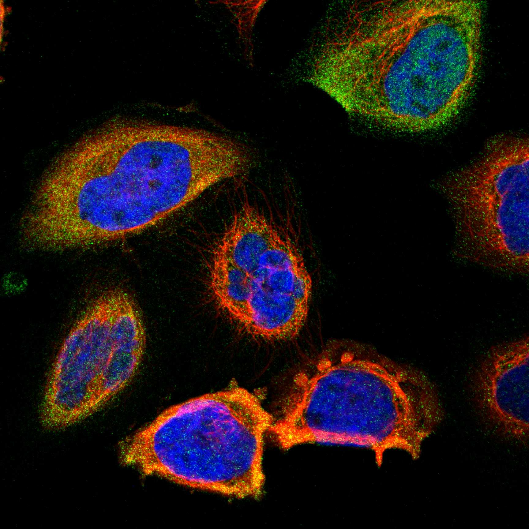

Comprehensive Human Protein Atlas IHC scoring per tissue (reliability: Enhanced — High consistency between antibody staining and RNA expression data.). Rows are taken directly from the HPA tissue chart — click any row's HPA link to view the source.
| Tissue | Cell type | Level | Evidence | Source |
|---|---|---|---|---|
| Adrenal gland | Glandular cells | High | Protein (IHC) | HPA → |
| Caudate | Neuronal cells | High | Protein (IHC) | HPA → |
| Cerebellum | GLUC cells - cytoplasm/membrane | High | Protein (IHC) | HPA → |
| Cerebral cortex | Neuronal cells | High | Protein (IHC) | HPA → |
| Hippocampus | Glial cells | High | Protein (IHC) | HPA → |
| Tissue | Cell type | Level | Evidence | Source |
|---|---|---|---|---|
| Appendix | Glandular cells | Not detected | Protein (IHC) | HPA → |
| Bone marrow | Hematopoietic cells | Not detected | Protein (IHC) | HPA → |
| Bronchus | Basal cells | Not detected | Protein (IHC) | HPA → |
| Cervix | Glandular cells | Not detected | Protein (IHC) | HPA → |
| Duodenum | Glandular cells | Not detected | Protein (IHC) | HPA → |
Troubleshoot CALB2 staining in paraffin sections by checking retrieval, controls, cellular location and scoring before interpreting chromogenic signal.
The catalog antibodies have paraffin-section IHC images from human cancer tissue and mouse or rat brain (A04255, PA1015, PB9248: IHC captions); PA1015 also has a mouse-brain IF image (PA1015: IF caption).
A04255 has IHC images from human rectal cancer and brain and mouse and rat brain; PA1015 has IHC images from human rectal cancer and mouse and rat brain, plus IF from mouse brain (A04255, PA1015: image captions). PB9248 has IHC images from mouse and rat brain and human intestinal cancer (PB9248: IHC captions).
Which to pick: For paraffin-section chromogenic IHC, A04255 and PA1015 have captions documenting EDTA retrieval at pH 8.0, goat-serum blocking and DAB detection; PB9248 also has paraffin IHC images, but its captions give fewer processing details (A04255, PA1015, PB9248: IHC captions). For IF, choose PA1015 because its own caption shows mouse-brain IF; for ICC, A04255 lists ICC as an application, but its supplied IF caption names a different SKU (PA1015: IF caption; A04255: applications and IF caption). For cross-species tissue IHC, A04255 and PA1015 each show human, mouse and rat samples (A04255, PA1015: IHC captions); the captions do not report the fixative (A04255, PA1015: IHC captions).