CAMKK2 / Calcium/calmodulin-dependent protein kinase kinase 2 · IHC design guide

Design Immunohistochemistry for CAMKK2

Plan CAMKK2 paraffin-section IHC using cytoplasmic staining as the tissue pattern (HPA tissue IHC). Compare cerebral cortex neurons and prostate glandular cells, both reported as high-staining, while accounting for low consistency between staining and RNA expression (HPA tissue IHC).

Evidence assembled Oct 2026 · For research use; verify linked source records and product datasheet before use
Immunohistochemistry protocol sheet for CAMKK2 (IHC for CAMKK2): expected localisation Predominantly cytoplasmic tissue staining (HPA tissue IHC), antibody A03048, validated IHC image, and IHC protocol steps
Printable CAMKK2 IHC protocol sheet — expected localisation Predominantly cytoplasmic tissue staining (HPA tissue IHC), antibody A03048, controls and protocol steps. Open the full CAMKK2 IHC guide →

CAMKK2 Immunohistochemistry Experimental Design Guide

Expected localisation, validated protocols, controls and antibodies — the at-a-glance facts below, then the full design guide.

Must know before staining
Expected localisation Predominantly cytoplasmic tissue staining (HPA tissue IHC)
Staining pattern Cytoplasmic staining in neurons and epithelial cells (HPA tissue IHC)
Antigen retrieval EDTA pH 8.0 HIER, heat-mediated (datasheet A03048)
Positive control ⓘ Cerebral cortex+4 more · see all
Negative control ⓘ Adipose tissue+4 more · see all
Important caveats
Reasons your staining may differ from the expected pattern.
Fixation Keep fixation consistent across sections. (standard IHC practice; not target-specific)
Caveat Staining has low consistency with RNA expression (HPA tissue IHC)
Regulation Higher expression in brain (UniProt)
Isoform / epitope 7 isoforms; epitope coverage is unverified (UniProt)
Section 1

Recommended CAMKK2 IHC & IF Protocols

Compare the catalog antibody’s IHC-P protocol (datasheet A03048) with published CAMKK2 staining protocols for gynecologic tumors (PMC10472110) and breast cancer tissue microarrays (PMC6547743).

Recommended immunohistochemistry (IHC-P) protocol parameters
SampleParaffin-embedded human colorectal adenocarcinoma tissue; fixative not specified (datasheet A03048)
FixationImage fixative and duration unreported (datasheet A03048); verify before use.
Sectioning4–5 µm sections on charged slides (standard)
DeparaffinisationXylene, graded ethanol series to water (standard)
Antigen retrievalHeat retrieval: EDTA pH 8.0 (datasheet A03048); 20 min, 95–100 °C (standard)
Peroxidase block3% H2O2, 10 min, room temperature (standard)
Blocking10% goat serum (datasheet A03048)
Primary antibodyRabbit anti-CAMKK2, 2-5 μg/ml (datasheet A03048)
Primary incubationOvernight at 4 °C (datasheet A03048)
DetectionHRP-conjugated secondary, DAB chromogen (datasheet A03048)
CounterstainHematoxylin, blue, dehydrate and mount (standard)
Expected resultCAMKK2-positive staining in neuronal cells of cerebral cortex (HPA tissue IHC: High). HPA tissue profile: General cytoplasmic expression. No signal in the no-primary control.
💡Decision noteStart with heat-mediated EDTA pH 8.0 for the catalog antibody (datasheet A03048). Both published protocols used citrate pH 6.0 with HPA017389 (PMC10472110; PMC6547743).
Section 2

What Is the Expected CAMKK2 Staining Pattern?

In paraffin tissue IHC, expect predominantly cytoplasmic CAMKK2 staining in neuronal cells of the cerebral cortex and glandular cells of the prostate, among other positive cell populations (HPA tissue IHC: High). Nuclear staining can be plausible because UniProt reports nuclear localization that varies with stimulation (UniProt Q96RR4: subcellular location). CAMKK2 has no transmembrane segment (UniProt Q96RR4: topology). Interpret intensity cautiously: HPA rates tissue staining Approved but reports low consistency with RNA data (HPA tissue IHC: reliability).

What am I looking at on my slide?
Clear cytoplasmic signal in cerebral cortex neurons or prostate glandular cells, with recognizable unstained neighboring structures.This matches two cell populations scored High by HPA (HPA tissue IHC). Score the intended cell population and compartment rather than treating the entire section as uniformly positive (general IHC practice). HPA’s Approved rating is accompanied by low staining–RNA consistency, so intensity alone does not prove specificity (HPA tissue IHC: reliability).
Predominantly crisp plasma-membrane outlines, extracellular deposits, or luminal staining replace intracellular signal.These patterns warrant an artefact or nonspecific-binding check: CAMKK2 is reported in cytoplasm and nucleus and lacks a transmembrane segment (UniProt Q96RR4: location and topology). Nuclear signal alone is not grounds for rejection; UniProt reports predominantly nuclear localization in unstimulated cells and redistribution after forskolin induction (UniProt Q96RR4: subcellular location).
Strong signal centers on adipocytes or skeletal muscle myocytes while the intended positive cells stain weakly.Those cell populations are scored Not detected in HPA tissue IHC (HPA tissue IHC: adipose tissue; skeletal muscle). Consider cross-reactivity or chromogenic endogenous activity before assigning CAMKK2 positivity (general IHC practice). A negative reference is a comparison for this staining setup, not proof that every specimen must lack signal.
Color covers stroma, empty spaces, and many cell types without a discernible intracellular boundary.Diffuse staining does not resemble HPA’s general cytoplasmic profile (HPA tissue IHC: profile). It may reflect background from antibody binding, detection reagents, or substrate development (general IHC practice). Compare matched controls and assess whether any signal remains specifically associated with the expected cells.
No discernible signal appears in cerebral cortex neurons or prostate glandular cells.These are HPA High populations, so an absent signal is an assay warning (HPA tissue IHC). Check slide integrity and the established IHC staining workflow before interpreting the specimen as negative (general IHC practice). HPA’s low staining–RNA consistency limits how confidently any single reference tissue predicts a result (HPA tissue IHC: reliability).
💡Expected CAMKK2 appearanceCall a result positive when distinct, chiefly cytoplasmic chromogen appears in the expected neuronal or glandular cells at a readily distinguishable intensity (HPA tissue IHC: High; HPA tissue IHC: general cytoplasmic expression); isolated nuclear signal may fit the reported localization (UniProt Q96RR4), whereas crisp surface outlines or broad cell-independent deposits warrant a false-positive check (UniProt Q96RR4: topology; general IHC practice).
How each factor affects the staining
Cell compartment and stimulation stateHPA describes general cytoplasmic tissue staining and supported cytosolic ICC-IF localization (HPA tissue IHC; HPA subcellular). UniProt also places CAMKK2 in the nucleus and reports movement into cytoplasm and neurites after forskolin induction (UniProt Q96RR4). Keep assay type and cell state in view when judging a nuclear component.
Tissue and cell selectionCerebral cortex neurons and prostate glandular cells are scored High, while adipocytes and skeletal muscle myocytes are Not detected (HPA tissue IHC). These comparisons help identify useful positive and low-signal reference populations; they do not define a universal intensity threshold (general IHC practice).
Antibody evidenceThe listed antibodies HPA017389 and HPA063713 are IHC Approved; only HPA017389 is ICC Supported (HPA antibodies). Tissue staining also carries a low staining–RNA consistency note (HPA tissue IHC: reliability). Verify which antibody generated a reference pattern before transferring that expectation to another reagent.
Isoforms and target structureUniProt lists 7 CAMKK2 isoforms, a kinase domain at residues 165–446, and no transmembrane segment or signal peptide (UniProt Q96RR4). Without an epitope map in the supplied evidence, these features cannot predict which isoforms a given antibody detects or justify a surface-staining interpretation.
Why is my staining missing, weak or wrong?
SituationLikely causeNext action
The known positive tissue has no signal.The staining workflow may have failed, or the antibody and tissue preparation may be incompatible (general IHC practice); HPA alone cannot identify which step failed.Review tissue preservation, the established IHC antigen-retrieval conditions, antibody preparation, detection reagents, and development against a working control (general IHC practice). Do not infer a CAMKK2-specific fixation effect: none is established by the supplied UniProt or HPA records.
A negative-reference cell population stains strongly.Cross-reactivity or endogenous chromogenic activity is possible (general IHC practice), especially when adipocytes or skeletal muscle myocytes are the stained cells (HPA tissue IHC: Not detected).Compare an appropriate no-primary control and the expected positive cell population; check the detection-system controls and blocking steps used for the chromogen (general IHC practice). Treat unexpected staining as unresolved until its source is separated from specific intracellular signal.
The whole section has diffuse brown background.Nonspecific binding, incomplete blocking, or excess chromogen development can obscure cell-level staining (general IHC practice).Check a no-primary control, review the established blocking and wash steps, and compare development with the positive reference (general IHC practice). Reassess only discrete intracellular staining; HPA describes a general cytoplasmic profile (HPA tissue IHC).
Only a strong membrane rim or extracellular deposit is visible.That distribution conflicts with reported cytoplasmic and nuclear locations and the absence of a transmembrane segment (UniProt Q96RR4).Inspect morphology and matched controls for edge artefact, trapped reagent, or nonspecific deposits (general IHC practice). Avoid scoring a membrane-only pattern as CAMKK2 without independent evidence.
Nuclear and cytoplasmic signals disagree between specimens.Cell state may matter: UniProt describes predominantly nuclear localization in unstimulated cells and redistribution after forskolin induction (UniProt Q96RR4). HPA tissue IHC reports general cytoplasmic expression (HPA tissue IHC).Record nuclear and cytoplasmic staining separately, compare like cell types and conditions, and avoid declaring nuclear signal false solely from the HPA tissue summary (UniProt Q96RR4; HPA tissue IHC).
Does IF/ICC require the same compartment call as tissue IHC?HPA reports supported cytosolic ICC-IF localization, while UniProt also reports nuclear and neuron-projection localization under specified contexts (HPA subcellular; UniProt Q96RR4).Use the separate IF/ICC guide for its assay workflow. Here, compare localization claims by assay and cell state; the supplied ICC-IF evidence supports cytosol but does not establish an IHC protocol or a universal absence of nuclear signal (HPA subcellular; UniProt Q96RR4).

Sample controls for CAMKK2 IHC & IF

🧪Run cerebral cortex first: neuronal cells should stain strongly (HPA: High in neuronal cells). Use adipose tissue as the negative tissue, where adipocytes are not detected (HPA: Not detected in adipocytes); on the cortex slide, neighboring cells without specific DAB staining can serve as an internal background reference, but their CAMKK2-negative status is unconfirmed.
Positive control tissue: Cerebral cortex (Neuronal cells, HPA High)
Negative control tissue: Adipose tissue (HPA Not detected)
ICC-IF cell lines (HPA subcellular resource): HPA ICC-IF images show CAMKK2 in A-431, U-251MG, U2OS, ASC52telo, hTERT-RPE1 (serum starved), with annotated localisation: Cytosol (supported) (HPA subcellular).
Technical controls: Include a no-primary, secondary-only control, a concentration-matched rabbit IgG isotype control matched to the primary antibody’s clonality, and a CAMKK2 knockout or validated peptide-block control (IHC caption: rabbit anti-CAMKK2 antibody). Quench endogenous peroxidase before HRP/DAB detection and inspect cerebral cortex for pigment that could be mistaken for chromogen (IHC caption: HRP/DAB detection; standard IHC practice).
⚠️Feasibility: A target-specific fixation window or fixation effect is unreported, and the selected A03048 tissue-IHC caption does not state the fixative (IHC caption: fixative not stated). Heat retrieval in EDTA at pH 8.0 was used for the paraffin-section image, but retrieval dependence has not been established (IHC caption: EDTA pH 8.0). The supplied evidence does not establish that frozen sections or tissue IF are easier; neuronal pigment may complicate cortex signal assessment (HPA: High in cerebral cortex neuronal cells; standard IHC/IF practice).

HPA tissue IHC evidence for CAMKK2

Comprehensive Human Protein Atlas IHC scoring per tissue (reliability: Approved — Low consistency between antibody staining and RNA expression data.). Rows are taken directly from the HPA tissue chart — click any row's HPA link to view the source.

Positive expression · recommended positive controls

TissueCell typeLevelEvidenceSource
Cerebral cortex Neuronal cells High Protein (IHC) HPA →
Esophagus Squamous epithelial cells High Protein (IHC) HPA →
Oral mucosa Squamous epithelial cells High Protein (IHC) HPA →
Parathyroid gland Glandular cells High Protein (IHC) HPA →
Prostate Glandular cells High Protein (IHC) HPA →

Undetected expression · recommended negative controls

TissueCell typeLevelEvidenceSource
Adipose tissue Adipocytes Not detected Protein (IHC) HPA →
Fallopian tube Glandular cells Not detected Protein (IHC) HPA →
Seminal vesicle Glandular cells Not detected Protein (IHC) HPA →
Skeletal muscle Myocytes Not detected Protein (IHC) HPA →
Soft tissue Fibroblasts Not detected Protein (IHC) HPA →
Section 3

Advanced CAMKK2 IHC Tips

Use the catalog antibody’s tissue protocol as a starting point, then verify staining against cell type, compartment and appropriate controls.

How should I adjust retrieval when CAMKK2 staining is weak?
Start with heat-mediated antigen retrieval in EDTA at pH 8.0 for paraffin sections (datasheet A03048). The catalog antibody produced a CAMKK2 image in a paraffin section after this retrieval, followed by 2 μg/ml primary antibody overnight at 4°C (datasheet A03048). If staining remains weak, compare retrieval duration across matched sections while keeping antibody concentration and detection constant (standard IHC practice). Include a neuronal-cell-rich cerebral cortex section as a positive reference and assess staining in the expected cells, since those cells show high tissue staining (HPA: High in cerebral cortex neuronal cells). Record whether stronger retrieval also increases diffuse background or damages tissue morphology (standard IHC practice).
Could fixation explain inconsistent CAMKK2 staining between paraffin blocks?
CAMKK2-specific sensitivity to fixation is unknown: the selected paraffin-section caption does not state a fixative, and the supplied evidence does not compare fixation conditions (datasheet A03048). Record each block’s fixative and fixation interval, then compare sections processed with the same EDTA pH 8.0 retrieval and staining run (datasheet A03048; standard IHC practice). Include a common positive reference section so changes in detection can be separated from differences among blocks (standard IHC practice). Assess preserved morphology alongside staining, because damaged or poorly preserved areas make cell-level interpretation unreliable (standard IHC practice). Do not assign a fixation effect to CAMKK2 without a controlled comparison (datasheet A03048: fixation unreported).
Should CAMKK2 staining appear nuclear or cytoplasmic in tissue sections?
Score cytoplasmic staining first, because the tissue IHC profile describes general cytoplasmic expression (HPA: tissue IHC profile). Nuclear staining is biologically plausible: CAMKK2 is predominantly nuclear in unstimulated cells and can move into cytoplasm and neurites after forskolin induction (UniProt Q96RR4: subcellular location). The supported subcellular imaging location is cytosol, so a nuclear-only tissue result merits independent confirmation (HPA: subcellular localisation). Compare nuclear and cytoplasmic signal within the same cell population rather than treating their combined intensity as one readout (standard IHC practice). Check whether the apparent nuclear signal follows intact nuclei across the section and persists when detection background is controlled (standard IHC practice).
Can this antibody distinguish active CAMKK2 isoforms in paraffin sections?
Do not infer isoform identity or kinase activity from staining unless the antibody’s epitope and isoform coverage have been established separately (UniProt Q96RR4: seven isoforms; datasheet A03048: epitope not supplied). CAMKK2 has 7 annotated isoforms and a kinase domain at residues 165–446 (UniProt Q96RR4: isoforms and domains). Isoforms 4–6 lack part of the calmodulin-binding domain and are inactive, so shared-epitope staining would not distinguish them from active isoforms (UniProt Q96RR4: function). Phosphorylated residues include serines 100, 114, 129 and 133, but total-protein staining does not establish their phosphorylation state (UniProt Q96RR4: modified residues; standard IHC practice). Use an independently validated isoform- or modification-specific assay if that distinction drives the conclusion (standard IHC practice).
How can I adapt the CAMKK2 tissue result for multiplex IF?
Treat IF as a separate optimisation: the selected antibody evidence describes peroxidase and DAB detection in a paraffin section, not fluorescence detection (datasheet A03048). Pair CAMKK2 with a marker identifying the expected neuronal cells when examining cerebral cortex, where neuronal staining is high (HPA: High in cerebral cortex neuronal cells). Choose spectrally separated fluorophores after inspecting unstained tissue autofluorescence, and include single-channel controls for bleed-through (standard IF practice). CAMKK2 has no transmembrane segment and is reported in cytosol and nucleus, so test permeabilisation that gives antibodies access to intracellular epitopes while preserving morphology (UniProt Q96RR4: topology and location; standard IF practice). Do not transfer the paraffin-section caption’s unstated fixation conditions into an IF protocol (datasheet A03048: fixative unreported).
What should I check when CAMKK2 DAB staining looks diffuse?
Compare the section with a primary-omission control to identify signal from the detection system or endogenous enzyme activity (standard IHC practice). The selected method uses a peroxidase-conjugated secondary and DAB, making a peroxidase block an appropriate general workflow control (datasheet A03048; standard IHC practice). If background rises across tissue and blank areas, review the 2 μg/ml overnight primary incubation, washing and detection exposure before changing retrieval (datasheet A03048; standard IHC practice). The illustrated section was blocked with 10% goat serum, which provides a documented starting condition for this antibody (datasheet A03048). Judge residual staining by cell morphology and compartment, since the tissue profile describes general cytoplasmic expression (HPA: tissue IHC profile).
How should I quantify CAMKK2 staining across tissue sections? ⚠ ANSWER MARKED FOR VERIFICATION
Define the cell population and compartment before scoring; CAMKK2 tissue IHC has a general cytoplasmic profile, with high staining reported in cerebral cortex neuronal cells and prostate glandular cells (HPA: tissue IHC profile). For chromogenic sections, report the percentage of positive target cells and an H-score using fixed intensity categories, or positive-cell density per mm² when cell abundance is the endpoint (standard IHC practice). Normalise to the number of eligible cells or analysed tissue area, and keep thresholds, retrieval and DAB development consistent across samples (standard IHC practice). Exclude damaged, necrotic and edge regions using the same prespecified rules for every section (standard IHC practice). Show nuclear and cytoplasmic scores separately if both compartments are being tested (UniProt Q96RR4: subcellular location; standard IHC practice).
How can I separate convincing CAMKK2 staining from artefact?
Look for reproducible cellular staining in an expected population, such as cerebral cortex neuronal cells, rather than relying on isolated intense pixels (HPA: High in cerebral cortex neuronal cells; standard IHC practice). Cytoplasmic signal fits the tissue IHC profile, while nuclear staining requires context because CAMKK2 can occupy the nucleus in unstimulated cells (HPA: tissue IHC profile; UniProt Q96RR4: subcellular location). Treat edge accentuation, necrotic deposits and staining that persists in a primary-omission control as potential artefacts (standard IHC practice). Check endogenous peroxidase contribution when interpreting DAB, because the selected method uses peroxidase detection (datasheet A03048; standard IHC practice). Interpret agreement with RNA cautiously: the tissue IHC profile is approved but has low consistency with RNA expression (HPA: reliability description).
Boster reagents

Best CAMKK2 / Calcium/calmodulin-dependent protein kinase kinase 2 IHC Antibodies

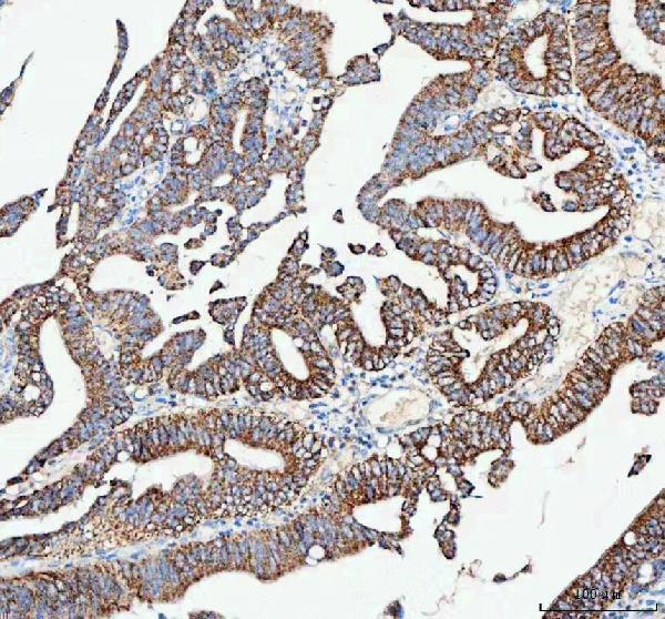
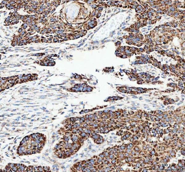
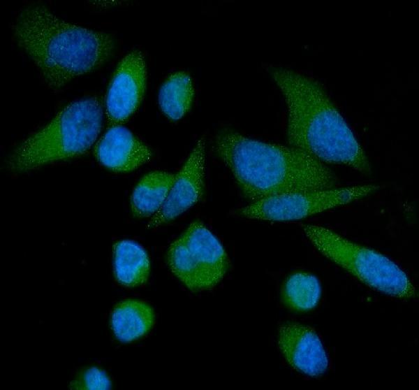
A03048 has IHC images from human paraffin sections and mouse brain, plus IF images from PC-3 and HeLa cells and a human paraffin section (catalog image captions).

Real IHC data IHC analysis of CAMKK2 using anti-CAMKK2 antibody (A03048). CAMKK2 was detected in a paraffin-embedded section of human colorectal adenocarcinoma tissue. Heat mediated antigen retrieval was performed in EDTA buffer (pH 8.0, epitope retrieval solution). The tissue section was blocked with 10% goat serum. The tissue section was then incubated with 2 μg/ml rabbit anti-CAMKK2 Antibody (A03048) overnight at 4°C. Peroxidase Conjugated Goat Anti-rabbit IgG was used as secondary antibody and incubated for 30 minutes at 37°C. The tissue section was developed using HRP Conjugated Rabbit IgG Super Vision Assay Kit (Catalog # SV0002) with DAB as the chromogen.
Anti-CAMKK2 Antibody ®
Cat # A03048

A03048 is listed for IHC and IF/ICC in human, mouse, and rat samples (catalog applications and reactivity). Its IHC images show human colorectal adenocarcinoma, esophageal squamous carcinoma, liver cancer, and mouse brain paraffin sections; its IF images show PC-3 cells, HeLa cells, and a human intestinal cancer paraffin section (A03048 image captions).

Which to pick: For tissue IHC, choose A03048 at 2–5 μg/ml: its IHC captions document staining in paraffin sections, but do not report the fixative (A03048 catalog dilution and IHC image captions). For IF/ICC, A03048 is listed at 5 μg/ml and has cell and paraffin-section IF images (A03048 catalog applications, dilution, and IF image captions). For work across species, A03048 lists human, mouse, and rat reactivity; its IHC images document human and mouse sections (A03048 catalog reactivity and IHC image captions).

Each figure is that product's own IHC / IF validation image from its datasheet.

References

  1. UniProt Consortium. UniProt entry Q96RR4 (KKCC2_HUMAN, Calcium/calmodulin-dependent protein kinase kinase 2).
  2. Human Protein Atlas. CAMKK2 tissue IHC expression (reliability: Approved).
  3. Human Protein Atlas. CAMKK2 subcellular location (ICC-IF): Localized to the cytosol..
  4. Human Protein Atlas. CAMKK2 antibody validation summary (2 antibodies).
  5. The prognostic value and immunological role of calcium/calmodulin dependent protein kinase kinase 2 (CAMKK2) in pan-cancer study. Medicine 2024 — PMC11479412.
  6. Targeting CaMKK2 Inhibits Actin Cytoskeletal Assembly to Suppress Cancer Metastasis. Cancer research 2023 — PMC10472110.
  7. Loss of CAMKK2 and iron-transport proteins-transferrin and its receptor-in the Alzheimer's disease hippocampus: link to tau pathology. Frontiers in cell and developmental biology 2026 — PMC12872923.
  8. CaMKK2 in myeloid cells is a key regulator of the immune-suppressive microenvironment in breast cancer. Nature communications 2019 — PMC6547743.
  9. PubMed PMID:11395482 — UniProt-cited evidence.
  10. PubMed PMID:9822657 — UniProt-cited evidence.
  11. PubMed PMID:12935886 — UniProt-cited evidence.