This website uses cookies to ensure you get the best experience on our website.


- Table of Contents
Plan CAMKV IHC in paraffin brain sections using the reported CNS staining patterns as a scoring reference (HPA tissue IHC; datasheet). This guide covers fixation, retrieval, chromogenic detection and antibody titration at 1:100–1:300 (datasheet; standard IHC practice).

Expected localisation, validated protocols, controls and antibodies — the at-a-glance facts below, then the full design guide.
| Expected localisation | Cerebellar granular-cell cytoplasm/membrane (HPA tissue IHC) | |
| Staining pattern | CNS: glia, granular-cell cytoplasm/membrane, neuropil (HPA tissue IHC) | |
| Antigen retrieval | Citrate pH 6.0 HIER, 95–98 °C, 20 min (rule: cytoplasmic / membrane antigen) | |
| Positive control | Caudate+3 more · see all | |
| Negative control | Adipose tissue+4 more · see all |
| Fixation | Keep fixation consistent across sections (standard IHC practice; not target-specific) | |
| Caveat | The stained cell type varies by CNS region (HPA tissue IHC) | |
| Regulation | CNS-selective expression (HPA tissue IHC) | |
| Isoform / epitope | 3 isoforms; epitope coverage is unspecified (UniProt; datasheet) |
This section pairs the catalog antibody’s IHC-P protocol (datasheet) with a published CAMKV tumor pathology IHC protocol (PMC11186719).
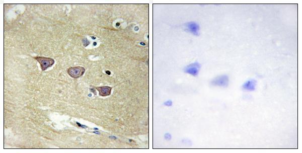
| Sample | Paraffin-embedded human brain tissue; fixative not specified (datasheet A30525) |
| Fixation | Image fixative and duration unreported (datasheet A30525); verify before use. |
| Sectioning | 4–5 µm sections on charged slides (standard) |
| Deparaffinisation | Xylene, graded ethanol series to water (standard) |
| Antigen retrieval | Heat-induced epitope retrieval in citrate buffer, pH 6.0, 20 min at 95–98 °C (standard rule: cytoplasmic / membrane antigen) |
| Peroxidase block | 3% H2O2, 10 min, room temperature (standard) |
| Blocking | 10% normal serum of the secondary host, 30 min, room temperature (standard) |
| Primary antibody | Rabbit anti-CAMKV, 1:100 - 1:300 (datasheet A30525) |
| Primary incubation | Overnight at 4 °C (standard) |
| Detection | HRP-polymer secondary, DAB chromogen 5–10 min (standard) |
| Counterstain | Hematoxylin, blue, dehydrate and mount (standard) |
| Expected result | CAMKV-positive staining in glial cells of caudate (HPA tissue IHC: Medium). HPA tissue profile: Selective expression in CNS. No signal in the no-primary control. |
CAMKV is associated with the cell membrane and cytoplasmic vesicle membrane, particularly around the soma and neurites; it has no transmembrane segment (UniProt Q8NCB2, subcellular location and topology). In paraffin IHC, expect selective CNS staining in glial cells, cerebellar granular cells, and cortical neuropil (HPA tissue IHC: Enhanced reliability). Interpret those tissue observations separately from HPA's nuclear ICC-IF pattern.
| Medium staining in caudate or hippocampal glial cells, cerebellar granular-cell cytoplasm/membrane, or cortical neuropil (HPA tissue IHC). | These are the reported positive patterns. Assess the named cells or structures within each section; a uniformly stained brain section would not reproduce HPA's selective CNS profile (HPA tissue IHC). |
| Predominantly nuclear staining in paraffin brain IHC, with little signal in the expected positive cells or neuropil. | This does not match the reported tissue IHC pattern (HPA tissue IHC). Check staining specificity and detection background before assigning CAMKV localisation; HPA reports nucleoplasm in ICC-IF, a separate application (HPA subcellular ICC-IF). |
| Strong staining in adipocytes or colon endothelial cells despite little staining in the expected CNS structures. | Those cell types were reported as not detected (HPA tissue IHC). Consider antibody cross-reactivity or endogenous detection activity (general IHC practice). The HPA result applies to the named cells, not every cell in those tissues. |
| Diffuse chromogen across cells, extracellular spaces, or most of the section, obscuring cellular boundaries. | An uninterpretable diffuse deposit cannot establish CAMKV positivity. Review blocking, antibody concentration, washes, and detection controls (general IHC practice); HPA reports selective CNS expression rather than a diffuse tissue-wide pattern (HPA tissue IHC). |
| No detectable staining in caudate glial cells, cerebellar granular cells, cortical neuropil, and hippocampal glial cells. | All four are reported at medium level (HPA tissue IHC), so first check section quality and the IHC detection workflow with appropriate controls (general IHC practice). One negative field alone does not establish CAMKV absence throughout a brain section. |
| Tissue and cell selection | HPA reports medium signal in four named CNS cell or structure contexts, but no detection in the listed adipocytes and colon endothelial cells (HPA tissue IHC). Compare the specified compartments rather than calling whole tissues positive or negative. |
| IHC antibody evidence | The HPA tissue profile has Enhanced reliability, and HPA007656 and CAB034931 each have Enhanced IHC validation (HPA tissue IHC; HPA antibodies). That supports the reported pattern, while an individual assay still needs its own controls (general IHC practice). |
| Membrane association and processing | CAMKV is associated with cell and cytoplasmic vesicle membranes despite having no transmembrane segment; no signal peptide or propeptide is annotated (UniProt Q8NCB2). These annotations do not predict a specific chromogen distribution in every CNS cell. |
| IF/ICC Q: Is nuclear signal expected? | A: HPA reports approved nucleoplasmic localisation, plus a cytokinetic bridge location, in ICC-IF; images are listed for CACO-2 and SH-SY5Y (HPA subcellular ICC-IF). Evaluate that finding in the separate IF/ICC guide; it does not replace the paraffin-IHC tissue pattern (HPA tissue IHC). |
| Isoforms and epitope coverage | Three isoforms are annotated (UniProt Q8NCB2). The payload gives no antibody epitope or isoform-specific IHC data, so staining cannot be assigned to a particular isoform or assumed to cover all three. |
| Situation | Likely cause | Next action |
|---|---|---|
| No chromogen in an expected positive brain region. | The selected field may lack the HPA-positive cells, or the IHC workflow may have failed (HPA tissue IHC; general IHC practice). | Inspect the specified cells or neuropil and verify section quality, detection controls, and the catalog antibody's IHC-P instructions (HPA tissue IHC; general IHC practice). |
| Signal is weak in an expected positive cell population. | The reported level is medium, so a faint result may be difficult to separate from background (HPA tissue IHC). | Compare a matched positive area with a detection control; review antigen retrieval and antibody dilution using the IHC-validated antibody's instructions (general IHC practice). No CAMKV-specific retrieval condition is supplied. |
| Most of the section shows diffuse brown staining. | Nonspecific binding or detection background can obscure the selective pattern (general IHC practice; HPA tissue IHC). | Review blocking, washes, antibody dilution, and detection controls; score CAMKV only where cells or neuropil can be resolved (general IHC practice). |
| Adipocytes or colon endothelial cells stain strongly. | This conflicts with the reported lack of detection in those cell types (HPA tissue IHC); cross-reactivity or endogenous activity is possible (general IHC practice). | Check a control that omits primary antibody and review the detection system. Reassess the exact cell identity before comparing with HPA (general IHC practice; HPA tissue IHC). |
| Brain IHC shows mainly nuclear staining. | HPA's approved nuclear localisation comes from ICC-IF, whereas the tissue IHC profile identifies glial cells, granular-cell cytoplasm/membrane, and neuropil (HPA subcellular ICC-IF; HPA tissue IHC). | Document the application and morphology, check detection controls, and compare the slide with the reported tissue-IHC pattern before interpreting the nuclear signal (general IHC practice; HPA tissue IHC). |


Comprehensive Human Protein Atlas IHC scoring per tissue (reliability: Enhanced — High consistency between antibody staining and RNA expression data.). Rows are taken directly from the HPA tissue chart — click any row's HPA link to view the source.
| Tissue | Cell type | Level | Evidence | Source |
|---|---|---|---|---|
| Caudate | Glial cells | Medium | Protein (IHC) | HPA → |
| Cerebellum | Granular cells - cytoplasm/membrane | Medium | Protein (IHC) | HPA → |
| Cerebral cortex | Neuropil | Medium | Protein (IHC) | HPA → |
| Hippocampus | Glial cells | Medium | Protein (IHC) | HPA → |
| Tissue | Cell type | Level | Evidence | Source |
|---|---|---|---|---|
| Adipose tissue | Adipocytes | Not detected | Protein (IHC) | HPA → |
| Appendix | Glandular cells | Not detected | Protein (IHC) | HPA → |
| Bone marrow | Hematopoietic cells | Not detected | Protein (IHC) | HPA → |
| Breast | Adipocytes | Not detected | Protein (IHC) | HPA → |
| Bronchus | Respiratory epithelial cells | Not detected | Protein (IHC) | HPA → |
Troubleshoot CAMKV staining in paraffin sections by checking retrieval, compartment, tissue context, and controls before comparing staining intensity.
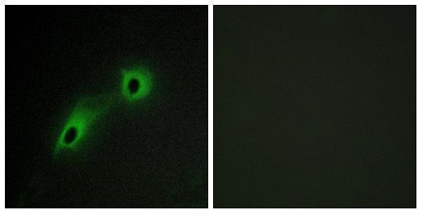
A30525 has IHC data from paraffin-embedded human brain tissue and IF data from HeLa cells; listed reactivity covers human, mouse, and rat (catalog IHC/IF image captions; reactivity).
A30525 is listed for IHC and shows staining of paraffin-embedded human brain tissue with a peptide-blocked comparison (catalog applications; IHC image caption). A30525 is also listed for IF/ICC and shows HeLa cell IF with a peptide-blocked comparison; listed reactivity is human, mouse, and rat (catalog applications/reactivity; IF image caption).
Which to pick: For tissue IHC, choose A30525: its own image shows paraffin-embedded human brain tissue, while the fixative is unreported (IHC image caption). For IF/ICC, choose A30525 because both applications are listed and its IF image shows HeLa cells (catalog applications; IF image caption). For cross-species work, A30525 is the listed rabbit polyclonal with human, mouse, and rat reactivity; the pictured IHC sample is human brain tissue (catalog host/clonality/reactivity; IHC image caption).