CAPN2 / Calpain-2 catalytic subunit · IHC design guide

Design Immunohistochemistry for CAPN2

Plan CAPN2 chromogenic IHC in paraffin sections with a starting antibody concentration of 0.5–1 μg/mL (datasheet: A03492 IHC-P). Use the documented cytoplasmic pattern across several cell types, including endothelium, to assess staining by cell type (HPA tissue IHC).

Evidence assembled Oct 2026 · For research use; verify linked source records and product datasheet before use
Immunohistochemistry protocol sheet for CAPN2 (IHC for CAPN2): expected localisation Cytoplasmic tissue staining (HPA tissue IHC); Ca²⁺-dependent membrane recruitment is a molecular expectation (UniProt), antibody A03492, validated IHC image, and IHC protocol steps
Printable CAPN2 IHC protocol sheet — expected localisation Cytoplasmic tissue staining (HPA tissue IHC); Ca²⁺-dependent membrane recruitment is a molecular expectation (UniProt), antibody A03492, controls and protocol steps. Open the full CAPN2 IHC guide →

CAPN2 Immunohistochemistry Experimental Design Guide

Expected localisation, validated protocols, controls and antibodies — the at-a-glance facts below, then the full design guide.

Must know before staining
Expected localisation Cytoplasmic tissue staining (HPA tissue IHC); Ca²⁺-dependent membrane recruitment is a molecular expectation (UniProt)
Staining pattern Cytoplasmic signal in several cell types, including endothelium (HPA tissue IHC)
Antigen retrieval Citrate pH 6 HIER, heat-mediated (datasheet A03492)
Positive control ⓘ Bronchus+4 more · see all
Negative control ⓘ Adipose tissue+4 more · see all
Important caveats
Reasons your staining may differ from the expected pattern.
Fixation Keep fixation consistent across sections. (standard IHC practice; not target-specific)
Caveat Cell-type variation can confound whole-section scoring (HPA tissue IHC)
Regulation Expression regulation is unreported (UniProt)
Isoform / epitope 2 isoforms; residues 2–19 form a propeptide (UniProt)
Section 1

Recommended CAPN2 IHC & IF Protocols

The catalog antibody has an IHC-P protocol (datasheet A03492); three published CAPN2 IHC methods provide additional paraffin-section conditions (PMC5716693; PMC5322456; PMC10022304).

Recommended immunohistochemistry (IHC-P) protocol parameters
SampleParaffin-embedded human intestinal cancer tissue; fixative not specified (datasheet A03492)
FixationImage fixative and duration unreported (datasheet A03492); verify before use.
Sectioning4–5 µm sections on charged slides (standard)
DeparaffinisationXylene, graded ethanol series to water (standard)
Antigen retrievalHeat retrieval: Citrate pH 6, 20 min (datasheet A03492)
Peroxidase block3% H2O2, 10 min, room temperature (standard)
Blocking10% goat serum (datasheet A03492)
Primary antibodyRabbit anti-CAPN2, 0.5-1μg/ml (datasheet A03492)
Primary incubationOvernight at 4 °C (datasheet A03492)
DetectionStreptavidin-biotin complex (SABC), DAB chromogen (datasheet A03492)
CounterstainHematoxylin, blue, dehydrate and mount (standard)
Expected resultCAPN2-positive staining in respiratory epithelial cells of bronchus (HPA tissue IHC: High). HPA tissue profile: Cytoplasmic expression in several different cell types, including endothelial cells. No signal in the no-primary control.
💡Decision noteStart with heat-mediated citrate retrieval at pH 6 (datasheet A03492); all three published methods also use citrate at pH 6 (PMC5716693; PMC5322456; PMC10022304).
Section 2

What Is the Expected CAPN2 Staining Pattern?

CAPN2 is mainly cytoplasmic, with possible plasma membrane localization after Ca(2+) binding; it has no transmembrane segment (UniProt P17655: localization and topology). In tissue IHC, expect staining in several cell types, including endothelial cells (HPA: tissue profile). Bronchial respiratory epithelial cells show high staining, while several other listed cell types show medium staining (HPA: tissue IHC). HPA rates its tissue staining Approved, with medium consistency against RNA data (HPA: reliability).

What am I looking at on my slide?
Cytoplasmic chromogen in bronchial respiratory epithelial cells is strong; neighboring cells may differ.This matches the high bronchial epithelial signal (HPA: High in respiratory epithelial cells). Compare staining within the identified cell population: HPA's tissue profile spans several cell types, so an evenly stained section is not the expected benchmark (HPA: tissue profile).
A cytoplasmic or peripheral pattern appears in a cell population with an HPA positive result.Cytoplasmic staining fits the tissue IHC profile (HPA: tissue profile). A peripheral component can fit CAPN2 translocation after Ca(2+) binding (UniProt P17655: localization), but a tissue section alone does not establish that calcium triggered it.
Predominantly nuclear staining appears without a convincing cytoplasmic component.Treat this as a compartment mismatch: the supplied localization evidence identifies cytoplasm or cytosol and possible membrane localization, not a nuclear pattern (UniProt P17655: localization; HPA: subcellular). Review controls and staining conditions before calling it CAPN2.
Strong signal appears in adipocytes, or in colon endothelial cells.These specific cell populations were not detected in the listed HPA tissue samples (HPA: adipose and breast adipocytes; colon endothelial cells). Check cell identity and controls; cross-reactivity or detection chemistry is possible, but one discrepant section cannot establish its cause.
Color spreads through tissue structures, or bronchial respiratory epithelium lacks signal.Widespread deposit obscures the expected cell and compartment pattern (HPA: tissue profile). Absent bronchial signal conflicts with the listed high result (HPA: bronchus), but does not by itself distinguish a failed stain from variation in the specimen.
💡Expected CAPN2 appearanceCall a result positive when cell-associated cytoplasmic staining is clear, especially high in bronchial respiratory epithelium; peripheral staining may occur, whereas dominant nuclear or structure-independent color is suspect (HPA: bronchus and tissue profile; UniProt P17655: localization).
How each factor affects the staining
Cell population and tissueHPA reports high bronchial respiratory epithelium, medium Bergmann glia and several glandular populations, and undetected adipocytes in its listed samples (HPA: tissue IHC). Score identified cells rather than assigning one intensity to a whole organ.
CompartmentCytoplasm is the tissue IHC expectation (HPA: tissue profile). CAPN2 can move to the plasma membrane upon Ca(2+) binding (UniProt P17655: localization); the protein has no transmembrane segment (UniProt P17655: topology). Membrane staining alone does not prove translocation.
Evidence behind an apparent negativeCAPN2 is described as ubiquitous, while HPA reports low tissue specificity and some undetected cell populations (UniProt P17655: tissue specificity; HPA: RNA specificity and tissue IHC). An undetected IHC result is a staining observation, not proof that a cell contains no CAPN2.
Antibody evidenceThe supplied HPA antibody, HPA024470, is Approved for IHC (HPA: antibody validation). The tissue profile has medium consistency with RNA data (HPA: reliability). Use those ratings as context for interpretation; they do not validate every new specimen or staining condition.
Forms and epitope coverageUniProt lists 2 isoforms and a processed chain spanning residues 20–700 (UniProt P17655: isoforms and processing). The supplied evidence gives no antibody epitope, so it cannot show whether both isoforms or all processed forms are detected.
Why is my staining missing, weak or wrong?
SituationLikely causeNext action
No signal in bronchial respiratory epithelium.This disagrees with the listed high staining (HPA: bronchus); antibody failure, detection failure, or specimen variation remains possible.Confirm cell identity, inspect a concurrent known-positive section, then review retrieval, antibody incubation and chromogen development as general IHC workflow checks.
Diffuse color prevents recognition of cell borders or cytoplasm.The result cannot be judged against the cell-associated cytoplasmic profile (HPA: tissue profile); nonspecific staining or detection background may contribute.Compare a no-primary control; review blocking, washes and chromogen development as general IHC workflow checks. Reassess only where the background permits cell-level scoring.
Nuclei dominate the stain.That compartment conflicts with the supplied cytoplasmic and cytosolic localization evidence (UniProt P17655: localization; HPA: subcellular).Check the no-primary control and counterstain, then verify whether cytoplasmic signal remains in an HPA positive cell population before interpreting the nuclear color.
Adipocytes or colon endothelial cells stain strongly.Those populations were not detected in the listed HPA samples (HPA: adipose and breast adipocytes; colon endothelial cells); the discrepancy has no established single cause.Verify the cell population and examine a no-primary control. If staining persists, report the discrepancy and assess antibody specificity before assigning it to CAPN2.
A membrane rim is the only apparent signal.Membrane association is possible after Ca(2+) binding (UniProt P17655: localization), but HPA describes a mainly cytoplasmic tissue pattern (HPA: tissue profile).Inspect an HPA positive population for accompanying cytoplasmic staining and compare controls. Describe the observed compartment without inferring calcium activation from the section.
A sample called negative conflicts with an expectation of broad CAPN2 expression.UniProt calls expression ubiquitous, yet HPA lists specific populations as undetected by IHC (UniProt P17655: tissue specificity; HPA: tissue IHC).Score the exact cell population and compare it with its HPA entry. Confirm that a positive control stained before treating an undetected result as evidence about protein abundance.

Sample controls for CAPN2 IHC & IF

🧪Run bronchus first: respiratory epithelial cells should stain strongly (HPA: High in respiratory epithelial cells). Use adipose tissue as a negative comparator, scoring adipocytes specifically (HPA: Not detected in adipocytes); on the bronchus slide, cells without visible target staining should remain at background, but HPA does not identify a bronchial cell type as an internal negative (HPA: bronchus row).
Positive control tissue: Bronchus (Respiratory epithelial cells, HPA High)
Negative control tissue: Adipose tissue (HPA Not detected)
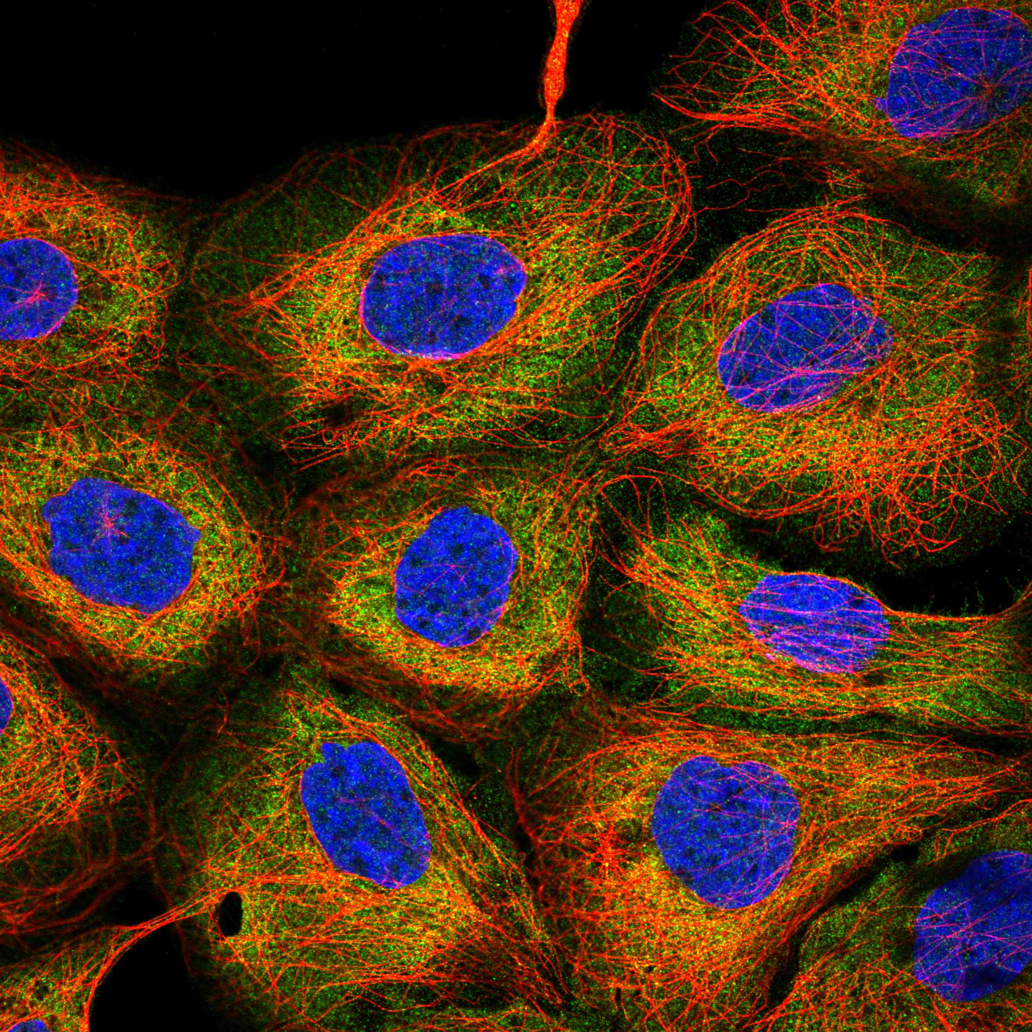
ICC-IF cell lines (HPA subcellular resource): HPA ICC-IF images show CAPN2 in A-431, U-251MG, U2OS, Sperm, with annotated localisation: Cytosol (supported), Connecting piece (approved), Mid piece (approved) (HPA subcellular).
Technical controls: Include a no-primary, secondary-only control and a rabbit IgG isotype control matched to the primary antibody’s clonality and concentration (selected-SKU caption: rabbit primary; standard IHC practice). Confirm specificity with CAPN2 knockout material or, if the immunizing peptide is available, peptide preabsorption; for bronchus DAB staining, block endogenous peroxidase and assess endogenous biotin because the reported detection uses a biotin-based system (selected-SKU caption: SABC with DAB; standard IHC practice).
⚠️Feasibility: The selected-SKU paraffin-section caption reports heat retrieval in citrate buffer at pH 6 for 20 minutes, but does not report the fixative or a target-specific fixation window or fixation effect (selected-SKU caption). HPA staining does not establish specimen processing, and the supplied evidence does not establish that frozen sections or IF are easier than the reported paraffin IHC workflow (HPA: tissue rows; selected-SKU caption). Bronchial endogenous peroxidase or biotin may add background to the reported DAB/SABC readout, so interpret epithelial staining against the matched controls (selected-SKU caption: SABC with DAB; standard IHC practice).

HPA tissue IHC evidence for CAPN2

Comprehensive Human Protein Atlas IHC scoring per tissue (reliability: Approved — Medium consistency between antibody staining and RNA expression data.). Rows are taken directly from the HPA tissue chart — click any row's HPA link to view the source.

Positive expression · recommended positive controls

TissueCell typeLevelEvidenceSource
Bronchus Respiratory epithelial cells High Protein (IHC) HPA →
Adrenal gland Glandular cells Medium Protein (IHC) HPA →
Appendix Glandular cells Medium Protein (IHC) HPA →
Cerebellum Bergmann glia - cytoplasm/membrane Medium Protein (IHC) HPA →
Fallopian tube Glandular cells Medium Protein (IHC) HPA →

Undetected expression · recommended negative controls

TissueCell typeLevelEvidenceSource
Adipose tissue Adipocytes Not detected Protein (IHC) HPA →
Breast Adipocytes Not detected Protein (IHC) HPA →
Caudate Glial cells Not detected Protein (IHC) HPA →
Colon Endothelial cells Not detected Protein (IHC) HPA →
Duodenum Glandular cells Not detected Protein (IHC) HPA →
Section 3

Advanced CAPN2 IHC Tips

Troubleshoot CAPN2 staining in paraffin sections by checking retrieval, compartment, cell type and controls before comparing staining intensity (UniProt P17655; HPA tissue IHC).

How should I retrieve CAPN2 in paraffin sections when staining is weak?
Start with heat-mediated retrieval in citrate buffer at pH 6 for 20 minutes (datasheet A03492). The selected paraffin-section example used those conditions before overnight incubation with 1 μg/mL primary antibody at 4°C (datasheet A03492). If staining remains weak, compare a modestly adjusted heating time on matched sections while keeping the same buffer, detection chemistry and control tissue (standard IHC practice). Record whether cytoplasmic staining improves in expected cells, since widespread edge staining or tissue damage can make an apparent gain misleading (HPA tissue IHC; standard IHC practice).
Could fixation explain uneven CAPN2 staining across my paraffin sections?
Target-specific CAPN2 sensitivity to fixation is unknown from the supplied evidence; the selected paraffin-section caption does not state a fixative (datasheet A03492). Compare sections with documented, consistent processing before attributing intensity differences to biology (standard IHC practice). Examine morphology alongside staining, because poor preservation and uneven processing can complicate cell-level interpretation in chromogenic IHC (standard IHC practice). Keep retrieval at citrate pH 6 for 20 minutes and primary incubation conditions constant during that comparison (datasheet A03492). Do not infer a fixation effect from CAPN2 localisation, modifications or tissue-staining patterns (UniProt P17655; HPA tissue IHC).
Is membrane staining credible when CAPN2 is usually cytoplasmic?
CAPN2 is annotated in the cytoplasm and can move to the plasma membrane upon calcium binding, despite having no transmembrane segment (UniProt P17655). Thus, cytoplasmic staining is an expected starting pattern, while membrane-enriched staining needs cell-level confirmation and suitable controls (UniProt P17655; standard IHC practice). The tissue atlas reports cytoplasmic expression across several cell types and cytoplasm or membrane staining in Bergmann glia (HPA tissue IHC). Compare membrane and cytoplasmic signal within intact cells at the same exposure and staining run, and exclude section edges before scoring a redistribution (standard IHC practice). Chromogenic localisation alone cannot establish calcium-dependent translocation (UniProt P17655; standard IHC practice).
Can this stain distinguish CAPN2 isoforms or a cleaved protein?
CAPN2 has 2 recorded isoforms, a propeptide at residues 2–19 and a processed catalytic chain at residues 20–700 (UniProt P17655). The supplied caption does not identify the antibody epitope, so its staining cannot assign an isoform or distinguish precursor from processed protein (datasheet A03492; UniProt P17655). Request or verify the immunogen or epitope information before interpreting a changed staining pattern as cleavage (standard IHC practice). Compare an independent epitope or an appropriate biological control where available, keeping retrieval and detection matched (standard IHC practice). CAPN2 has no annotated glycosylation sites, which alone does not establish epitope accessibility in sections (UniProt P17655).
How can I check CAPN2 localisation by IF alongside cell identity?
For a separate IF experiment, pair CAPN2 with a validated marker of the expected cell type; the tissue atlas reports cytoplasmic CAPN2 in several cell types, including endothelial cells (HPA tissue IHC; standard IF practice). Choose fluorophores and acquisition channels after checking tissue autofluorescence with an unstained control, and assess channel bleed-through with single-stain controls (standard IF practice). Use permeabilisation appropriate for a cytosolic epitope, then verify that it preserves cell boundaries before interpreting membrane enrichment (UniProt P17655 localisation; standard IF practice). CAPN2 has no transmembrane segment, and the antibody epitope side is unspecified, so optimise permeabilisation empirically (UniProt P17655; datasheet A03492). HPA subcellular images provide localisation context, not validation of this catalog antibody for IF (HPA subcellular; datasheet A03492).
What should I change when CAPN2 DAB staining appears diffuse?
The selected paraffin-section workflow used 10% goat serum, a biotinylated secondary antibody, a streptavidin–biotin complex and DAB (datasheet A03492). Run a primary-omission control and inspect whether diffuse colour persists, then check blocking, washes and secondary-antibody specificity (standard IHC practice). A peroxidase block and assessment of endogenous biotin are relevant to this detection chemistry, but they are general workflow controls rather than evidence of a CAPN2-specific interference (datasheet A03492; standard IHC practice). Compare intact-cell cytoplasmic signal with acellular deposits, edges and damaged regions before adjusting the 1 μg/mL primary concentration or overnight 4°C incubation (datasheet A03492; HPA tissue IHC; standard IHC practice).
How should I score CAPN2 across mixed cell populations? ⚠ ANSWER MARKED FOR VERIFICATION
Define the cell population and compartment before scoring, because CAPN2 staining varies among cell types and is commonly cytoplasmic in tissue IHC (HPA tissue IHC). For chromogenic sections, report an H-score from intensity categories and their cell percentages, or report the percentage of positive cells within each defined population (standard IHC practice). For spatial sampling, positive-cell density per mm² can supplement those measures when the sampled area is recorded (standard IHC practice). Normalise counts to evaluable cells or tissue area within the same compartment, and apply one threshold, retrieval run and imaging setup across groups (standard IHC practice). Document excluded necrotic and edge regions (standard IHC practice).
How do I separate genuine CAPN2 positivity from staining artefact?
Look first for signal within intact cells, predominantly in the cytoplasm; membrane-associated staining is biologically plausible but needs corroboration (UniProt P17655; HPA tissue IHC). Check cell identity against the sampled tissue: respiratory epithelial cells in bronchus were reported high, whereas adipocytes in adipose tissue were not detected in the atlas dataset (HPA tissue IHC). Edge-only colour, necrotic regions and staining that persists without primary antibody favour artefact (standard IHC practice). Review a primary-omission control and peroxidase blocking when DAB signal is unexpected, since endogenous enzyme activity can contribute colour independently of antibody binding (standard IHC practice). Treat discordance with atlas patterns as a prompt to investigate, given its reported medium staining–RNA consistency (HPA tissue IHC).
Boster reagents

Best CAPN2 / Calpain-2 catalytic subunit IHC Antibodies

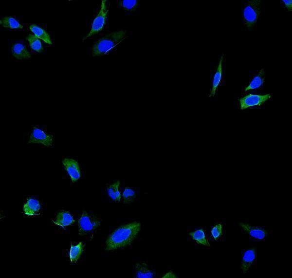
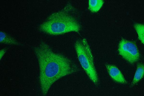
Both anti-CAPN2 antibodies have paraffin-section IHC images from human tissue and IF/ICC images from human cells (catalog image captions); their listed reactivity also covers other species (catalog reactivity).

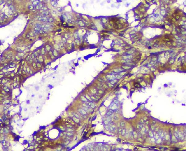
Real IHC data IHC analysis of Calpain 2 using anti-Calpain 2 antibody (A03492). Calpain 2 was detected in paraffin-embedded section of human intestinal cancer tissue. Heat mediated antigen retrieval was performed in citrate buffer (pH6, epitope retrieval solution) for 20 mins. The tissue section was blocked with 10% goat serum. The tissue section was then incubated with 1μg/ml rabbit anti-Calpain 2 Antibody (A03492) overnight at 4°C. Biotinylated goat anti-rabbit IgG was used as secondary antibody and incubated for 30 minutes at 37°C. The tissue section was developed using Strepavidin-Biotin-Complex (SABC)(Catalog # SA1022) with DAB as the chromogen.
Anti-Calpain 2/CAPN2 Antibody ®
Cat # A03492
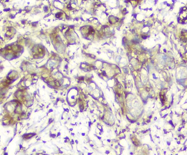
Real IHC data IHC analysis of Calpain 2 using anti-Calpain 2 antibody (M03492). Calpain 2 was detected in paraffin-embedded section of human intestinal cancer tissue. Heat mediated antigen retrieval was performed in EDTA buffer (pH8.0, epitope retrieval solution). The tissue section was blocked with 10% goat serum. The tissue section was then incubated with 1μg/ml mouse anti-Calpain 2 Antibody (M03492) overnight at 4°C. Biotinylated goat anti-mouse IgG was used as secondary antibody and incubated for 30 minutes at 37°C. The tissue section was developed using Strepavidin-Biotin-Complex (SABC) (Catalog # SA1021) with DAB as the chromogen.
Anti-Calpain 2 CAPN2 Antibody ® (monoclonal, 8I6)
Cat # M03492

A03492 has IHC images from human intestinal and lung cancer tissue and rat small intestine, plus an IF/ICC image from HeLa cells (A03492 image captions). M03492 has IHC images from human intestinal and mammary cancer tissue and mouse lung, plus IF/ICC images from A549 and U251 cells (M03492 image captions).

Which to pick: For paraffin-section IHC, choose A03492 for the documented citrate pH 6 retrieval or M03492 for the documented EDTA pH 8 retrieval and monoclonal clone 8I6 (respective IHC image captions; M03492 catalog clone). For IF/ICC, both have cell images; A03492 was shown in HeLa, while M03492 was shown in A549 and U251 (respective IF image captions). For cross-species work, A03492 lists human, mouse and rat reactivity; M03492 lists human, monkey and mouse (catalog reactivity); the IHC captions do not report the fixative (respective IHC image captions).

Each figure is that product's own IHC / IF validation image from its datasheet.

References

  1. UniProt Consortium. UniProt entry P17655 (CAN2_HUMAN, Calpain-2 catalytic subunit).
  2. Human Protein Atlas. CAPN2 tissue IHC expression (reliability: Approved).
  3. Human Protein Atlas. CAPN2 subcellular location (ICC-IF): Mainly localized to the cytosol, connecting piece and mid piece. In addition localized to the principal piece..
  4. Human Protein Atlas. CAPN2 antibody validation summary (1 antibodies).
  5. Overexpression of CAPN2 promotes cell metastasis and proliferation via AKT/mTOR signaling in renal cell carcinoma. Oncotarget 2017 — PMC5716693.
  6. Silencing CAPN2 Expression Inhibited Castration-Resistant Prostate Cancer Cells Proliferation and Invasion via AKT/mTOR Signal Pathway. BioMed research international 2017 — PMC5322456.
  7. CAPN2 promotes apalutamide resistance in metastatic hormone-sensitive prostate cancer by activating protective autophagy. Journal of translational medicine 2024 — PMC11155045.
  8. Changes in calpain-2 expression during glioblastoma progression predisposes tumor cells to temozolomide resistance by minimizing DNA damage and p53-dependent apoptosis. Cancer cell international 2023 — PMC10022304.
  9. PubMed PMID:2852952 — UniProt-cited evidence.
  10. PubMed PMID:10944468 — UniProt-cited evidence.
  11. PubMed PMID:14702039 — UniProt-cited evidence.