This website uses cookies to ensure you get the best experience on our website.


- Table of Contents
Plan CARD11 IHC-P with the catalog antibody’s starting concentration of 10 μg/mL (datasheet: A00740). Assess cytoplasmic staining in lymphoid cell subsets, using lymph node, tonsil, spleen or appendix as positive tissue references (HPA tissue IHC).

Expected localisation, validated protocols, controls and antibodies — the at-a-glance facts below, then the full design guide.
| Expected localisation | Cytoplasm in subsets of lymphoid cells (HPA tissue IHC) | |
| Staining pattern | Subset lymphoid cells show cytoplasmic staining (HPA tissue IHC) | |
| Antigen retrieval | Citrate pH 6.0 HIER, 95–98 °C, 20 min (rule: cytoplasmic / membrane antigen) | |
| Positive control | Appendix+3 more · see all | |
| Negative control | Adipose tissue+4 more · see all |
| Fixation | Keep fixation conditions consistent across paraffin sections. (standard IHC practice; not target-specific) | |
| Caveat | Presumed off-target staining was observed and disregarded (HPA tissue IHC) | |
| Regulation | No expression change specified (UniProt) | |
| Isoform / epitope | 0 isoforms; one full-length chain; epitope site unreported (UniProt) |
The catalog antibody IHC-P protocol is accompanied by published CARD11 methods for kidney cancer tissue microarrays (PMC8568653) and avian organ sections (PMC9969321).
| Sample | Tissue sections; selected-image fixative not specified (standard IHC workflow) |
| Fixation | Image fixative and duration unreported (datasheet A00740); verify before use. |
| Sectioning | 4–5 µm sections on charged slides (standard) |
| Deparaffinisation | Xylene, graded ethanol series to water (standard) |
| Antigen retrieval | Heat-induced epitope retrieval in citrate buffer, pH 6.0, 20 min at 95–98 °C (standard rule: cytoplasmic / membrane antigen) |
| Peroxidase block | 3% H2O2, 10 min, room temperature (standard) |
| Blocking | 10% normal serum of the secondary host, 30 min, room temperature (standard) |
| Primary antibody | Rabbit anti-CARD11, 10 μg/mL (datasheet A00740) |
| Primary incubation | Overnight at 4 °C (standard) |
| Detection | HRP-polymer secondary, DAB chromogen 5–10 min (standard) |
| Counterstain | Hematoxylin, blue, dehydrate and mount (standard) |
| Expected result | CARD11-positive staining in lymphoid tissue of appendix (HPA tissue IHC: Medium). HPA tissue profile: Cytoplasmic expression in subsets of lymphoid cells in several tissues. No signal in the no-primary control. |
CARD11 should stain the cytoplasm of subsets of lymphoid cells in paraffin sections (HPA tissue IHC: Approved; cytoplasmic expression in lymphoid subsets). UniProt also places CARD11 in membrane rafts and reports no transmembrane segment, so a continuous cell-surface outline is not required (UniProt Q9BXL7: subcellular location and topology). HPA notes presumed off-target binding, which should temper interpretation of unexpected staining (HPA tissue IHC: reliability description).
| Cytoplasmic staining in some germinal-center cells of lymph node or tonsil, with unstained neighboring cells. | This fits the reported cell pattern. HPA grades germinal-center staining Medium in both tissues and describes expression in subsets of lymphoid cells; a mixed positive and negative field is therefore plausible (HPA tissue IHC: lymph node, tonsil, profile). |
| A strong, uniform nuclear signal dominates cells expected to contain CARD11. | Treat this as an unexpected compartment pattern and check controls and morphology before scoring it as CARD11. The supplied localization is cytoplasm and membrane raft; HPA describes cytoplasmic tissue staining, without a nuclear pattern (UniProt Q9BXL7: subcellular location; HPA tissue IHC: profile). |
| Broad staining appears in a cell population reported as unstained, such as adipocytes in adipose tissue. | Consider nonspecific binding or detection-system activity, then inspect the negative control. HPA reports adipocytes as Not detected and explicitly notes presumed off-target binding in its antibody assessment; a positive reaction here needs independent support (HPA tissue IHC: adipose tissue, reliability description). |
| Diffuse color covers tissue and empty spaces without resolving individual cytoplasms. | This is background until controls show a cell-associated pattern. Review blocking, washes, chromogen development and the no-primary control as general IHC practice. HPA's interpretable CARD11 pattern is cytoplasmic staining in lymphoid subsets, not a field-wide haze (HPA tissue IHC: profile). |
| No cytoplasmic signal is seen in lymph node germinal-center cells or spleen white-pulp cells. | First confirm that the relevant cells are present and that the run worked with a suitable positive control. HPA reports Medium staining in these specific populations, but its Approved rating is not a guarantee that every section or run will show signal (HPA tissue IHC: lymph node, spleen, reliability). |
| Cell population and tissue selection | Appendix lymphoid tissue, lymph node and tonsil germinal centers, and spleen white pulp are reported at Medium; score the named cells rather than the entire section (HPA tissue IHC: positive tissues, profile). |
| Subcellular context | Cytoplasm is the primary expected compartment. Membrane-raft association can contribute localized signal, but the record gives no IHC requirement for a membrane rim (UniProt Q9BXL7: subcellular location, topology; HPA tissue IHC: profile). |
| Strength of antibody evidence | The listed antibody HPA052984 has IHC status Approved, and HPA reports presumed off-target binding. Unexpected staining needs control-based interpretation; the supplied record does not label this antibody IHC Enhanced (HPA antibodies: HPA052984; HPA tissue IHC: reliability description). |
| Activation-related organization | UniProt describes CARD11 polymerization after T-cell or B-cell receptor activation and a membrane-raft complex. These facts explain possible spatial heterogeneity, but the supplied IHC observations do not establish a reproducible activation-specific stain pattern (UniProt Q9BXL7: function, subunit; HPA tissue IHC: profile). |
| Situation | Likely cause | Next action |
|---|---|---|
| Known-positive lymphoid tissue is blank. | The relevant cell population may be absent from the section, or the IHC run may have failed; the record gives no CARD11-specific retrieval or dilution setting (HPA tissue IHC: positive tissues). | Confirm germinal-center or white-pulp cells on morphology, check a concurrently stained positive control, then review retrieval and detection against the validated IHC workflow as general IHC practice. |
| A faint reaction is hard to distinguish from background. | HPA reports staining in subsets at Medium, so the expected signal is cell-restricted; diffuse color cannot be assigned to CARD11 from intensity alone (HPA tissue IHC: profile, positive tissues). | Compare the same cell population with a no-primary control and assess cytoplasmic localization at higher magnification; review blocking and wash steps as general IHC practice. |
| Adipocytes or another reported negative population stain strongly. | HPA reports adipocytes as Not detected and acknowledges presumed off-target binding; endogenous detection activity or nonspecific antibody binding are general IHC possibilities (HPA tissue IHC: adipose tissue, reliability description). | Run no-primary and detection-only controls, inspect where the color develops, and seek an independent antibody or orthogonal result before assigning the signal to CARD11 (general IHC practice). |
| Signal appears predominantly nuclear or as a continuous surface rim. | Neither pattern is the reported tissue appearance. UniProt places CARD11 in cytoplasm and membrane rafts and lists no transmembrane segment (HPA tissue IHC: profile; UniProt Q9BXL7: subcellular location, topology). | Check compartment assignment against the counterstain and cell boundaries, then compare a positive lymphoid control; do not score the unexpected pattern as confirmed CARD11 without independent support (general IHC practice). |
| All cells in a lymphoid region stain equally. | HPA describes cytoplasmic expression in subsets of lymphoid cells, so uniform staining may reflect background or an overdeveloped reaction; the record does not define a universal positive-cell fraction (HPA tissue IHC: profile). | Recheck exposure to chromogen, washes and no-primary control as general IHC practice; score identifiable cells and compartments rather than the overall darkness of the region. |
| Can an ICC-IF image or IF protocol be inferred from this IHC pattern? | HPA lists no ICC-IF image-bearing cell lines or subcellular location call for CARD11, and the listed antibody has no ICC validation status (HPA subcellular: image list, location; HPA antibodies: HPA052984). | Use this section only to set a cautious localization expectation. Consult the separate IF/ICC guide for assay design; this IHC evidence does not establish an IF staining result or protocol (HPA tissue IHC: profile; HPA subcellular: image list). |
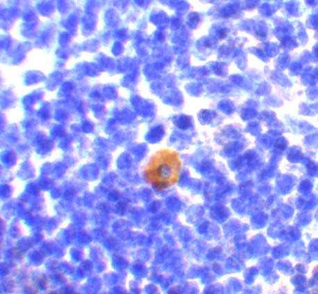
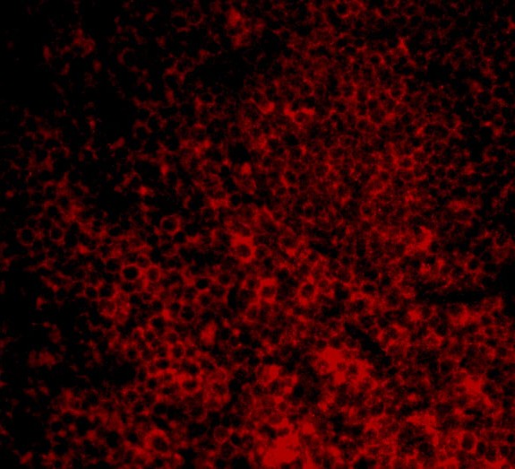


Comprehensive Human Protein Atlas IHC scoring per tissue (reliability: Approved — Presumed off target binding observed and disregarded. External characterization data supports antibody staining. Antibody staining in cells/structures not annotated, view images.). Rows are taken directly from the HPA tissue chart — click any row's HPA link to view the source.
| Tissue | Cell type | Level | Evidence | Source |
|---|---|---|---|---|
| Appendix | Lymphoid tissue | Medium | Protein (IHC) | HPA → |
| Lymph node | Germinal center cells | Medium | Protein (IHC) | HPA → |
| Spleen | Cells in white pulp | Medium | Protein (IHC) | HPA → |
| Tonsil | Germinal center cells | Medium | Protein (IHC) | HPA → |
| Tissue | Cell type | Level | Evidence | Source |
|---|---|---|---|---|
| Adipose tissue | Adipocytes | Not detected | Protein (IHC) | HPA → |
| Adrenal gland | Glandular cells | Not detected | Protein (IHC) | HPA → |
| Bone marrow | Hematopoietic cells | Not detected | Protein (IHC) | HPA → |
| Breast | Adipocytes | Not detected | Protein (IHC) | HPA → |
| Bronchus | Respiratory epithelial cells | Not detected | Protein (IHC) | HPA → |
Troubleshoot CARD11 staining in paraffin sections by checking retrieval, cell identity, cytoplasmic localisation, and controls before comparing staining intensity.
A00740 has mouse thymus IHC and mouse spleen cell IF images (catalog image captions); human and mouse reactivity is listed (catalog: reactivity).
The rendered card is A00740, listed for IHC-P and IF (catalog: applications). Its images show mouse thymus IHC at 10 μg/mL and mouse spleen cell IF at 20 μg/mL (catalog image captions).
Which to pick: Choose A00740 for paraffin-section IHC because IHC-P is listed and its own IHC image shows mouse thymus staining (catalog: applications; A00740 IHC image caption); the fixative is unreported (A00740 IHC image caption). For IF/ICC, A00740 has an IF image of mouse spleen cells, while M00740 is listed for ICC/IF without a supplied image (catalog: A00740 IF image caption; M00740 applications/image alts). Both list human and mouse reactivity, and M00740 is a rabbit monoclonal; only A00740 lists IHC-P (catalog: reactivity/host/clone/applications).