CASP7 / Caspase-7 · IHC design guide

Design Immunohistochemistry for CASP7

Plan CASP7 paraffin IHC around cytoplasmic staining with additional nuclear signal in gastrointestinal glandular cells (HPA tissue IHC). The catalog antibody has an IHC range of 2–5 μg/mL (datasheet A01044-2); interpret staining with CASP7 cleavage and epitope location in mind (UniProt).

Evidence assembled Oct 2026 · For research use; verify linked source records and product datasheet before use
Immunohistochemistry protocol sheet for CASP7 (IHC for CASP7): expected localisation Cytoplasmic with additional nuclear staining (HPA tissue IHC), antibody A01044-2, validated IHC image, and IHC protocol steps
Printable CASP7 IHC protocol sheet — expected localisation Cytoplasmic with additional nuclear staining (HPA tissue IHC), antibody A01044-2, controls and protocol steps. Open the full CASP7 IHC guide →

CASP7 Immunohistochemistry Experimental Design Guide

Expected localisation, validated protocols, controls and antibodies — the at-a-glance facts below, then the full design guide.

Must know before staining
Expected localisation Cytoplasmic with additional nuclear staining (HPA tissue IHC)
Staining pattern GI glandular cells: cytoplasmic with some nuclear staining (HPA tissue IHC)
Antigen retrieval EDTA pH 8.0 HIER, heat-mediated (datasheet A01044-2)
Positive control ⓘ Appendix+4 more · see all
Negative control ⓘ Adipose tissue+4 more · see all
Important caveats
Reasons your staining may differ from the expected pattern.
Fixation Keep fixation consistent across sections. (standard IHC practice; not target-specific)
Caveat Cleavage may change which CASP7 forms the antibody detects (UniProt)
Regulation Cleavage activates CASP7 (UniProt)
Isoform / epitope 4 isoforms; cleavage yields p20 and p11, so check the epitope (UniProt)
Section 1

Recommended CASP7 IHC & IF Protocols

The catalog antibody protocol (datasheet A01044-2) appears alongside two published CASP7 IHC workflows (PMC4673127; PMC7907442).

Recommended immunohistochemistry (IHC-P) protocol parameters
SampleParaffin-embedded human breast cancer tissue; fixative not specified (datasheet A01044-2)
FixationImage fixative and duration unreported (datasheet A01044-2); verify before use.
Sectioning4–5 µm sections on charged slides (standard)
DeparaffinisationXylene, graded ethanol series to water (standard)
Antigen retrievalHeat retrieval: EDTA pH 8.0 (datasheet A01044-2); 20 min, 95–100 °C (standard)
Peroxidase block3% H2O2, 10 min, room temperature (standard)
Blocking10% goat serum (datasheet A01044-2)
Primary antibodyRabbit anti-CASP7, 2-5μg/ml (datasheet A01044-2)
Primary incubationOvernight at 4 °C (datasheet A01044-2)
DetectionStreptavidin-biotin complex (SABC), DAB chromogen (datasheet A01044-2)
CounterstainHematoxylin, blue, dehydrate and mount (standard)
Expected resultCASP7-positive staining in glandular cells of appendix (HPA tissue IHC: High). HPA tissue profile: General cytoplasmic with additional nuclear expression, most abundant in gastrointestinal tract. No signal in the no-primary control.
💡Decision noteStart with EDTA pH 8.0 heat retrieval (datasheet A01044-2); use citrate pH 6.0 for the porcine endometrium protocol (PMC7907442).
Section 2

What Is the Expected CASP7 Staining Pattern?

In paraffin-section IHC, expect CASP7 mainly in the cytoplasm, with nuclear staining in some cells (HPA tissue IHC; UniProt P55210 localization). Glandular cells of the gastrointestinal tract are useful positive examples (HPA: High in appendix, colon, duodenum, rectum and stomach). CASP7 has no transmembrane segment (UniProt P55210 topology). HPA rates its tissue IHC reliability Enhanced, while reporting medium consistency between staining and RNA expression (HPA tissue IHC).

What am I looking at on my slide?
Cytoplasmic staining, sometimes with nuclear staining, in gastrointestinal glandular cells (HPA tissue IHC).This fits the reported tissue pattern; HPA scores these cells High in appendix, colon, duodenum, rectum and stomach (HPA tissue IHC). Compare staining within the same section so that cell identity and compartment remain visible.
Predominantly sharp membrane outlines or staining confined to an unexpected compartment.Recheck localization and staining controls before assigning the signal to CASP7: tissue IHC is generally cytoplasmic with additional nuclear expression (HPA tissue IHC). Plasma-membrane localization is reported in ICC-IF, so a membrane signal alone cannot establish an IHC artefact (HPA subcellular).
Strong staining in cells expected to be unstained, such as adipocytes or skeletal myocytes (HPA tissue IHC).Consider cross-reactivity or endogenous chromogenic detection activity, then inspect a no-primary control. HPA reports CASP7 as Not detected in these cell types (HPA tissue IHC); that observation is a comparator, not proof that every positive cell is nonspecific.
Diffuse colour over tissue, empty spaces or the whole section, with poor cell boundaries.Treat this as background until controls and cellular morphology support a specific pattern. In chromogenic IHC, nonspecific antibody binding, endogenous detection activity or incomplete washing can obscure localization (standard IHC practice).
No staining in gastrointestinal glandular cells that should provide a positive comparator (HPA tissue IHC).Check section quality, detection and antibody conditions before interpreting other negative cells. HPA reports High staining in several gastrointestinal glandular populations, but its Enhanced rating also carries a medium-consistency caveat against assuming every specimen will match (HPA tissue IHC).
💡Expected CASP7 appearanceCall a result positive when gastrointestinal glandular cells show clear cytoplasmic, sometimes nuclear, staining consistent with their High HPA levels; widespread diffuse colour without cell boundaries is suspect background (HPA tissue IHC; standard IHC practice).
How each factor affects the staining
Tissue and cell selectionHPA reports High glandular-cell staining in appendix, colon, duodenum, rectum and stomach, but Not detected in adipocytes and skeletal myocytes (HPA tissue IHC). Use identified cell populations as comparators; a whole-organ label can hide different cell-level results.
Compartment and topologyTissue IHC is generally cytoplasmic with additional nuclear expression (HPA tissue IHC). UniProt places CASP7 in cytosol and nucleus and annotates no transmembrane segment (UniProt P55210 localization and topology). Judge staining against cell boundaries rather than treating every nuclear signal as erroneous.
Processing and antibody epitopeCASP7 has annotated propeptides and p20/p11 chains after processing (UniProt P55210 processing). The supplied record does not map this antibody’s epitope, so staining alone cannot distinguish precursor from cleaved CASP7 or establish protease activation.
Extracellular signalUniProt describes secretion after cleavage and activation through GSDMD or perforin pores (UniProt P55210 localization). Extracellular colour could merit investigation in a relevant specimen, but tissue IHC provides no basis here to call an extracellular deposit activated CASP7 without controls.
Source disagreementUniProt describes high expression in heart and skeletal muscle and no brain expression, while HPA tissue IHC reports cardiomyocytes and myocytes Not detected and Purkinje cells High (UniProt P55210 tissue specificity; HPA tissue IHC). Preserve the cell-level HPA observations when choosing an IHC comparator.
IF/ICC Q&A: should nuclear signal be expected?Yes. HPA ICC-IF places CASP7 mainly in nucleoplasm and nucleoli fibrillar center, with additional plasma-membrane localization (HPA subcellular). Those ICC-IF observations inform localization; they do not specify an IHC staining intensity or an IF/ICC protocol.
Why is my staining missing, weak or wrong?
SituationLikely causeNext action
Positive-control gastrointestinal glands are unstained (HPA tissue IHC).The cause is unresolved; section quality, antibody conditions or the detection workflow may have failed (standard IHC practice).Review tissue preservation and run controls; verify the chosen antibody’s IHC-P instructions and detection steps before interpreting a negative result (standard IHC practice).
The entire section has diffuse chromogenic colour.Nonspecific binding, endogenous enzyme activity or residual detection reagents can create background (standard IHC practice).Compare a no-primary control; review blocking, washes and detection chemistry, then score only staining with identifiable cellular boundaries (standard IHC practice).
Unexpectedly strong signal appears in adipocytes or skeletal myocytes (HPA tissue IHC).HPA reports these cells Not detected; cross-reactivity or endogenous detection activity is possible, though a single discordant slide cannot identify the cause (HPA tissue IHC; standard IHC practice).Check a no-primary control and a known positive glandular population on the same run; reassess cell identity before scoring (HPA tissue IHC; standard IHC practice).
Only membrane-like staining is visible in tissue IHC.That differs from HPA’s general cytoplasmic and additional nuclear tissue pattern; ICC-IF separately reports some plasma-membrane localization (HPA tissue IHC; HPA subcellular).Inspect morphology and controls, and record the compartment as observed. Do not infer cleavage or activation from membrane-like staining alone (UniProt P55210 processing; standard IHC practice).
A brain section stains despite an expectation of no brain expression.UniProt says no brain expression, whereas HPA reports High staining in cerebellar Purkinje cells and Not detected in several glial populations (UniProt P55210 tissue specificity; HPA tissue IHC).Identify the stained cell type and compare it with the specific HPA cell-level observation before calling the result discordant (HPA tissue IHC).
Nuclear staining raises concern about specificity.Nuclear staining is compatible with HPA tissue IHC and UniProt localization; HPA ICC-IF also reports nucleoplasmic and nucleolar localization (HPA tissue IHC; UniProt P55210 localization; HPA subcellular).Score nuclear and cytoplasmic compartments separately, and use morphology and controls to assess whether the signal follows identifiable cells (standard IHC practice).

Sample controls for CASP7 IHC & IF

🧪Run colon first and expect its glandular cells to stain (HPA: High in colon glandular cells). Use adipose tissue adipocytes as the biological negative (HPA: Not detected in adipocytes); on the positive slide, score neighboring cells as internal negatives only where they remain unstained after inspection.
Positive control tissue: Appendix (Glandular cells, HPA High)
Negative control tissue: Adipose tissue (HPA Not detected)
ICC-IF cell lines (HPA subcellular resource): HPA ICC-IF images show CASP7 in HEK293, Rh30, U2OS, with annotated localisation: Nucleoplasm (approved), Nucleoli fibrillar center (approved) (HPA subcellular).
Technical controls: Include a no-primary, secondary-only control and a concentration-matched rabbit isotype control alongside the catalog antibody (caption: rabbit primary antibody). Confirm target dependence with CASP7 knockout material; for the colon DAB assay, block endogenous peroxidase and check endogenous biotin background because the reported detection uses a biotinylated secondary and streptavidin–biotin complex (caption: SABC with DAB).
⚠️Feasibility: A target-specific fixation window or fixation effect is unreported, and the selected tissue-IHC caption does not state the fixative (caption: fixative not stated). The reported paraffin-section procedure uses heat retrieval in EDTA at pH 8.0; whether CASP7 staining depends on that retrieval condition is unreported (caption: EDTA heat retrieval). Frozen-section performance and whether IF is easier are unreported; in colon, assess glandular background with the no-primary control before scoring (HPA: High in colon glandular cells).

HPA tissue IHC evidence for CASP7

Comprehensive Human Protein Atlas IHC scoring per tissue (reliability: Enhanced — Medium consistency between antibody staining and RNA expression data.). Rows are taken directly from the HPA tissue chart — click any row's HPA link to view the source.

Positive expression · recommended positive controls

TissueCell typeLevelEvidenceSource
Appendix Glandular cells High Protein (IHC) HPA →
Cerebellum Purkinje cells High Protein (IHC) HPA →
Colon Glandular cells High Protein (IHC) HPA →
Duodenum Glandular cells High Protein (IHC) HPA →
Rectum Glandular cells High Protein (IHC) HPA →

Undetected expression · recommended negative controls

TissueCell typeLevelEvidenceSource
Adipose tissue Adipocytes Not detected Protein (IHC) HPA →
Bone marrow Hematopoietic cells Not detected Protein (IHC) HPA →
Caudate Glial cells Not detected Protein (IHC) HPA →
Cerebral cortex Glial cells Not detected Protein (IHC) HPA →
Heart muscle Cardiomyocytes Not detected Protein (IHC) HPA →
Section 3

Advanced CASP7 IHC Tips

Troubleshoot CASP7 staining in paraffin sections by checking retrieval, compartment patterns, antibody specificity, and scoring against appropriate controls.

Which retrieval conditions should I try first for CASP7 staining in paraffin sections?
Start with heat mediated retrieval in EDTA pH 8.0 for paraffin sections (datasheet A01044-2). The selected tissue image used this condition before an overnight primary incubation at 4°C, so keep that sequence as the initial comparison (datasheet A01044-2). If staining is weak, vary heating duration on adjacent sections while holding antibody concentration, detection chemistry, and development time constant (standard IHC practice). Compare signal in intact cells with no primary controls and watch for tissue damage or diffuse background after stronger heating (standard IHC practice). Retrieval can expose different epitopes differently, and the antibody’s precise epitope is not supplied (datasheet A01044-2).
How should I troubleshoot variable CASP7 staining between paraffin blocks?
The selected image describes paraffin embedded human breast cancer tissue but does not state its fixative or fixation duration (datasheet A01044-2). CASP7 specific sensitivity to fixation is therefore unknown from the supplied evidence; do not assign a particular weak or strong pattern to a CASP7 fixation effect (datasheet A01044-2). Record the fixative, duration, processing history, section age, and retrieval conditions for each block before comparing staining (standard IHC practice). Run adjacent sections through the same EDTA pH 8.0 retrieval and detection sequence, with a no primary control for each batch (datasheet A01044-2; standard IHC practice). Score only intact, comparably processed regions when assessing block variation (standard IHC practice).
Is nuclear CASP7 staining credible when I expected a cytoplasmic signal?
Yes: CASP7 is annotated in the cytosol and nucleus, and tissue IHC shows general cytoplasmic staining with additional nuclear expression (UniProt P55210 subcellular; HPA tissue IHC). Nuclear signal is also compatible with the reported nucleoplasm and nucleoli fibrillar center locations in cell imaging (HPA subcellular). Score nuclear and cytoplasmic compartments separately, then compare each with a no primary control and nearby intact cells (standard IHC practice). Do not treat a bright nuclear rim or staining restricted to torn section edges as localisation without confirming cell boundaries and morphology (standard IHC practice). Extracellular CASP7 is context dependent after proteolytic activation, so diffuse extracellular chromogen alone is insufficient evidence for secretion (UniProt P55210 subcellular).
Can this antibody distinguish CASP7 isoforms or activated fragments in tissue?
The supplied caption establishes tissue staining with A01044-2 but gives no epitope or cleavage specificity, so its staining cannot by itself identify an isoform or activated CASP7 (datasheet A01044-2). CASP7 has 4 annotated isoforms and is processed into a p20 chain at residues 24–198 and a p11 chain at 207–303 (UniProt P55210 isoforms; UniProt P55210 processing). Document the immunogen or mapped epitope before interpreting which forms the antibody can detect (standard antibody validation practice). For an activation claim, pair tissue staining with an independently validated cleavage specific reagent or orthogonal functional readout in matched samples (standard IHC practice). Phosphorylation at annotated CASP7 residues adds another reason to avoid assuming uniform epitope accessibility across conditions (UniProt P55210 modified residues).
How should I adapt CASP7 localisation questions to multiplex IF?
Use the separate IF/ICC workflow to multiplex CASP7 with a marker identifying the cell population being scored; gastrointestinal glandular cells are a plausible reference population because they show high tissue IHC staining (HPA tissue IHC; standard IF practice). Select spectrally separated fluorophores and place the weaker signal in a channel with low measured tissue autofluorescence, using single stain controls to check bleed through (standard IF practice). CASP7 has no transmembrane segment and has cytosolic and nuclear annotations, so choose permeabilisation for the intracellular epitope rather than assuming an extracellular antibody binding site (UniProt P55210 topology; UniProt P55210 subcellular; standard IF practice). If testing extracellular signal, assess it separately from intracellular staining because secretion is linked to cleavage and pore formation in specific contexts (UniProt P55210 subcellular).
What should I change when CASP7 DAB staining is diffuse or widespread?
The selected IHC image used 10% goat serum blocking, 2 µg/ml primary antibody overnight at 4°C, a biotinylated secondary, and DAB development (datasheet A01044-2). Reproduce those documented conditions before changing one variable at a time, and include a no primary control to identify detection background (datasheet A01044-2; standard IHC practice). Quench endogenous peroxidase before chromogen development and check whether biotin based detection contributes signal in control sections (standard chromogenic IHC practice). Reduce primary concentration or DAB development time if the control is clean but tissue staining obscures cell boundaries (standard IHC practice). Expect interpretable cellular staining primarily in cytoplasmic and sometimes nuclear compartments, while assessing any extracellular signal in its biological context (HPA tissue IHC; UniProt P55210 subcellular).
How can I quantify CASP7 IHC without confusing abundance and cell composition? ⚠ ANSWER MARKED FOR VERIFICATION
Define the cell population and tissue region before scoring, since the tissue profile shows high glandular staining at several gastrointestinal sites and lower staining in other cell populations (HPA tissue IHC). Report the percentage of positive cells and an H-score for nuclear and cytoplasmic staining separately, using a fixed intensity scale and threshold across batches (standard IHC practice; HPA tissue IHC). For sparse positive cells, report positive cell density per mm² of viable tissue as a complementary measure (standard IHC practice). Normalise counts to the number of evaluable cells or viable tissue area within the same annotated compartment, and exclude folds and necrosis (standard IHC practice). CASP7 abundance scores alone do not establish protease activation (UniProt P55210 processing).
How do I distinguish meaningful CASP7 staining from tissue artefact?
Assess whether signal outlines intact cells in plausible cytoplasmic or nuclear compartments, which are supported by CASP7 annotations and tissue IHC observations (UniProt P55210 subcellular; HPA tissue IHC). Check the identity of positive cells: gastrointestinal glandular cells have high reported staining, while several muscle cell populations are reported as undetected by tissue IHC (HPA tissue IHC). Compare suspicious staining at section edges, folds, and necrotic regions with intact central tissue and a no primary control (standard IHC practice). If DAB appears in the control, investigate endogenous peroxidase or detection reagent background before assigning CASP7 positivity (standard chromogenic IHC practice). Strong staining indicates detected antigen under these conditions; activation requires separate cleavage specific or functional evidence (datasheet A01044-2; UniProt P55210 processing).
Boster reagents

Best CASP7 / Caspase-7 IHC Antibodies

Three anti-CASP7 antibodies have human paraffin-section IHC figures (catalog IHC captions); A01044-2 also lists human IF/ICC, although no IF figure is supplied (A01044-2 catalog applications and image records).

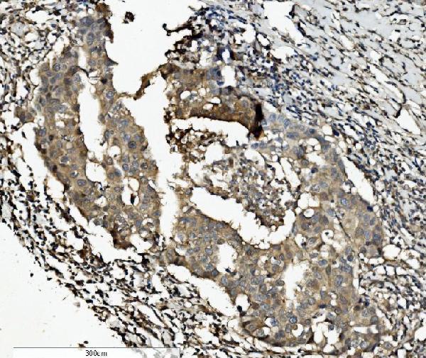
Real IHC data IHC analysis of Caspase-7/CASP7 using anti-Caspase-7/CASP7 antibody (A01044-2). Caspase-7/CASP7 was detected in paraffin-embedded section of human breast cancer tissue. Heat mediated antigen retrieval was performed in EDTA buffer (pH8.0, epitope retrieval solution). The tissue section was blocked with 10% goat serum. The tissue section was then incubated with 2μg/ml rabbit anti-Caspase-7/CASP7 Antibody (A01044-2) overnight at 4°C. Biotinylated goat anti-rabbit IgG was used as secondary antibody and incubated for 30 minutes at 37°C. The tissue section was developed using Strepavidin-Biotin-Complex (SABC) (Catalog # SA1022) with DAB as the chromogen.
Anti-Caspase-7/CASP7 Antibody ®
Cat # A01044-2
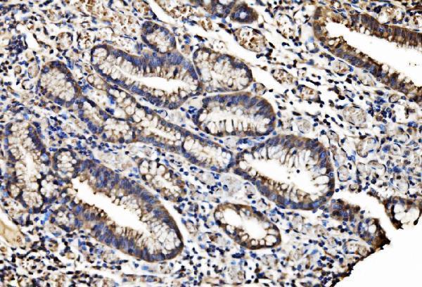
Real IHC data Anti-Caspase-7(P11) antibody, PA1442, IHC(P) IHC(P): Human Intestinal Cancer Tissue
Anti-Caspase-7(P11)/CASP7 Antibody ®
Cat # PA1442
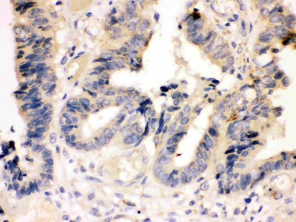
Real IHC data Anti-Caspase-7 vendor antibody, PB9369,IHC(P) IHC(P): Human Mammary Cancer Tissue
Anti-Caspase-7/CASP7 Antibody ®
Cat # PB9369

A01044-2 shows human breast, gastric, lung and rectal cancer paraffin sections (A01044-2 IHC captions); its listed reactivity is human and rat (catalog). PA1442 shows human intestinal cancer tissue (PA1442 IHC caption), while PB9369 shows human mammary cancer tissue (PB9369 IHC caption); both list IHC and human reactivity (catalog).

Which to pick: For tissue IHC, A01044-2 has the most documented paraffin-section examples, including EDTA retrieval at pH 8.0 and 2 μg/ml primary antibody (A01044-2 IHC captions); the fixative is unreported (A01044-2 IHC captions). For IF/ICC, choose A01044-2 because those applications are listed at 5 μg/ml, although no IF image is supplied (A01044-2 catalog applications, dilution and image records). For work across species, PA1442 lists human, mouse and rat reactivity, but its IHC figure documents human tissue only (PA1442 catalog reactivity and IHC caption); clonality is unreported for all three (catalog clone fields).

Each figure is that product's own IHC / IF validation image from its datasheet.

References

  1. UniProt Consortium. UniProt entry P55210 (CASP7_HUMAN, Caspase-7).
  2. Human Protein Atlas. CASP7 tissue IHC expression (reliability: Enhanced).
  3. Human Protein Atlas. CASP7 subcellular location (ICC-IF): Mainly localized to the nucleoplasm and nucleoli fibrillar center. In addition localized to the plasma membrane..
  4. Human Protein Atlas. CASP7 antibody validation summary (2 antibodies).
  5. MicroRNA-224 is implicated in lung cancer pathogenesis through targeting caspase-3 and caspase-7. Oncotarget 2015 — PMC4673127.
  6. Spatiotemporal characterization of extracellular matrix maturation in human artificial stromal-epithelial tissue substitutes. BMC biology 2024 — PMC11575135.
  7. Expression of Caspases in the Pig Endometrium Throughout the Estrous Cycle and at the Maternal-Conceptus Interface During Pregnancy and Regulation by Steroid Hormones and Cytokines. Frontiers in veterinary science 2021 — PMC7907442.
  8. miR-224 Is Significantly Upregulated and Targets Caspase-3 and Caspase-7 During Colorectal Carcinogenesis. Translational oncology 2019 — PMC6240712.
  9. PubMed PMID:8521391 — UniProt-cited evidence.
  10. PubMed PMID:8576161 — UniProt-cited evidence.
  11. PubMed PMID:8567622 — UniProt-cited evidence.