This website uses cookies to ensure you get the best experience on our website.
- Table of Contents
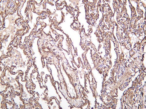
1 Citations 5 Q&As
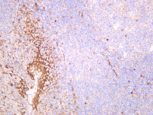
4 Citations 18 Q&As
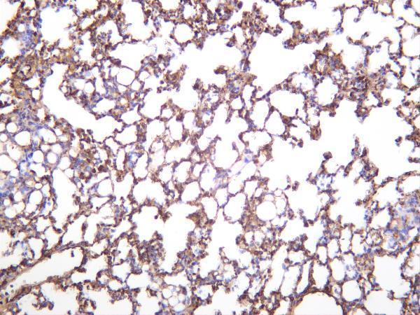
2 Citations 15 Q&As


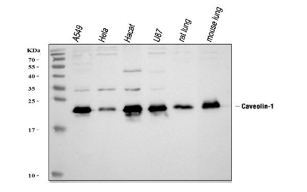
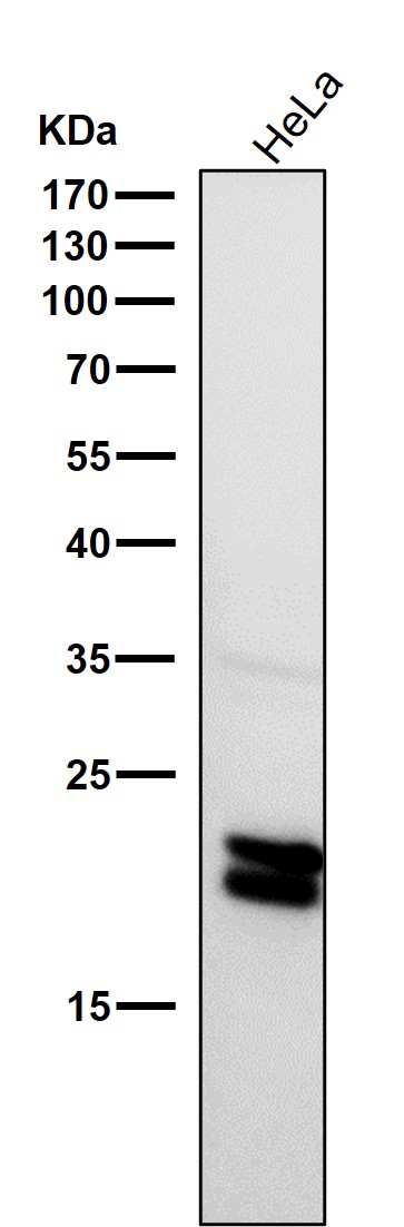
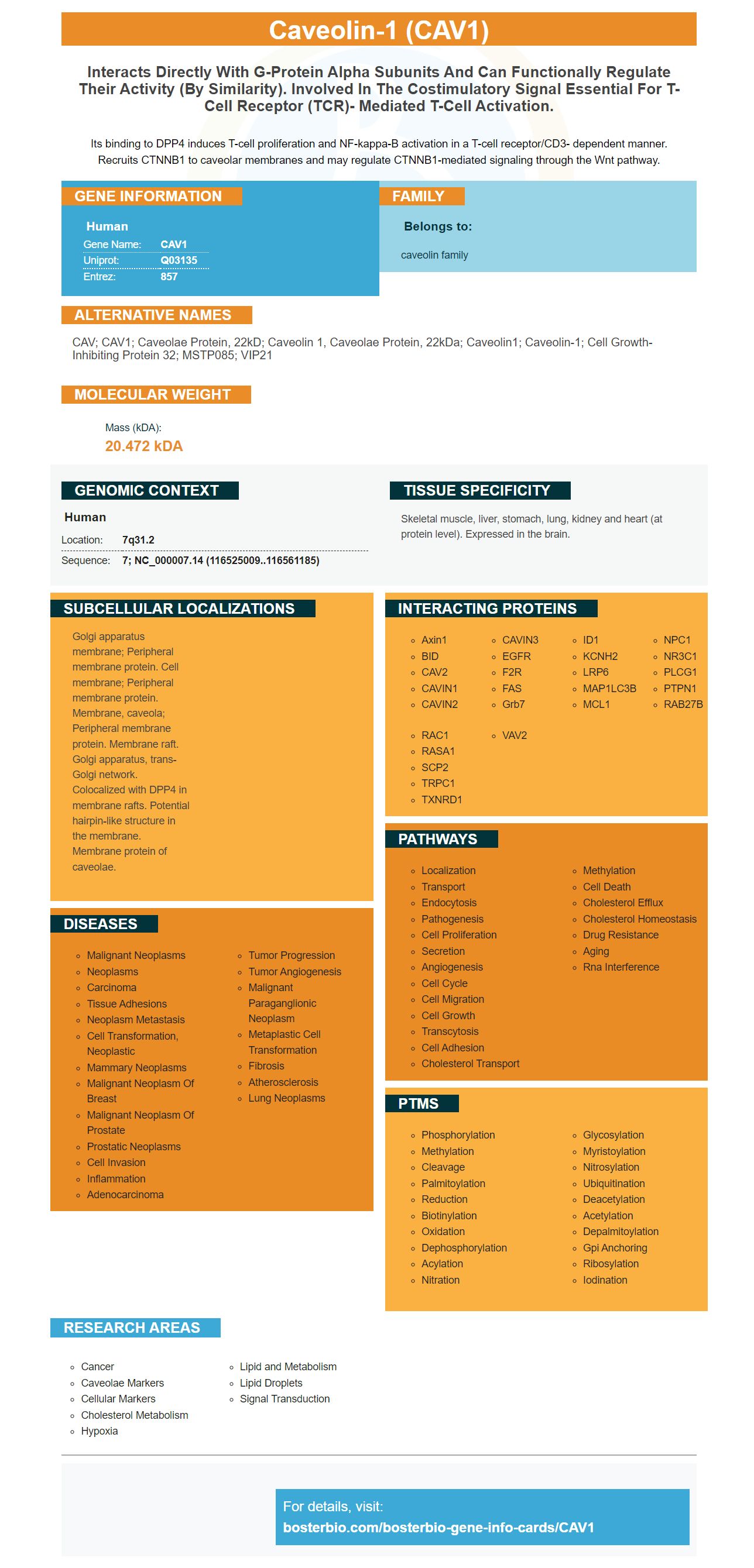
Facts about Caveolin-1.

Its binding to DPP4 induces T-cell proliferation and NF-kappa-B activation in a T-cell receptor/CD3- dependent manner. Recruits CTNNB1 to caveolar membranes and may regulate CTNNB1-mediated signaling through the Wnt pathway.
| Human | |
|---|---|
| Gene Name: | CAV1 |
| Uniprot: | Q03135 |
| Entrez: | 857 |

| Belongs to: |
|---|
| caveolin family |

CAV; CAV1; caveolae protein, 22kD; caveolin 1, caveolae protein, 22kDa; Caveolin1; Caveolin-1; cell growth-inhibiting protein 32; MSTP085; VIP21
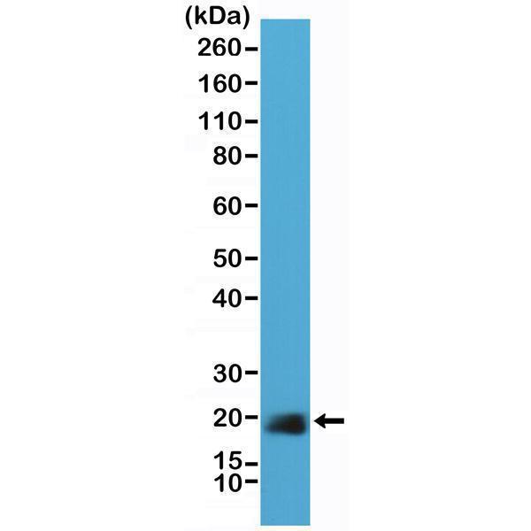
Mass (kDA):
20.472 kDA

| Human | |
|---|---|
| Location: | 7q31.2 |
| Sequence: | 7; NC_000007.14 (116525009..116561185) |
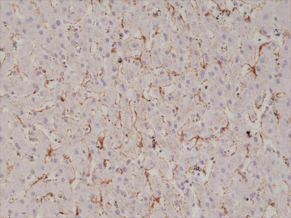
Skeletal muscle, liver, stomach, lung, kidney and heart (at protein level). Expressed in the brain.
Golgi apparatus membrane; Peripheral membrane protein. Cell membrane; Peripheral membrane protein. Membrane, caveola; Peripheral membrane protein. Membrane raft. Golgi apparatus, trans-Golgi network. Colocalized with DPP4 in membrane rafts. Potential hairpin-like structure in the membrane. Membrane protein of caveolae.


PMID: 1360410 by Glenney J.R. Jr.; The sequence of human caveolin reveals identity with VIP21, a component of transport vesicles.
PMID: 10086342 by Hurlstone A.F., et al. Analysis of the CAVEOLIN-1 gene at human chromosome 7q31.1 in primary tumours and tumour-derived cell lines.
*More publications can be found for each product on its corresponding product page