This website uses cookies to ensure you get the best experience on our website.


- Table of Contents
Plan CAVIN1 staining in paraffin sections using its reported tissue pattern and molecular localisation (HPA tissue IHC; UniProt). The guide covers fixation, controls and scoring around prominent vascular and adipocyte staining (HPA tissue IHC).

Expected localisation, validated protocols, controls and antibodies — the at-a-glance facts below, then the full design guide.
| Expected localisation | Caveolar membrane; cytosol possible (UniProt) | |
| Staining pattern | Staining mainly in blood vessels and adipocytes (HPA tissue IHC) | |
| Antigen retrieval | EDTA pH 8.0 HIER, heat-mediated (datasheet A31732-2) | |
| Positive control | Adipose tissue+4 more · see all | |
| Negative control | Adrenal gland+4 more · see all |
| Fixation | Keep fixation consistent across sections. (standard IHC practice; not target-specific) | |
| Caveat | Vascular and adipocyte staining can complicate scoring (HPA tissue IHC) | |
| Regulation | Tissue expression regulation is not annotated (UniProt) | |
| Isoform / epitope | 3 isoforms; epitope coverage is unknown (UniProt) |
Use the catalog antibody’s IHC-P protocol (datasheet: A31732-2) alongside two published chromogenic CAVIN1 methods (PMC8107102; PMC3636273).
| Sample | Paraffin-embedded human glioblastoma tissue; fixative not specified (datasheet A31732-2) |
| Fixation | Image fixative and duration unreported (datasheet A31732-2); verify before use. |
| Sectioning | 4–5 µm sections on charged slides (standard) |
| Deparaffinisation | Xylene, graded ethanol series to water (standard) |
| Antigen retrieval | Heat retrieval: EDTA pH 8.0 (datasheet A31732-2); 20 min, 95–100 °C (standard) |
| Peroxidase block | 3% H2O2, 10 min, room temperature (standard) |
| Blocking | 10% goat serum (datasheet A31732-2) |
| Primary antibody | Rabbit anti-CAVIN1, 1-2 μg/ml (datasheet A31732-2) |
| Primary incubation | Overnight at 4 °C (datasheet A31732-2) |
| Detection | HRP-conjugated secondary, DAB chromogen (datasheet A31732-2) |
| Counterstain | Hematoxylin, blue, dehydrate and mount (standard) |
| Expected result | CAVIN1-positive staining in adipocytes of adipose tissue (HPA tissue IHC: High). HPA tissue profile: Expression mainly in blood vessels and adipocytes. No signal in the no-primary control. |
CAVIN1 is associated with caveolae and the cell membrane; UniProt also lists cytosolic, nuclear and other locations, and reports no transmembrane segment (UniProt Q6NZI2). In tissue IHC, expect staining mainly in blood vessels and adipocytes, with high staining reported in several defined cell populations (HPA: tissue IHC). HPA rates the tissue pattern Supported, with medium consistency between staining and RNA data (HPA: tissue IHC reliability).
| Staining outlines adipocytes or follows vascular structures, while nearby cells differ in intensity. | This fits the broad tissue pattern of expression mainly in adipocytes and blood vessels (HPA: tissue IHC). Judge the stained cells within each structure; a vessel in a tissue with a negative listed cell population does not, by itself, contradict that population's result. HPA describes the tissue evidence as Supported but notes medium consistency with RNA data and staining in some cells or structures that it has not annotated (HPA: tissue IHC reliability). |
| A membrane-associated pattern appears in expected cells, with some intracellular staining. | Membrane association is consistent with the caveolar and cell-membrane locations in UniProt and the mainly plasma-membrane ICC-IF pattern in HPA (UniProt Q6NZI2 subcellular location; HPA: subcellular ICC-IF). Intracellular signal alone is not proof of artefact: UniProt also lists cytosol, endoplasmic reticulum, microsomes, mitochondria and nucleus, while HPA reports additional vesicular localization in ICC-IF. Assess cell identity, control tissue and background together. |
| Predominantly nuclear or uniformly intracellular staining appears throughout the section. | This is less persuasive as the expected tissue result because HPA describes expression mainly in blood vessels and adipocytes, and its ICC-IF summary places the main signal at the plasma membrane (HPA: tissue IHC; HPA: subcellular ICC-IF). UniProt includes nuclear and other intracellular locations, so compartment alone cannot establish an artefact (UniProt Q6NZI2 subcellular location). Compare the pattern with a known-positive tissue and appropriate staining controls before interpreting it. |
| Strong chromogen appears in cells reported as not detected, or it also appears in a no-primary control. | For example, HPA lists adrenal gland glandular cells and bone-marrow hematopoietic cells as not detected; first confirm which cells actually carry the signal (HPA: tissue IHC). A positive no-primary control points to detection-system or endogenous-activity background as a general IHC interpretation. If the no-primary control is clear, unexpected staining can reflect antibody cross-reactivity or a pattern requiring further validation; HPA's Supported rating does not validate every stained cell. |
| The slide looks uniformly hazy, or a known-positive adipose sample has no discernible cellular signal. | Diffuse haze makes localization and cell-based interpretation unreliable; check background controls before scoring (general IHC practice). Adipocytes are listed as High in adipose tissue, so absent staining there calls for a technical review, including section integrity, retrieval and detection controls, rather than an immediate biological absence call (HPA: High in adipocytes; general IHC practice). HPA does not provide target-specific fixation or retrieval behavior in this record. |
| Cell population and comparison tissue | HPA reports expression mainly in blood vessels and adipocytes, with High staining in adipocytes, breast myoepithelial cells, kidney Bowman's capsule, lung type I alveolar cells and smooth muscle cells (HPA: tissue IHC). Its negative calls name particular cells, not entire tissue sections: adrenal glandular cells, for example, are listed as Not detected. Select and score the specified cell population rather than treating every cell in a section as one control. |
| Compartment and topology | UniProt places CAVIN1 at caveolae and the cell membrane and reports no transmembrane segment; it also lists several intracellular locations (UniProt Q6NZI2). HPA's plasma-membrane and vesicle observations come from ICC-IF, so they inform localization but do not set a mandatory chromogenic IHC shape (HPA: subcellular ICC-IF). Interpret compartment alongside cell identity and controls. |
| Antibody evidence and isoforms | HPA049838 is listed as Supported for IHC and ICC, whereas HPA074213 is listed as Supported for ICC with no IHC status shown (HPA: antibodies). UniProt lists 3 isoforms; the supplied record gives no antibody epitope or isoform-coverage information, so an isoform-specific staining claim cannot be made (UniProt Q6NZI2 isoforms; HPA: antibodies). |
| IF/ICC Q&A | Q: Does IF/ICC evidence define the expected paraffin-section IHC pattern? A: It supports mainly plasma-membrane localization with additional vesicles, but it comes from ICC-IF images and should be read alongside tissue IHC observations for this guide (HPA: subcellular ICC-IF; HPA: tissue IHC). HPA lists HPA049838 and HPA074213 as ICC Supported; only HPA049838 has an IHC Supported status in the supplied antibody list (HPA: antibodies). |
| Fixation and retrieval evidence | The supplied UniProt and HPA records give no CAVIN1-specific fixation sensitivity or antigen-retrieval condition (UniProt Q6NZI2; HPA: tissue IHC). Retrieval and detection checks can be part of general paraffin-IHC troubleshooting, but these sources do not justify predicting how formalin exposure changes CAVIN1 signal or declaring a particular retrieval setting optimal. |
| Situation | Likely cause | Next action |
|---|---|---|
| No staining in adipocytes used as the positive comparison. | Adipocytes are reported High, so a completely blank result may reflect a failed staining run or an unsuitable section; the record does not identify a CAVIN1-specific fixation cause (HPA: High in adipocytes). | Verify tissue identity and preservation, then check primary-antibody application, general paraffin-IHC retrieval steps and detection-system performance using run controls. Reassess the adipocytes before reporting absence (general IHC practice). |
| Only faint or patchy staining is visible in an expected cell population. | HPA gives cell-level observations rather than a required intensity for every specimen, and its tissue reliability is Supported with medium RNA–staining consistency (HPA: tissue IHC reliability). Technical variation also needs exclusion (general IHC practice). | Compare like cell populations across sections, review section quality and run controls, and score the observed localization and intensity without turning a weak result into a categorical negative (general IHC practice). |
| Signal is widespread in glandular cells listed as not detected. | The named negative call applies to those cells; nearby vessels can be a separate stained population because HPA describes expression mainly in blood vessels (HPA: adrenal glandular cells Not detected; HPA: tissue IHC profile). If the glandular cells themselves stain, specificity needs checking. | Identify the stained cells morphologically, inspect a no-primary control and compare a known-positive sample from the same run. Treat persistent unexpected cell staining as unresolved until independently validated (general IHC practice). |
| The no-primary control shows brown deposits or a section-wide haze. | Detection-system background or endogenous activity can produce signal without primary antibody; this is a general chromogenic-IHC control interpretation, not a CAVIN1-specific property (general IHC practice). | Resolve the control background and review blocking, washing and detection steps before scoring CAVIN1. A signal that persists without primary antibody cannot establish target localization (general IHC practice). |
| The main signal is nuclear despite an otherwise clean slide. | Nucleus is a listed UniProt location, but HPA's main ICC-IF localization is plasma membrane; neither source makes nuclear staining alone a tissue-IHC specificity test (UniProt Q6NZI2; HPA: subcellular ICC-IF). | Check whether expected adipocytes or vascular-associated cells also stain, compare positive and no-primary controls, and report the observed compartment without assigning it to caveolae solely from nuclear signal (HPA: tissue IHC; general IHC practice). |
| An ICC-supported antibody produces an ambiguous paraffin IHC result. | ICC support does not establish IHC support: HPA074213 has ICC Supported and no IHC status shown, while HPA049838 has IHC Supported (HPA: antibodies). | For tissue-IHC interpretation, favor an IHC-validated antibody and require expected cell-population staining plus clean controls; keep the ICC-IF localization evidence as context rather than a substitute for paraffin IHC validation (HPA: antibodies; HPA: tissue IHC). |




Comprehensive Human Protein Atlas IHC scoring per tissue (reliability: Supported — Medium consistency between antibody staining and RNA expression data. Antibody staining in cells/structures not annotated, view images.). Rows are taken directly from the HPA tissue chart — click any row's HPA link to view the source.
| Tissue | Cell type | Level | Evidence | Source |
|---|---|---|---|---|
| Adipose tissue | Adipocytes | High | Protein (IHC) | HPA → |
| Breast | Myoepithelial cells | High | Protein (IHC) | HPA → |
| Kidney | Bowman's capsule | High | Protein (IHC) | HPA → |
| Lung | Alveolar cells type I | High | Protein (IHC) | HPA → |
| Smooth muscle | Smooth muscle cells | High | Protein (IHC) | HPA → |
| Tissue | Cell type | Level | Evidence | Source |
|---|---|---|---|---|
| Adrenal gland | Glandular cells | Not detected | Protein (IHC) | HPA → |
| Appendix | Endocrine cells | Not detected | Protein (IHC) | HPA → |
| Bone marrow | Hematopoietic cells | Not detected | Protein (IHC) | HPA → |
| Caudate | Glial cells | Not detected | Protein (IHC) | HPA → |
| Cerebellum | Cells in granular layer | Not detected | Protein (IHC) | HPA → |
These answers focus on chromogenic CAVIN1 IHC in paraffin sections; IF/ICC is addressed separately in one entry (caption A31732-2; HPA subcellular).
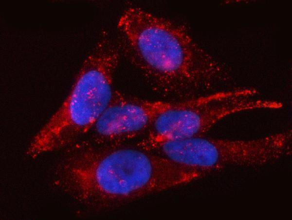
A31732-2 has IHC images from human paraffin sections and an IF/ICC image from HeLa cells (catalog image captions); its listed reactivity is human, mouse and rat (catalog applications/reactivity).
A31732-2 has IHC images from human glioblastoma, thyroid cancer, placenta and breast cancer paraffin sections (catalog IHC captions). The same SKU has an IF/ICC image from HeLa cells and lists human, mouse and rat reactivity (catalog IF caption; catalog applications/reactivity).
Which to pick: For paraffin-section IHC, choose A31732-2: its IHC captions document EDTA retrieval at pH 8.0 and primary antibody at 2 μg/ml; the fixative is unreported (catalog IHC captions). For IF/ICC, A31732-2 has a HeLa-cell image at 5 μg/ml (catalog IF caption). For mouse or rat work, A31732-2 lists both species as reactive, but the supplied IHC and IF images show human samples; clonality is unreported (catalog applications/reactivity; catalog image captions; catalog clone field).