This website uses cookies to ensure you get the best experience on our website.
- Table of Contents
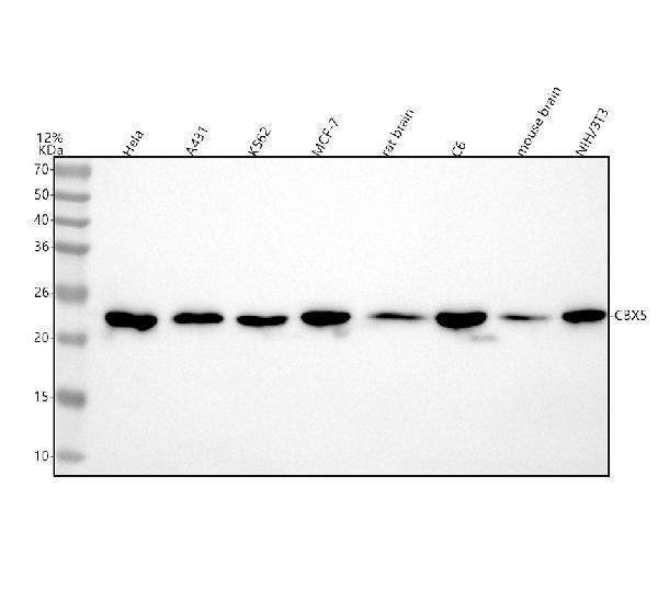
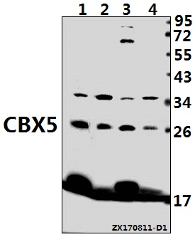
Facts about Chromobox protein homolog 5.
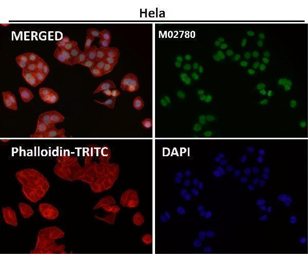
Can interact with lamin-B receptor (LBR). This interaction can lead to the institution of the heterochromatin with the inner nuclear membrane.
Mouse | |
---|---|
Gene Name: | Cbx5 |
Uniprot: | Q61686 |
Entrez: | 12419 |
Belongs to: |
---|
No superfamily |
Antigen p25; chromobox homolog 5 (Drosophila HP1 alpha); chromobox homolog 5 (HP1 alpha homolog, Drosophila); chromobox homolog 5; chromobox protein homolog 5; Heterochromatin protein 1 homolog alpha; heterochromatin protein 1-alpha; HP1 alpha homolog; HP1 alpha; HP1; HP1A; HP1-ALPHA; HP1Hs alpha; HP1Hs-alpha
Mass (kDA):
22.186 kDA
Mouse | |
---|---|
Location: | 15|15 F3 |
Sequence: | 15; |
If you are in the market for a new antibody to detect the CBX5 protein, then you have come to the right place. In this article, we will cover the Best Uses of the Anti-HP1 alpha/CBX5 Marker, as well as discuss its Validation on WB, ICC, and Immunofluorescence. We will also go over its validation on ICC and immunofluorescence, which is necessary to properly use the anti-CBX5 Marker.
Using the Anti-HP1 alpha/CBx5 Marker in Boster Bio allows for a precise and sensitive analysis of HP1alpha and CBX5 protein expression in human cells. HP1a has been shown to be a di-nucleosome crosslinker, as confirmed by Machida et al. in the 2018 Cell journal. Recent methodological developments have allowed for visualization of chromatin regions, and Ou et al. found that mesh density increased in euchromatic and heterochromatic regions.
Anti-HP1 alpha/CBx5 antibodies are available from several suppliers, including Boster Bio. HP1 alpha is a 191 amino acid protein with a mass of 22,225 daltons. HP1 alpha is predicted to be located in the nucleus. Boster Bio uses rabbit and mouse to develop its antibodies. These antibodies recognize histone H3 tails methylated at Lys-9 (H3K9me), leading to epigenetic repression. H3Y41ph excludes Cbx5 from chromatin.
The H3.3 variant is enriched at the PCH after sperm decondensation. This enrichment is replication-independent and suggests a different mechanism of chromatin modification. Both HP1 alpha and CBX5 play a role in the regulation of gene expression and cell proliferation. Interestingly, this CBX2 variant binds to both oocytes and chromosomes in the same cell type.
The HP1alpha/CBX5 marker in Boster Bio has recently been used for chromosome decondensation. HP1alpha contributes to the mechanical rigidity of mitotic chromosomes, and its depletion "strongly impairs" the mechanics. However, this effect size is too small to have any biological relevance.
HP1alpha regulates the shape of the nucleus, and HP1alpha depletion results in softened interphase nuclei and chromosome missegregation. This leads to aberrant nuclear morphologies. In addition, HP1alpha depletion decreases nuclear solidity and increases curvature. To assess the role of HP1alpha in cellular processes, HP1alpha should be evaluated as a critical factor in determining the level of HP1alpha expression in human cells.
HP1a-AID-sfGFP was found to have a similar function in parental and AID-sfGFP-modified cells. However, the HP1a-AID-sfGFP-tagged cell lines exhibited a significant difference in HP1a function when they are degraded by auxin. Various measurables of HP1a function were determined, including the general distribution of HP1a and its H3K9me2,3 levels, as well as nuclear shape, mitotic fidelity and nuclear dynamics.
While WB is not the only method of antibody validation, it is still an important tool in evaluating the performance of antibodies. Studies with antibodies that have little to no WB validation may have inaccurate data. To minimize this problem, use an antibody that has been validated on WB. However, many studies use antibodies that have not been validated and therefore are prone to bias. A good rule of thumb is to always perform immunoblotting on antibodies before use in other studies.
The terms ICC and reliability are two important concepts in clinical practice. The former describes how reliable a measurement is. The latter is a measurement's reliability when a single rater or one measurement performs the same task. The two terms are not the same, and this difference is the basis for the ICC definition. The two definitions are often used interchangeably, but the former is the more common. In this article, we will discuss the ICC definition and how it can be applied to reliability studies.
The target protein is tagged with a fluorescent or affinity tag. The tagged pattern is then compared with the signal of the antibody, confirming that the antibody recognizes the target protein. ICC-IF also allows validation by tagged proteins. The size of the protein stained by the antibody is compared to the pattern obtained by the cell line using a positive control. The antibodies that are validated using this method will be introduced in the ICC-IF phenotype.
ICC judges assess whether a state has a willingness to carry out genuine proceedings. They consider the evidence provided by the state with jurisdiction and the information obtained during national investigations. ICC judges also consider if the state is unwilling to prosecute the case or unable to do so. If the state refuses to prosecute, the case will be moved to a national court. However, this does not mean that the state cannot prosecute the case.
ICC-ES reports are an excellent way to determine whether a new building product is compliant with code requirements. ICC-ES Reports also provide valuable information on installation, product identification, and evaluation criteria. These reports are based on the expertise of ICC-ES engineers. In addition to the ICC-ES reports, you can trust their findings. The validation process is simple. You can complete the form online or download the PDF version.
A single-target antibody is validated using three different methods. First, it is evaluated for consistency by comparing the staining pattern of the antibody to the target protein's mRNA levels in up to 46 normal tissues. Then, the staining patterns of the two antibodies are compared in representative images of high and low-expression cells. Then, they are compared with the same staining pattern in a third tissue, which is typically the eukaryotic cell.
In immunofluorescence, cellular proteins are labeled with specific antibodies and fluororochrome-conjugated secondary antibodies. Most fluorescence microscopes can also analyze protein subcellular localization, expression level, and activation state in multiple cells. Multiplexed analyses are beneficial for scientists because they eliminate the need for consecutive sections. This also saves reagents and time. If you are looking for an immunofluorescence antibody, you can trust the researchers at Fortis Life Sciences.
To validate IF/ICC antibodies, you should look for a company with a large antigen library. Sino Biological, Inc. has extensive experience in determining the subcellular localization of therapeutic antibodies using nature cells. The company has tested their antibodies on numerous tumor cell lines and stem cells. This provides a high-quality, reliable product. Further, it has more than 1700 quality Immunofluorescence/ICC antibodies.
Several methods are required for multiplex IHC validation. One of these is motif enrichment. EnCODE accepts data where a motif is at least fourfold enriched in the bands. A further validation method is required for histone antibody to be accepted by ENCODE. This webinar will describe a simplified approach to optimize antibodies for multiplex immunofluorescence and immune checkpoint assays. You will learn about the benefits of optimizing monoclonal antibodies in multiplex immunofluorescence assays.
The other level of validation is peptide competition assays. If the target histone accounts for at least 80% of the immunoprecipitated sample, the antibody is valid. The antibody must also be able to identify mutants defective in histone modifying proteins by demonstrating an antibody signal below ten percent of wild-type signals. Finally, you should look for overlaps between localization of histone modifications and the localization of histone proteins in previous ChIP-seq datasets.
PMID: 8978696 by le Douarin B., et al. A possible involvement of TIF1 alpha and TIF1 beta in the epigenetic control of transcription by nuclear receptors.
PMID: 11107181 by Li Y.-J., et al. Contiguous arrangement of p45 NFE2, HnRNP A1, and HP1 alpha on mouse chromosome 15 and human chromosome 12: evidence for suppression of these genes due to retroviral integration within the Fli-2 locus.