This website uses cookies to ensure you get the best experience on our website.


- Table of Contents
Plan chromogenic IHC on paraffin sections for CCDC134 using the IHC-validated antibody at a starting concentration of 2.5 μg/mL (datasheet). This guide highlights the cytoplasmic tissue pattern and respiratory epithelial staining (HPA tissue IHC).

Expected localisation, validated protocols, controls and antibodies — the at-a-glance facts below, then the full design guide.
| Expected localisation | Cytoplasmic in most tissues (HPA tissue IHC) | |
| Staining pattern | Cytoplasmic staining in respiratory epithelial cells (HPA tissue IHC) | |
| Antigen retrieval | Tris-EDTA pH 9.0 HIER, 95–98 °C, 20 min (rule: nuclear antigen) | |
| Positive control | Bronchus+4 more · see all | |
| Negative control | Bone marrow+4 more · see all |
| Fixation | Keep fixation consistent across sections (standard IHC practice; not target-specific); target-specific effects are unknown. | |
| Caveat | Secretion can make tissue protein and RNA patterns differ (HPA tissue IHC) | |
| Regulation | Higher expression in activated CD8+ T cells (UniProt) | |
| Isoform / epitope | 0 isoforms; mature chain starts at residue 23 after signal peptide cleavage (UniProt) |
The catalog antibody’s IHC-P protocol and one published CCDC134 IHC procedure (PMC5746300) provide starting conditions for paraffin sections.
| Sample | Tissue sections; selected-image fixative not specified (standard IHC workflow) |
| Fixation | Image fixative and duration unreported (datasheet A14619); verify before use. |
| Sectioning | 4–5 µm sections on charged slides (standard) |
| Deparaffinisation | Xylene, graded ethanol series to water (standard) |
| Antigen retrieval | Heat-induced epitope retrieval in Tris-EDTA buffer, pH 9.0, 20 min at 95–98 °C (standard rule: nuclear antigen) |
| Peroxidase block | 3% H2O2, 10 min, room temperature (standard) |
| Blocking | 10% normal serum of the secondary host, 30 min, room temperature (standard) |
| Primary antibody | Rabbit anti-CCDC134, 2.5 μg/mL (datasheet A14619) |
| Primary incubation | Overnight at 4 °C (standard) |
| Detection | HRP-polymer secondary, DAB chromogen 5–10 min (standard) |
| Counterstain | Hematoxylin, blue, dehydrate and mount (standard) |
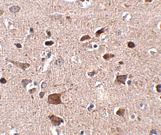
| Expected result | CCDC134-positive staining in respiratory epithelial cells of bronchus (HPA tissue IHC: High). HPA tissue profile: Cytoplasmic expression in most tissues. No signal in the no-primary control. |
CCDC134 is mainly in the endoplasmic reticulum lumen and has no transmembrane segment; it is also reported as secreted, cytoplasmic and nuclear (UniProt Q9H6E4). In paraffin tissue sections, expect predominantly cytoplasmic staining in many tissues (HPA: IHC profile, Approved). Respiratory epithelial cells in bronchus and nasopharynx are useful high-staining examples (HPA: High). Secretion means tissue RNA and protein locations may differ (HPA: reliability description).
| Cytoplasmic staining in bronchial or nasopharyngeal respiratory epithelial cells. | This matches the reported tissue profile and high staining level (HPA: cytoplasmic expression in most tissues; High in these cells). Score the relevant epithelial cells separately from surrounding tissue (general IHC practice). |
| Predominantly nuclear staining across an otherwise routine tissue section. | Treat this as a compartment mismatch against the usual cytoplasmic IHC profile (HPA: tissue IHC). Nuclear accumulation is reported after UV irradiation (UniProt Q9H6E4), so context matters; check the control and staining distribution before interpreting it. |
| Strong staining in esophageal or oral squamous epithelial cells. | These cell types were not detected in the cited tissue IHC observations (HPA: esophagus; oral mucosa). Unexpected staining warrants a check for cross-reactivity or endogenous detection activity (general IHC practice); it alone does not identify the cause. |
| Uniform color over cells, extracellular space and the negative control. | A diffuse field does not resemble the cell-associated cytoplasmic profile (HPA: tissue IHC). Consider nonspecific background or detection chemistry and compare controls before assigning CCDC134 positivity (general IHC practice). |
| No signal in respiratory epithelium of a bronchus or nasopharynx control. | Both are reported as high-staining sites (HPA: bronchus; nasopharynx). Review tissue integrity, retrieval, antibody incubation and chromogenic detection as general IHC checks; a failed control prevents a confident negative call in the test section. |
| Secretion and tissue distribution | CCDC134 is secreted and lacks a transmembrane segment (UniProt Q9H6E4). Its protein location need not follow tissue RNA location (HPA: reliability description); assess stained cell types directly. |
| Intracellular compartment | The ER lumen is the main location reported by UniProt, while HPA tissue IHC describes a cytoplasmic pattern (UniProt Q9H6E4; HPA: tissue IHC). Routine chromogenic IHC may show this as cytoplasmic staining. |
| Processing and glycosylation | The annotated signal peptide spans residues 1–22, the mature chain spans 23–229, and one glycosylation site is annotated at 148 (UniProt Q9H6E4). These annotations do not establish epitope accessibility after retrieval. |
| Evidence across assays | IHC is Approved for HPA003936; ICC is Supported for HPA075348 (HPA: antibodies). IF/ICC Q&A: should the image match paraffin IHC exactly? No: HPA reports cytosol with additional vesicles in ICC-IF, while tissue IHC is broadly cytoplasmic (HPA: subcellular; tissue IHC). |
| Situation | Likely cause | Next action |
|---|---|---|
| Positive control is blank. | Bronchial and nasopharyngeal respiratory epithelial cells are reported High (HPA: tissue IHC); a blank control suggests an assay or specimen problem, without identifying which. | Inspect section quality and confirm retrieval, antibody incubation and detection steps (general IHC practice). Resolve the control before scoring test tissue. |
| Signal appears mainly in nuclei. | This differs from the usual cytoplasmic IHC profile (HPA: tissue IHC), although UV-associated nuclear accumulation is reported (UniProt Q9H6E4). | Check exposure context if known, then compare with the positive and negative controls and review whether nuclear color is cell-specific (general IHC practice). |
| Unexpected squamous epithelial staining dominates. | Esophageal and oral squamous epithelial cells were not detected in HPA tissue IHC (HPA: esophagus; oral mucosa); cross-reactivity or endogenous activity are possibilities (general IHC practice). | Compare a negative control and the expected respiratory epithelial positive control; investigate detection background before calling this target-specific staining (general IHC practice). |
| Diffuse color obscures cell boundaries. | The reported pattern is cytoplasmic expression in most tissues (HPA: tissue IHC); widespread color may reflect nonspecific staining or detection background (general IHC practice). | Review blocking, washes, antibody concentration and chromogen development against controls (general IHC practice). Reassess only when cellular staining is distinguishable. |
| Low or absent signal in one tissue is treated as a universal negative. | Expression varies by cell type: liver hepatocytes are Low, while respiratory epithelial cells are High (HPA: tissue IHC). A single tissue does not establish assay sensitivity. | Include a documented high-staining respiratory epithelial control and score the relevant cell population, not the whole section (HPA: tissue IHC; general IHC practice). |
| IF/ICC appears vesicular while IHC appears broadly cytoplasmic. | HPA reports supported cytosolic and additional approved vesicular ICC-IF localization, alongside a cytoplasmic tissue IHC profile (HPA: subcellular; tissue IHC). | Interpret each assay against its own reported pattern and controls; do not use the ICC-IF image alone to redefine a paraffin IHC positive call (HPA: subcellular; tissue IHC). |
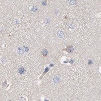
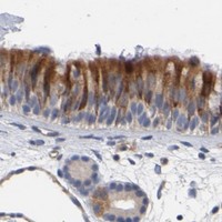


Comprehensive Human Protein Atlas IHC scoring per tissue (reliability: Approved — Secreted protein, tissue location of RNA and protein is expected to differ. Caution, Splice and/or transcript discrepancy exists.). Rows are taken directly from the HPA tissue chart — click any row's HPA link to view the source.
| Tissue | Cell type | Level | Evidence | Source |
|---|---|---|---|---|
| Bronchus | Respiratory epithelial cells | High | Protein (IHC) | HPA → |
| Nasopharynx | Respiratory epithelial cells | High | Protein (IHC) | HPA → |
| Adipose tissue | Adipocytes | Medium | Protein (IHC) | HPA → |
| Adrenal gland | Glandular cells | Medium | Protein (IHC) | HPA → |
| Appendix | Glandular cells | Medium | Protein (IHC) | HPA → |
| Tissue | Cell type | Level | Evidence | Source |
|---|---|---|---|---|
| Bone marrow | Hematopoietic cells | Not detected | Protein (IHC) | HPA → |
| Cerebral cortex | Endothelial cells | Not detected | Protein (IHC) | HPA → |
| Esophagus | Squamous epithelial cells | Not detected | Protein (IHC) | HPA → |
| Lymph node | Germinal center cells | Not detected | Protein (IHC) | HPA → |
| Oral mucosa | Squamous epithelial cells | Not detected | Protein (IHC) | HPA → |
Troubleshoot CCDC134 staining in paraffin sections by checking retrieval, compartment, cell type and controls before comparing chromogenic signal across samples.
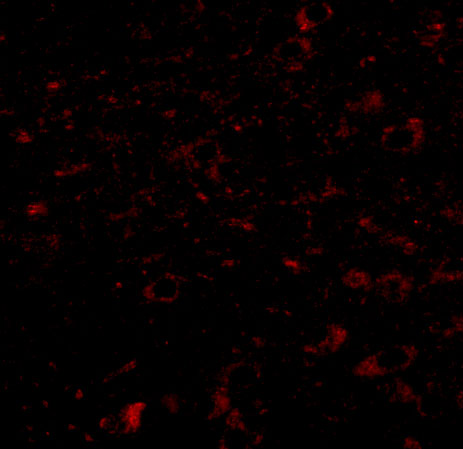
A14619 has real IHC data and an IF image in human brain tissue (catalog: figure tag and image captions). Its listed reactivity covers human, mouse and rat (catalog: reactivity).
A14619 is listed for IHC-P and IF, with human, mouse and rat reactivity (catalog: applications and reactivity). Its human brain images show IHC at 2.5 μg/mL and IF at 20 μg/mL (A14619: IHC and IF image captions).
Which to pick: For paraffin-section IHC, choose A14619: IHC-P is listed, and its own IHC image shows human brain staining at 2.5 μg/mL (catalog: applications; A14619 IHC image caption); the fixative is unreported (A14619 IHC image caption). For IF, choose A14619, whose human brain IF image uses 20 μg/mL; separate ICC validation is not listed (A14619 IF image caption; catalog: applications). For mouse or rat samples, A14619 has listed reactivity, but both supplied tissue images are human; clonality is unspecified (catalog: reactivity and clone; A14619 image captions).