This website uses cookies to ensure you get the best experience on our website.


- Table of Contents
Plan CCDC69 IHC in paraffin sections with the IHC-validated antibody at 1–5 μg/mL (datasheet: A17261). Expect cytoplasmic staining in most cell types, but interpret intensity cautiously because tissue staining has low agreement with RNA expression (HPA tissue IHC).

Expected localisation, validated protocols, controls and antibodies — the at-a-glance facts below, then the full design guide.
| Expected localisation | Tissue: cytoplasm (HPA tissue IHC); mitosis: spindle/midbody (UniProt) | |
| Staining pattern | Cytoplasmic staining in most cell types (HPA tissue IHC) | |
| Antigen retrieval | Citrate pH 6.0 HIER, 95–98 °C, 20 min (rule: cytoplasmic / membrane antigen) | |
| Positive control | Adrenal gland+4 more · see all | |
| Negative control | Adipose tissue |
| Fixation | Keep fixation consistent across sections (standard IHC practice; not target-specific) | |
| Caveat | Staining has low agreement with RNA expression (HPA tissue IHC) | |
| Regulation | High in duodenum, pancreas and prostate (UniProt) | |
| Isoform / epitope | 0 annotated isoforms; epitope site unspecified (UniProt) |
Compare the catalog antibody’s IHC-P protocol with three published CCDC69 IHC workflows (PMC7922363; PMC11992169; PMC10571395).
| Sample | Tissue sections; selected-image fixative not specified (standard IHC workflow) |
| Fixation | Image fixative and duration unreported (datasheet A17261); verify before use. |
| Sectioning | 4–5 µm sections on charged slides (standard) |
| Deparaffinisation | Xylene, graded ethanol series to water (standard) |
| Antigen retrieval | Heat-induced epitope retrieval in citrate buffer, pH 6.0, 20 min at 95–98 °C (standard rule: cytoplasmic / membrane antigen) |
| Peroxidase block | 3% H2O2, 10 min, room temperature (standard) |
| Blocking | 10% normal serum of the secondary host, 30 min, room temperature (standard) |
| Primary antibody | Chicken anti-CCDC69, 1-5 μg/mL (datasheet A17261) |
| Primary incubation | Overnight at 4 °C (standard) |
| Detection | HRP-polymer secondary, DAB chromogen 5–10 min (standard) |
| Counterstain | Hematoxylin, blue, dehydrate and mount (standard) |
| Expected result | CCDC69-positive staining in glandular cells of adrenal gland (HPA tissue IHC: Medium). HPA tissue profile: Cytoplasmic expression in most cell types. No signal in the no-primary control. |
In paraffin-section IHC, expect predominantly cytoplasmic staining in many cell types, including glandular cells and respiratory epithelium (HPA: tissue IHC). CCDC69 also localizes to the spindle and midbody during cell division (UniProt A6NI79: subcellular location). It has no transmembrane segment (UniProt A6NI79: topology). Interpret any pattern cautiously: HPA rates tissue IHC reliability Uncertain because staining has low consistency with RNA expression (HPA: tissue IHC reliability).
| Cytoplasmic signal in glandular cells or respiratory epithelium. | This fits the broad tissue profile: glandular cells stain at Medium level in adrenal gland, appendix, breast and cervix; bronchial respiratory epithelial cells stain at Medium level (HPA: tissue IHC). It is a plausible positive pattern, but HPA's Uncertain reliability limits how confidently staining alone identifies CCDC69 (HPA: tissue IHC reliability). |
| Predominantly nuclear signal, or a membrane-only pattern, in paraffin sections. | Reassess the result against the reported cytoplasmic tissue-IHC profile and UniProt spindle/midbody localization (HPA: tissue IHC; UniProt A6NI79: subcellular location). A different compartment can indicate nonspecific staining or a preparation-dependent pattern (general IHC practice). HPA reports plasma-membrane localization in ICC-IF, but that assay does not establish a membrane-only tissue-IHC pattern (HPA: subcellular ICC-IF). |
| Strong staining in adipocytes, while the expected cells show little signal. | HPA reports adipocytes as Not detected in adipose tissue (HPA: tissue IHC). Check whether the signal follows the cells or the detection chemistry; off-target antibody binding or endogenous detection activity can produce misleading cell-specific staining (general IHC practice). This tissue comparison is provisional because HPA rates CCDC69 tissue IHC Uncertain (HPA: tissue IHC reliability). |
| Broad haze, precipitate, or similar color across cells and surrounding tissue. | Treat an indiscriminate pattern as background until controls show cell-associated staining (general IHC practice). Review blocking, washes and chromogen development, and inspect a no-primary control for detection-system background (general IHC practice). HPA's cytoplasmic profile concerns cellular staining; it does not validate diffuse background as CCDC69 (HPA: tissue IHC). |
| No signal in glandular cells of a tissue listed as Medium by HPA. | First verify that the sampled section contains the listed cell type and that the detection run worked (HPA: tissue IHC; general IHC practice). Then review antibody dilution, retrieval and detection controls using the validated IHC procedure (general IHC practice). A single negative section cannot settle expression because HPA's tissue-IHC reliability is Uncertain (HPA: tissue IHC reliability). |
| Tissue-IHC evidence quality | HPA describes cytoplasmic expression in most cell types but assigns Uncertain reliability owing to low agreement between antibody staining and RNA expression (HPA: tissue IHC). Use its Medium and Not detected entries as comparisons, not absolute pass/fail standards. |
| Cell and tissue selection | Listed Medium examples include adrenal, appendix, breast and cervical glandular cells and bronchial respiratory epithelium; adipocytes are Not detected (HPA: tissue IHC). UniProt's high tissue-expression list is a separate observation and does not supply cell-level IHC scores (UniProt A6NI79: tissue specificity). |
| Assay-specific localization | HPA tissue IHC reports cytoplasmic staining, whereas HPA ICC-IF approves plasma-membrane localization with additional Golgi and actin-filament localization (HPA: tissue IHC; HPA: subcellular ICC-IF). Judge paraffin-section chromogenic results against tissue-IHC observations; investigate discordance before assigning an IF compartment to IHC. |
| Topology and mitotic context | CCDC69 has no annotated transmembrane segment (UniProt A6NI79: topology). UniProt describes movement along interpolar microtubules in anaphase and concentration at the midbody in telophase (UniProt A6NI79: subcellular location). Such structures depend on finding cells in the relevant division stage (general IHC practice). |
| Antibody validation scope | HPA043648 is listed as IHC Uncertain, while HPA052896 is listed as ICC Approved with no IHC status supplied (HPA: antibodies). An ICC approval alone cannot validate a paraffin-section result; assess the IHC antibody and its tissue controls in the intended assay (general IHC practice). |
| Situation | Likely cause | Next action |
|---|---|---|
| Expected glandular cells are blank. | The section may lack the scored cell population, or the staining run may have failed (HPA: tissue IHC; general IHC practice). | Confirm cell identity and inspect a working positive control; then check retrieval, antibody dilution and detection against the IHC-validated procedure (general IHC practice). |
| Only nuclei or tissue edges are intensely colored. | The distribution departs from HPA's cytoplasmic tissue profile; edge effects or nonspecific signal are possible (HPA: tissue IHC; general IHC practice). | Compare interior cells with edges and a no-primary control; review section handling, washes and chromogen development (general IHC practice). |
| Adipocytes appear strongly positive. | HPA lists adipocytes as Not detected; endogenous detection activity or off-target binding may explain apparent color (HPA: tissue IHC; general IHC practice). | Check a no-primary control and cell morphology, then compare staining in the listed Medium cell populations within a controlled run (general IHC practice; HPA: tissue IHC). |
| Color obscures cell boundaries across the section. | Excess detection product or insufficient washing can create diffuse background (general IHC practice). | Inspect the no-primary control, shorten chromogen development if excessive, and review blocking and wash steps (general IHC practice). |
| Two tissue sections give different apparent intensities. | The sampled cell types may differ, and HPA's tissue-IHC profile has Uncertain reliability (HPA: tissue IHC). | Score comparable cells with the same detection conditions and controls; report compartment, cell type and intensity separately (general IHC practice). |
| Does ICC-IF membrane staining establish a positive IHC result? | HPA approves a mainly plasma-membrane ICC-IF location, while its tissue-IHC profile is cytoplasmic and Uncertain (HPA: subcellular ICC-IF; HPA: tissue IHC). | Evaluate the paraffin section with tissue-IHC controls and its own compartment readout; consult the separate IF/ICC guide for fluorescence assay design (general IHC/IF practice). |


Comprehensive Human Protein Atlas IHC scoring per tissue (reliability: Uncertain — Low consistency between antibody staining and RNA expression data. Pending external verification.). Rows are taken directly from the HPA tissue chart — click any row's HPA link to view the source.
| Tissue | Cell type | Level | Evidence | Source |
|---|---|---|---|---|
| Adrenal gland | Glandular cells | Medium | Protein (IHC) | HPA → |
| Appendix | Glandular cells | Medium | Protein (IHC) | HPA → |
| Breast | Glandular cells | Medium | Protein (IHC) | HPA → |
| Bronchus | Respiratory epithelial cells | Medium | Protein (IHC) | HPA → |
| Caudate | Glial cells | Medium | Protein (IHC) | HPA → |
| Tissue | Cell type | Level | Evidence | Source |
|---|---|---|---|---|
| Adipose tissue | Adipocytes | Not detected | Protein (IHC) | HPA → |
Troubleshoot CCDC69 staining by checking retrieval, compartment patterns and controls before comparing chromogenic IHC scores (UniProt A6NI79; HPA: tissue IHC reliability uncertain).
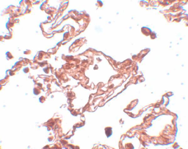
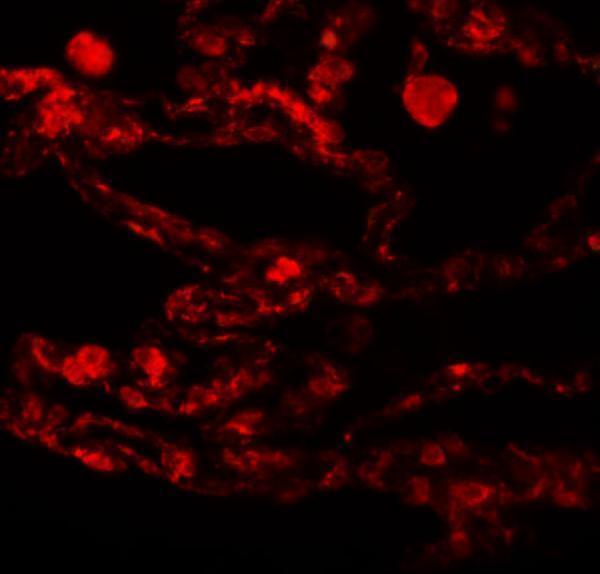
A17261 has IHC and IF images from human lung tissue (catalog image captions). Human, mouse and rat reactivity is listed, but IHC and IF validation is reported only for human samples (datasheet).
A17261 has an IHC image of human lung tissue at 5 μg/mL (IHC image caption). A17261 also has an IF image of human lung tissue at 20 μg/mL (IF image caption).
Which to pick: For tissue IHC, choose A17261: it is listed for IHC-P, with a human lung IHC image at 5 μg/mL; the image caption does not report the fixative (catalog applications; IHC image caption). For IF, choose A17261 based on its human lung IF image; ICC is not listed as an application (IF image caption; catalog applications). For mouse or rat work, A17261 lists both species as reactive, but its datasheet reports IHC and IF validation only in human samples (catalog reactivity; datasheet).