This website uses cookies to ensure you get the best experience on our website.


- Table of Contents
Plan chromogenic CCT8 IHC in paraffin sections using the catalog antibody’s 0.5–1 μg/mL range (datasheet PB9993). Choose controls from HPA’s tissue staining data and assess cytoplasmic staining in the appropriate cells (HPA tissue IHC).

Expected localisation, validated protocols, controls and antibodies — the at-a-glance facts below, then the full design guide.
| Expected localisation | Cytoplasmic staining in tissue (HPA tissue IHC) | |
| Staining pattern | General cytoplasmic staining across tissue cells (HPA tissue IHC) | |
| Antigen retrieval | EDTA pH 8.0 HIER, heat-mediated (datasheet PB9993) | |
| Positive control | Adrenal gland+4 more · see all | |
| Negative control | Smooth muscle+1 more · see all |
| Fixation | Keep fixation consistent across sections (standard IHC practice; not target-specific) | |
| Caveat | Antibody staining and RNA show medium consistency (HPA tissue IHC) | |
| Regulation | Specific expression regulators are not annotated (UniProt) | |
| Isoform / epitope | 3 isoforms; epitope coverage is unknown (UniProt) |
The catalog antibody’s IHC protocol is followed by published CCT8 protocols for colorectal cancer tissue (PMC8642402) and lung adenocarcinoma tissue microarrays (PMC10622983).
| Sample | Paraffin-embedded mouse kidney tissue; fixative not specified (datasheet PB9993) |
| Fixation | Image fixative and duration unreported (datasheet PB9993); verify before use. |
| Sectioning | 4–5 µm sections on charged slides (standard) |
| Deparaffinisation | Xylene, graded ethanol series to water (standard) |
| Antigen retrieval | Heat retrieval: EDTA pH 8.0 (datasheet PB9993); 20 min, 95–100 °C (standard) |
| Peroxidase block | 3% H2O2, 10 min, room temperature (standard) |
| Blocking | 10% goat serum (datasheet PB9993) |
| Primary antibody | Rabbit anti-CCT8, 0.5-1μg/ml (datasheet PB9993) |
| Primary incubation | Overnight at 4 °C (datasheet PB9993) |
| Detection | Streptavidin-biotin complex (SABC), DAB chromogen (datasheet PB9993) |
| Counterstain | Hematoxylin, blue, dehydrate and mount (standard) |
| Expected result | CCT8-positive staining in glandular cells of adrenal gland (HPA tissue IHC: High). HPA tissue profile: General cytoplasmic expression. No signal in the no-primary control. |
CCT8 is a cytoplasmic chaperonin with no transmembrane segment (UniProt P50990). In paraffin section IHC, expect general cytoplasmic staining (HPA: tissue IHC), including high staining in adrenal glandular cells, bronchial respiratory epithelial cells, and placental cytotrophoblasts (HPA: High). HPA rates the tissue pattern Enhanced, with medium consistency between antibody staining and RNA expression (HPA: tissue IHC reliability).
| Distinct cytoplasmic chromogen in adrenal glandular cells, bronchial respiratory epithelium, or placental cytotrophoblasts. | This matches the reported compartment and high-staining cell populations (HPA: general cytoplasmic expression; High in these cells). Judge intensity within the identified cells; neighboring cell types need their own HPA reference. |
| Predominantly nuclear, surface-restricted, or extracellular staining with little cytoplasmic signal. | Recheck localisation before calling this CCT8: tissue IHC is generally cytoplasmic (HPA: tissue IHC), and CCT8 lacks a transmembrane segment (UniProt P50990). Nuclear localisation in ICC-IF is marked uncertain (HPA: subcellular); it does not validate nuclear IHC. |
| Strong staining in smooth muscle cells or chondrocytes while expected positive cells also stain. | These cells were not detected in the HPA tissue survey (HPA: smooth muscle cells; chondrocytes). The discrepancy warrants review for antibody cross-reactivity or endogenous detection activity (general IHC practice); it alone does not identify the cause. |
| Diffuse chromogen over many structures, including spaces between cells, obscures cell boundaries. | This is difficult to score as the reported cytoplasmic pattern (HPA: tissue IHC). Assess no-primary and detection controls for background, and review blocking and washes (general IHC practice) before interpreting intensity. |
| No staining in an otherwise interpretable adrenal glandular cell or bronchial epithelial region. | Both are high-staining populations in the HPA survey (HPA: adrenal gland; bronchus). Check section identity, antibody and detection performance, and antigen retrieval conditions (general IHC practice); an absent signal alone does not establish absent CCT8. |
| Tissue and cell selection | Use an identified high-staining population to judge assay performance (HPA: adrenal glandular cells; bronchial respiratory epithelial cells; placental cytotrophoblasts, High). Smooth muscle cells and chondrocytes are useful low-expectation comparators, not absolute negative controls (HPA: Not detected). |
| Evidence strength | The tissue profile is rated Enhanced, while antibody staining and RNA have medium consistency (HPA: tissue IHC reliability). Three listed antibodies have Enhanced IHC status (HPA: HPA018520, HPA021051, HPA029426). Treat an unexpected pattern as a result to investigate, not as proof of a new localisation. |
| Cellular location and processing | CCT8 is annotated in cytoplasm, centrosome, and cilium basal body; it has no signal peptide or transmembrane segment (UniProt P50990). The expected tissue-level readout remains general cytoplasmic staining (HPA: tissue IHC); the annotations do not promise resolvable centrosomal puncta in chromogenic sections. |
| IF/ICC Q: should nuclear signal be expected? | A: HPA lists nucleoplasm as uncertain and cytosol as supported in ICC-IF, with approved sperm-piece locations (HPA: subcellular). Its IHC pattern is generally cytoplasmic (HPA: tissue IHC). Interpret IF/ICC on its own guide; these observations supply no IF protocol option here. |
| Situation | Likely cause | Next action |
|---|---|---|
| A high-staining reference region is blank. | The result conflicts with the reported high staining, but does not distinguish specimen from assay failure (HPA: adrenal glandular cells; bronchial epithelium, High). | Confirm the cell population and run an appropriate positive section; then review retrieval, primary incubation, and detection controls (general IHC practice). |
| Colour appears mainly in nuclei. | This differs from general cytoplasmic tissue staining (HPA: tissue IHC); nucleoplasm is uncertain in ICC-IF (HPA: subcellular). | Compare a high-staining reference section and a no-primary control; review morphology and detection background before assigning nuclear CCT8 (general IHC practice). |
| Smooth muscle cells or chondrocytes stain strongly. | Both were not detected in the HPA survey (HPA: smooth muscle cells; chondrocytes); cross-reactivity or endogenous detection activity is possible (general IHC practice). | Check a no-primary control and an alternative detection condition, then compare staining with identified high-staining cells (general IHC practice; HPA: tissue IHC). |
| Background makes cytoplasmic borders unreadable. | Diffuse deposit cannot be reliably matched to the reported cytoplasmic profile (HPA: tissue IHC); nonspecific binding or detection background may contribute (general IHC practice). | Review blocking, washes, primary concentration, and no-primary controls; score only cells whose localisation remains interpretable (general IHC practice). |
| Weak staining is seen in oral mucosa or liver cholangiocytes. | Low staining is reported for those populations (HPA: oral mucosa squamous epithelial cells; liver cholangiocytes, Low). | Compare with a high-staining reference population before treating a weak signal as assay failure (HPA: adrenal glandular cells, High; general IHC practice). |
| Puncta near nuclei are the only apparent signal. | Centrosome localisation is annotated (UniProt P50990), but the tissue survey describes general cytoplasmic expression (HPA: tissue IHC). | Review a high-staining tissue region for cytoplasmic signal and check no-primary background; do not score puncta alone as a validated chromogenic pattern (general IHC practice; HPA: tissue IHC). |


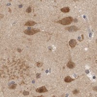


Comprehensive Human Protein Atlas IHC scoring per tissue (reliability: Enhanced — Medium consistency between antibody staining and RNA expression data.). Rows are taken directly from the HPA tissue chart — click any row's HPA link to view the source.
| Tissue | Cell type | Level | Evidence | Source |
|---|---|---|---|---|
| Adrenal gland | Glandular cells | High | Protein (IHC) | HPA → |
| Bronchus | Respiratory epithelial cells | High | Protein (IHC) | HPA → |
| Hippocampus | Neuronal cells | High | Protein (IHC) | HPA → |
| Ovary | Follicle cells | High | Protein (IHC) | HPA → |
| Placenta | Cytotrophoblasts | High | Protein (IHC) | HPA → |
Troubleshoot CCT8 staining in paraffin sections by checking retrieval, cellular location, controls and scoring before interpreting chromogenic signal.
PB9993 has real IHC images from paraffin-embedded mouse kidney, rat testis and human mammary cancer tissue, plus an IF image from A549 cells (PB9993 image captions).
PB9993 is listed for IHC, IHC-F, IF and ICC in human, mouse and rat (PB9993 catalog applications and reactivity). Its images show paraffin-section IHC in mouse kidney, rat testis and human mammary cancer, and IF in A549 cells (PB9993 image captions).
Which to pick: For tissue IHC, choose PB9993: its IHC captions document paraffin-embedded sections, but do not report the fixative (PB9993 IHC image captions). For IF/ICC, PB9993 is listed for both applications and has an A549 cell IF image (PB9993 catalog applications; PB9993 IF image caption). For cross-species work, PB9993 lists human, mouse and rat reactivity; its clonality is unreported (PB9993 catalog reactivity and clone field).