This website uses cookies to ensure you get the best experience on our website.


- Table of Contents
Plan chromogenic CD109 IHC on paraffin sections using parathyroid glandular cells and hair follicle external root sheath cells as positive controls (HPA tissue IHC). This guide covers fixation consistency, membranous staining assessment (HPA tissue IHC), and epitope planning for 4 annotated isoforms (UniProt).

Expected localisation, validated protocols, controls and antibodies — the at-a-glance facts below, then the full design guide.
| Expected localisation | Membrane in parathyroid gland and hair follicle (HPA tissue IHC) | |
| Staining pattern | Membranous in glandular and external root sheath cells (HPA tissue IHC) | |
| Antigen retrieval | Citrate pH 6.0 HIER, 95–98 °C, 20 min (rule: cytoplasmic / membrane antigen) | |
| Positive control | Hair+1 more · see all | |
| Negative control | Adipose tissue+4 more · see all |
| Fixation | Keep fixation consistent across paraffin sections. (standard IHC practice; not target-specific) | |
| Caveat | Endothelial cell expression may add tissue signal (UniProt) | |
| Regulation | Expressed in activated platelets (UniProt) | |
| Isoform / epitope | 4 isoforms; mature chain 22–1420; map the epitope (UniProt) |
Compare the catalog antibody's IHC-P protocol (catalog datasheet) with 4 published CD109 IHC methods (PMC6639710; PMC7926025; PMC5929441; PMC5808841).
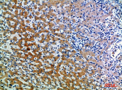
| Sample | Paraffin-embedded human liver tissue; fixative not specified (datasheet A03248) |
| Fixation | Image fixative and duration unreported (datasheet A03248); verify before use. |
| Sectioning | 4–5 µm sections on charged slides (standard) |
| Deparaffinisation | Xylene, graded ethanol series to water (standard) |
| Antigen retrieval | Heat-induced epitope retrieval in citrate buffer, pH 6.0, 20 min at 95–98 °C (standard rule: cytoplasmic / membrane antigen) |
| Peroxidase block | 3% H2O2, 10 min, room temperature (standard) |
| Blocking | 10% normal serum of the secondary host, 30 min, room temperature (standard) |
| Primary antibody | Rabbit anti-CD109, 1:50 recommended; image 1:200 (datasheet A03248) |
| Primary incubation | Overnight at 4 °C (standard) |
| Detection | HRP-polymer secondary, DAB chromogen 5–10 min (standard) |
| Counterstain | Hematoxylin, blue, dehydrate and mount (standard) |
| Expected result | CD109-positive staining in cells in external root sheath of hair (HPA tissue IHC: High). HPA tissue profile: Membranous expression in parathyroid gland and hair follicle. No signal in the no-primary control. |
CD109 is a cell-surface protein without a transmembrane segment (UniProt Q6YHK3 topology; UniProt Q6YHK3: cell membrane). In paraffin-section IHC, expect predominantly membranous staining in parathyroid glandular cells and cells of the hair follicle external root sheath (HPA tissue IHC: High; membranous profile). HPA rates the tissue pattern Enhanced, citing consistency between antibody staining and RNA expression (HPA tissue IHC: Enhanced).
| Clear cell-edge staining in parathyroid glandular cells or the hair follicle external root sheath. | This matches the reported membranous pattern in two High-staining cell populations (HPA tissue IHC: profile; High in both populations). Judge the relevant cells and compartment together; a dark deposit elsewhere does not establish the expected CD109 pattern. |
| Predominantly nuclear or broadly cytoplasmic staining, with little staining at cell edges. | Reassess this as an IHC result because HPA describes membranous tissue staining (HPA tissue IHC: membranous profile). Cytosolic localization is additionally reported by ICC-IF, so cytoplasmic signal alone is not proof of artefact (HPA subcellular: Cytosol, approved). |
| Strong staining in adipocytes or respiratory epithelial cells instead of the expected positive cells. | These cell types were Not detected in the listed HPA tissue images (HPA tissue IHC: adipose adipocytes; bronchus respiratory epithelium). Check antibody specificity and endogenous detection activity before calling them CD109-positive; an unexpected stain alone cannot distinguish those causes (general IHC practice). |
| Haze across cells and tissue spaces obscures their edges. | The compartment cannot be scored reliably against the membranous reference pattern (HPA tissue IHC: membranous profile). Background may arise from nonspecific antibody binding or the detection system; compare controls before interpreting faint cell-associated color (general IHC practice). |
| No visible staining in parathyroid glandular cells or the hair follicle external root sheath. | This conflicts with the reported High staining in those cells (HPA tissue IHC: High in both populations). First check that the expected cells are present, then assess the staining run; one negative section does not establish absent CD109 expression (general IHC practice). |
| Surface location and topology | A cell-edge pattern fits CD109's cell-membrane annotation and absence of a transmembrane segment (UniProt Q6YHK3: cell membrane; topology). Its GPI-anchor keyword supports a surface-associated interpretation but does not specify how it will appear after paraffin processing (UniProt Q6YHK3: GPI-anchor keyword). |
| Processed protein and epitope | UniProt annotates a signal peptide at residues 1–21, a CD109 chain at 22–1420, and a propeptide at 1421–1445 (UniProt Q6YHK3: processing). Those boundaries matter when interpreting a known antibody epitope; no epitope position is supplied here, so reactivity with each processed form cannot be predicted. |
| Isoforms | Four isoforms are listed; isoform 1 is reported in keratinocytes and placenta, and isoform 4 in placenta (UniProt Q6YHK3: isoforms; tissue specificity). These expression notes do not establish which isoforms the IHC antibody recognizes or guarantee staining in every cell of those tissues. |
| Tissue evidence and antibody validation | The HPA tissue pattern is Enhanced, and HPA015723 has an Enhanced IHC designation (HPA tissue IHC: reliability; HPA antibodies: HPA015723 IHC Enhanced). Use the reported cell-level pattern as the comparison; the designation does not validate an unexpected compartment or cell type in a new specimen. |
| Antigen retrieval | Retrieval conditions are an assay variable for paraffin-section IHC (general IHC practice). No CD109-specific retrieval or fixation response is supplied by UniProt or HPA; optimize against a reported positive cell population without claiming that a particular treatment exposes a CD109 epitope. |
| IF/ICC Q&A: where should CD109 appear? | Mainly at the plasma membrane, with an additional cytosolic location (HPA subcellular: Plasma membrane, supported; Cytosol, approved). This ICC-IF observation can inform localization, but it does not replace the separate membranous tissue-IHC reference pattern (HPA tissue IHC: profile). |
| Situation | Likely cause | Next action |
|---|---|---|
| The known-positive tissue gives no signal. | The expected cells may be absent from the section, or the staining run may have failed; HPA reports High staining only for the specified cell populations (HPA tissue IHC: High in parathyroid glandular cells and external root sheath cells). | Confirm those cells are present and compare a run control. Review primary-antibody application, retrieval settings, and detection steps as general IHC checks; no CD109-specific retrieval setting is established here (general IHC practice). |
| Color appears mainly inside nuclei. | Nuclear dominance conflicts with the reported membranous IHC profile (HPA tissue IHC: membranous profile). The appearance alone does not identify which staining step caused it. | Compare the same run with its controls and inspect whether cell-edge staining is also present. Recheck the detection and counterstain steps before interpreting nuclear color as CD109 (general IHC practice). |
| A HPA-listed Not detected cell type stains strongly. | Unexpected staining may reflect antibody cross-reactivity or endogenous detection activity; the slide alone cannot separate them (HPA tissue IHC: Not detected cell types; general IHC practice). | Compare with a no-primary control and, where applicable, an endogenous-activity control. Confirm cell identity and compartment before assigning CD109 positivity (general IHC practice). |
| Diffuse color makes cell borders hard to distinguish. | Nonspecific binding or detection background may obscure the membranous pattern (general IHC practice; HPA tissue IHC: membranous profile). | Check no-primary and run controls, then review blocking, primary-antibody concentration, washes, and chromogen development within the assay workflow (general IHC practice). Score localization only when cell edges can be resolved. |
| Cytoplasmic staining accompanies weak membrane staining. | HPA reports an additional cytosolic location by ICC-IF, while its tissue-IHC summary emphasizes membrane staining (HPA subcellular: Cytosol, approved; HPA tissue IHC: membranous profile). The two assay observations should be interpreted in their own contexts. | Record membrane and cytoplasmic staining separately, compare positive tissue and controls, and avoid treating cytoplasmic color alone as the expected paraffin-section pattern (HPA tissue IHC: profile; general IHC practice). |
| A tissue described as expressing CD109 by UniProt is IHC-negative. | UniProt's broad tissue-expression description and HPA's cell-specific IHC observations answer different questions (UniProt Q6YHK3: tissue specificity; HPA tissue IHC: cell-level results). Expression in a tissue does not guarantee a visible stain in every sampled cell. | Identify the cells actually present and compare them with HPA's named positive populations. Verify assay performance using one of those populations before interpreting the negative result (HPA tissue IHC: High in parathyroid glandular cells and external root sheath cells; general IHC practice). |


Comprehensive Human Protein Atlas IHC scoring per tissue (reliability: Enhanced — High consistency between antibody staining and RNA expression data.). Rows are taken directly from the HPA tissue chart — click any row's HPA link to view the source.
| Tissue | Cell type | Level | Evidence | Source |
|---|---|---|---|---|
| Hair | Cells in external root sheath | High | Protein (IHC) | HPA → |
| Parathyroid gland | Glandular cells | High | Protein (IHC) | HPA → |
| Tissue | Cell type | Level | Evidence | Source |
|---|---|---|---|---|
| Adipose tissue | Adipocytes | Not detected | Protein (IHC) | HPA → |
| Adrenal gland | Glandular cells | Not detected | Protein (IHC) | HPA → |
| Appendix | Glandular cells | Not detected | Protein (IHC) | HPA → |
| Bone marrow | Hematopoietic cells | Not detected | Protein (IHC) | HPA → |
| Breast | Adipocytes | Not detected | Protein (IHC) | HPA → |
Troubleshoot CD109 chromogenic IHC in paraffin sections using the catalog antibody’s tissue image, the page retrieval setting, and independent localisation evidence.
A03248 has real IHC data from paraffin-embedded human liver (catalog IHC image caption). IF is listed for human and mouse, with no IF image supplied (catalog applications, reactivity and IF image list).
A03248 will render with an IHC image of paraffin-embedded human liver (catalog IHC image caption). It lists IHC and IF applications and human and mouse reactivity (catalog applications and reactivity).
Which to pick: For tissue IHC, pick A03248: its own image shows a paraffin-embedded human liver section, and the fixative is unreported (catalog IHC image caption). For IF, A03248 is listed, but no IF image or ICC application is supplied (catalog applications and IF image list). For human and mouse work, A03248 lists reactivity with both species; its shown IHC section is human, and clonality is unreported (catalog reactivity, IHC image caption and clone field).