This website uses cookies to ensure you get the best experience on our website.
- Table of Contents
Facts about C-type lectin domain family 4 member K.
Binds to sulfated as well as mannosylated glycans, keratan sulfate (KS) and beta-glucans. Facilitates uptake of antigens and is involved in the routing and/or processing of antigen for presentation to T cells.
Human | |
---|---|
Gene Name: | CD207 |
Uniprot: | Q9UJ71 |
Entrez: | 50489 |
Belongs to: |
---|
No superfamily |
CD207 antigen; CD207 antigen, langerin; CD207 molecule, langerin; CD207; CLEC4KLangerhans cell specific c-type lectin; C-type lectin domain family 4 member K; C-type lectin domain family 4, member K; Langerin
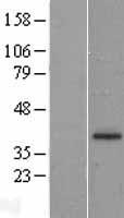
Mass (kDA):
36.725 kDA
Human | |
---|---|
Location: | 2p13.3 |
Sequence: | 2; NC_000002.12 (70825248..70860787, complement) |
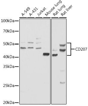
Exclusively expressed by Langerhans cells. Expressed in astrocytoma and malignant ependymoma, but not in normal brain tissues.
Membrane; Single-pass type II membrane protein. Found in Birbeck granules (BGs), which are organelles consisting of superimposed and zippered membranes.
You have reached the right place if you are looking for information on Boster Bio's Anti-Macrosialin CD68 Antibody. I will give you an overview and explain the CD207 Marker. This antibody can detect CD207 markers in macrophages. It will also measure the IFN g release. This information can be used by any scientist around the world.
This Rabbit Polyclonal antibody against CD68 recognizes Human antigen. It has been validated for Western Blot and immunohistochemistry applications. Boster Bio Anti-Macrosialin CD68 Antibody uses CD207 marker. In addition to its high affinity, this antibody can also detect proteins in immunohistochemistry. It is highly recommended for use in immunohistochemistry or Western blot due to its reliability and ease of use.
It is possible to detect CD207 in macrophages, which can be used to diagnose inflammatory conditions. DCs with this marker do not express the major DC markers such as CD141 or CD1c. The tCD207 DCs, which are unusually located in the tonsil lymphoid system, lack EpCAM expression. These cells are likely to represent the tonsil epithelium.
It is important to note that DCs with CD207 expression are closely related to myeloid DCs. These DCs are highly polarized in Th2 and Th27, and lack Th2 responses. However, it is not clear if these cells have a different function than conventional CD1cDCs. To determine if the tCD207 DC population has a different role than conventional CD1c DCs, further studies are necessary.
The detection of CD207 Marker (CD206), in macrophages can help differentiate between these two types. Its reactivity has been shown to be very high in isolated dendritic cells. In contrast, the reactivity with CD207 in macrophages is low, and the resultant results were consistent with the diagnosis of cutaneous JXG.
The anti CD207-mAb visualizes CD8+DCs better. It labels a subset of CD11chigh DCs, whereas the anti-CD205 mAb labels only a subset of CD8+ DCs. Langerin outperforms the other markers in FACS. Langerin is expressed as CD8+ DCs.
The CD207 Marker is a vital tool for identifying other activated macrophages. The M2 marker can serve many purposes. The T2 macrophage performs many functions and contributes in various ways to tissue homeostasis, immunity, and tissue integrity. Researchers can better understand how macrophages function by detecting CD207. These studies are timely.
The role of CD207 in tumor-associated macrophages is unclear. Its high expression in tumors is a sign of the severity of the disease. This marker plays a variety of roles in the immune system. It influences both normal and pathological processes. For more information, see the related article on the function of CD207 in macrophages.
While DCs and macrophages are of distinct origins, their biology overlaps. Despite their similarities, their roles in disease progression are not fully understood. It has been suggested these cells can undergo transdifferentiation to respond to environmental cues. Their phenotypes can also be affected by lineage commitments, which affect their ability to respond locally to microenvironmental factors.
We used a assay that measures IFN-g release using cytokines. These data are the means and standard deviations of at least three independent experiments. We used the BosterBio CD207 marker to determine how many cells had a high IFN–g reaction when using autologous PBMCs.
The culture medium containing DCs derived from skin was able to produce higher levels IFN-g than those derived directly from lymph nodes. This indicates that skin-derived DCs are responsible to CD8+ T cells priming and activation. In contrast, MNs failed to release IFN-g despite transdermal delivery of NPs.
Three different anti HCMV combinations were given to huDEC205–Tg mice. The vaccines were administered to the mice fourteen days later. After the vaccines had been administered, the mice were sacrificed. Bulk splenocytes of the mice were then taken out and stimulated using a 1mg/ml EBNA1/HCMV pp65 peptide. After stimulation, IFN-1g release was measured using ICS on CD4+ gated cell cultures. Data were gathered from two independent experiments, with at most five mice per group.
The shockwave device primed the immune systems, in contrast to other forms. It also increased the levels of immune markers. The animals vaccinated with shockwave devices had higher levels than those who received the standard oral route. These results are encouraging as they highlight the importance of shockwaves in stimulating immunity.
The researchers also measured cytokines released by mice after vaccine administration by injecting the beads into the skin. The vaccine beads that were injected with the device attracted Langerhans cells. This is critical for the immune response. The results of this study indicated that the vaccines contained sufficient IFN-g levels to induce an effective immune response.
PMID: 10661407 by Valladeau J., et al. Langerin, a novel C-type lectin specific to Langerhans cells, is an endocytic receptor that induces the formation of Birbeck granules.
PMID: 12626394 by Stambach N.S., et al. Characterization of carbohydrate recognition by langerin, a C-type lectin of Langerhans cells.