CD209 / CD209 antigen · IHC design guide

Design Immunohistochemistry for CD209

CD209 is a membrane receptor with an extracellular region spanning residues 59–404 (UniProt). This guide uses paraffin-section IHC evidence to plan controls and interpret selective staining, including high staining in placental cytotrophoblasts (HPA tissue IHC).

Evidence assembled Sep 2026 · For research use; verify linked source records and product datasheet before use
Immunohistochemistry protocol sheet for CD209 (IHC for CD209): expected localisation Cell membrane expected; tissue-IHC compartment unreported (UniProt), antibody M01025-1, validated IHC image, and IHC protocol steps
Printable CD209 IHC protocol sheet — expected localisation Cell membrane expected; tissue-IHC compartment unreported (UniProt), antibody M01025-1, controls and protocol steps. Open the full CD209 IHC guide →

CD209 Immunohistochemistry Experimental Design Guide

Expected localisation, validated protocols, controls and antibodies — the at-a-glance facts below, then the full design guide.

Must know before staining
Expected localisation Cell membrane expected; tissue-IHC compartment unreported (UniProt)
Staining pattern Selective monocyte-derived cells; compartment unreported (HPA tissue IHC)
Antigen retrieval Citrate pH 6.0 HIER, 95–98 °C, 20 min (rule: cytoplasmic / membrane antigen)
Positive control ⓘ Placenta+3 more · see all
Negative control ⓘ Adipose tissue+4 more · see all
Important caveats
Reasons your staining may differ from the expected pattern.
Fixation Use 10% NBF; prolonged fixation may mask epitopes (standard IHC practice)
Caveat Secreted CD209 may produce staining beyond cell surfaces (UniProt)
Regulation Predominant expression in dendritic cells (UniProt)
Isoform / epitope 12 isoforms; extracellular versus cytoplasmic epitopes matter (UniProt)
Section 1

Recommended CD209 IHC & IF Protocols

The catalog antibody’s IHC-P protocol is accompanied by published CD209 methods for myocardium, porcine testis, and left atrial wall (PMC3614732; PMC4723225; PMC6159861).

Recommended immunohistochemistry (IHC-P) protocol parameters
SampleFFPE tissue sections (standard)
Fixation10% neutral-buffered formalin, 24 h, standard processing (standard)
Sectioning4–5 µm sections on charged slides (standard)
DeparaffinisationXylene, graded ethanol series to water (standard)
Antigen retrievalHeat-induced epitope retrieval in citrate buffer, pH 6.0, 20 min at 95–98 °C (standard rule: cytoplasmic / membrane antigen)
Peroxidase block3% H2O2, 10 min, room temperature (standard)
Blocking10% normal serum of the secondary host, 30 min, room temperature (standard)
Primary antibodyMouse monoclonal (clone Clone: 5D7) anti-CD209, 5 - 10 μg/mL (datasheet M01025-1)
Primary incubationOvernight at 4 °C (standard)
DetectionHRP-polymer secondary, DAB chromogen 5–10 min (standard)
CounterstainHematoxylin, blue, dehydrate and mount (standard)
Expected resultCD209-positive staining in cytotrophoblasts of placenta (HPA tissue IHC: High). HPA tissue profile: Selective expression in subsets of monocyte-derived cells. No signal in the no-primary control.
💡Decision noteStart with pH 6.0 citrate retrieval at 95–98 °C for 20 min (page antigen-retrieval rule); test boiling Tris-EDTA for porcine testis sections (PMC4723225).
Section 2

What Is the Expected CD209 Staining Pattern?

CD209 is a membrane receptor with an extracellular domain (UniProt Q9NNX6 topology). In paraffin sections, expect selective staining of placental cytotrophoblasts and lung macrophages (HPA: High and Medium, respectively; reliability Supported).

What am I looking at on my slide?
Membrane-associated staining in placental cytotrophoblastsExpected positive pattern (HPA: cytotrophoblasts High; UniProt Q9NNX6 topology).
Nuclear-only stainingSuspect an artefact; CD209 is annotated at the membrane (UniProt Q9NNX6 subcellular).
Uniform adipocyte stainingConsider cross-reactivity or endogenous activity (HPA: adipocytes Not detected).
Diffuse chromogen across the sectionSuspect background from detection or blocking (standard IHC practice).
No signal in placental cytotrophoblastsCheck assay sensitivity before scoring negative (HPA: cytotrophoblasts High).
💡Expected CD209 appearanceCall positive when placental cytotrophoblasts show clear membrane-associated staining (HPA: High; UniProt Q9NNX6 topology); nuclear-only signal is suspect (UniProt Q9NNX6 subcellular).
How each factor affects the staining
Fixation and cross-linkingOverfixation can mask epitopes and weaken signal (standard IHC practice).
Antigen retrievalRetrieval conditions can change signal and tissue integrity (standard IHC practice).
Epitope locationExtracellular region is glycosylated at 80; epitope accessibility may vary (UniProt Q9NNX6).
Isoforms12 isoforms are listed; confirm the antibody's target region (UniProt Q9NNX6).
Secreted annotationInterpret extracellular signal cautiously (UniProt Q9NNX6 subcellular).
Endogenous peroxidaseCan produce chromogen background; use a peroxide block and control (standard IHC practice).
Species reactivityConfirm reactivity for the specimen species before extrapolating (standard IHC practice).
IF/ICC: expected location?Membrane (HPA: subcellular summary); no ICC-IF images are listed (HPA: ICC-IF).
Why is my staining missing, weak or wrong?
SituationLikely causeNext action
No placental cytotrophoblast signal (HPA: High)Epitope masking from fixation (standard IHC practice).Optimize retrieval and check the positive control (standard IHC practice).
Weak membrane signal (UniProt Q9NNX6 topology)Insufficient retrieval or antibody concentration (standard IHC practice).Titrate retrieval and antibody dilution (standard IHC practice).
Nuclear-only signalNonspecific staining is possible (UniProt Q9NNX6 subcellular).Review controls and antibody dilution (standard IHC practice).
Diffuse chromogen across the sectionInadequate blocking or washing (standard IHC practice).Improve blocking and washes; inspect the no-primary control (standard IHC practice).
Uniform adipocyte signal (HPA: Not detected)Cross-reactivity or endogenous activity is possible (standard IHC practice).Check no-primary control and antibody specificity (standard IHC practice).
Signal without primary antibodyEndogenous enzyme or detection background (standard IHC practice).Check the peroxide block and detection reagents (standard IHC practice).

Sample controls for CD209 IHC & IF

🧪Run placenta first and look for staining in cytotrophoblasts (High; HPA: placenta cytotrophoblasts). Use adrenal gland as the negative tissue and score its glandular cells (HPA: adrenal gland glandular cells not detected); on the placenta slide, cells outside the cytotrophoblast compartment should show only background staining unless independently verified as CD209-positive.
Positive control tissue: Placenta (Cytotrophoblasts, HPA High)
Negative control tissue: Adipose tissue (HPA Not detected)
ICC-IF cell lines (HPA subcellular resource): HPA carries no ICC-IF cell line for CD209; derive a cell-line control from the positive tissue's cell type (Cytotrophoblasts) and confirm it by RNA or western blot first.
Technical controls: Include no-primary (secondary-only), an isotype control matched to the primary antibody’s host species and monoclonal subclass or polyclonal IgG, and a CD209 knockout control or peptide block if a validated blocking peptide is available. Check endogenous peroxidase and biotin background in placenta for chromogenic IHC, or tissue autofluorescence for IF.
⚠️Feasibility: Keep fixation conditions consistent across sections and validate antigen retrieval with the IHC-validated antibody, since FFPE processing can mask epitopes. CD209 has an extracellular domain spanning residues 59–404 (UniProt Q9NNX6 topology), so a frozen section or IF may help assess membrane staining if FFPE staining is weak; the separate IF/ICC guide covers that application. Interpret placental signal by cell morphology and control staining because the positive HPA call is specific to cytotrophoblasts (HPA: placenta cytotrophoblasts High).

HPA tissue IHC evidence for CD209

Comprehensive Human Protein Atlas IHC scoring per tissue (reliability: Supported — External characterization data supports antibody staining. Antibody staining in cells/structures not annotated, view images.). Rows are taken directly from the HPA tissue chart — click any row's HPA link to view the source.

Positive expression · recommended positive controls

TissueCell typeLevelEvidenceSource
Placenta Cytotrophoblasts High Protein (IHC) HPA →
Bone marrow Hematopoietic cells Medium Protein (IHC) HPA →
Lung Macrophages Medium Protein (IHC) HPA →
Testis Elongated or late spermatids Medium Protein (IHC) HPA →

Undetected expression · recommended negative controls

TissueCell typeLevelEvidenceSource
Adipose tissue Adipocytes Not detected Protein (IHC) HPA →
Adrenal gland Glandular cells Not detected Protein (IHC) HPA →
Appendix Glandular cells Not detected Protein (IHC) HPA →
Breast Adipocytes Not detected Protein (IHC) HPA →
Bronchus Respiratory epithelial cells Not detected Protein (IHC) HPA →
Section 3

Advanced CD209 IHC Tips

Troubleshoot CD209 staining in paraffin sections by checking retrieval, tissue context, membrane localisation and cell-specific scoring.

How should I retrieve CD209 in paraffin sections when membrane staining is weak?
Start with citrate pH 6.0 heat-induced retrieval at 95–98 °C for 20 min (page retrieval specification). Keep section thickness, cooling time and detection conditions constant while comparing a weak section with a previously positive control (standard IHC practice). Because CD209 has an extracellular region spanning residues 59–404, retrieval can affect access to its antibody epitope (UniProt Q9NNX6 topology). If staining remains weak, test a longer retrieval time on adjacent sections while watching for damaged tissue or stronger nonspecific staining (standard IHC practice). Record the retrieval condition alongside the control result before comparing staining across samples (standard IHC practice).
Could fixation explain absent CD209 staining despite intact tissue morphology?
Yes: prolonged formalin fixation can mask epitopes even when tissue architecture remains recognizable (standard IHC practice). Compare sections from blocks with documented fixation histories using the same citrate pH 6.0, 95–98 °C, 20 min retrieval (page retrieval specification). Include a known positive section in the same staining run, since CD209 expression is selective among monocyte-derived cell subsets (HPA: tissue IHC profile). If the control stains but the study section does not, review fixation, tissue sampling and cell composition before increasing antibody concentration (standard IHC practice). If both fail, check retrieval and detection reagents with their respective controls before interpreting the sample as negative (standard IHC practice).
What staining pattern should I expect for CD209 in a paraffin section?
Look first for staining at cell borders: CD209 is a cell-membrane protein with a transmembrane segment at residues 38–58 (UniProt Q9NNX6 topology). Intracellular signal can also be plausible because CD209 participates in endocytosis and returns to the surface after internalisation (UniProt Q9NNX6 function). Assess that signal within identifiable cells rather than treating diffuse stain over extracellular material as localisation evidence (standard IHC practice). In lung, macrophages provide a reported staining context, although expression is selective across monocyte-derived cell subsets (HPA: lung macrophages, Medium; HPA: tissue IHC profile). Compare the border and intracellular patterns with adjacent negative cells and a positive control before assigning either to CD209 (standard IHC practice).
How can isoforms or epitope placement change a CD209 IHC result?
CD209 has 12 listed isoforms, so determine which sequence the antibody recognises before comparing stains across specimens (UniProt Q9NNX6 isoforms). Its cytoplasmic region occupies residues 1–37, while residues 59–404 are extracellular (UniProt Q9NNX6 topology). If the immunogen or epitope is documented, map it to those regions and check whether the sequence is present in the isoforms relevant to the study (standard IHC practice). Glycosylation at residue 80 may matter when interpreting an extracellular epitope near that position (UniProt Q9NNX6 glycosylation). Without an epitope map, do not infer isoform coverage or the reason for a negative stain from localisation alone (standard IHC practice).
How should I check a CD209 pattern by multiplex immunofluorescence?
Use the separate IF/ICC guide for that assay, and pair CD209 with a validated marker for the expected cell type in the specimen (standard IF practice). Select fluorophores after checking tissue autofluorescence and reserve the clearest channel for the weaker signal (standard IF practice). CD209 is membrane-associated, with an extracellular region at residues 59–404 and a cytoplasmic region at residues 1–37 (UniProt Q9NNX6 topology). Match permeabilisation to the mapped epitope: an extracellular epitope may need none, whereas access to a cytoplasmic epitope generally requires it (standard IF practice). Confirm channel separation and cell-level overlap with single-stain controls before comparing fluorescence with chromogenic IHC (standard IF practice).
How can I reduce diffuse or cell-independent CD209 chromogen staining?
First compare the stained section with a no-primary control to identify signal from the detection system or tissue itself (standard IHC practice). Block endogenous peroxidase before chromogenic detection, and inspect pigment or precipitate under the same illumination used for scoring (standard IHC practice). Titrate the IHC-validated antibody and detection reagent on adjacent sections while keeping citrate pH 6.0, 95–98 °C, 20 min retrieval fixed (page retrieval specification; standard IHC practice). CD209 is reported at the cell membrane and in membrane rafts, making broad, cell-independent deposits inconsistent with its listed localisation (UniProt Q9NNX6 subcellular location). Require a reproducible cell-associated pattern in a positive control before accepting a faint specimen signal (standard IHC practice).
How should I score CD209 staining when positive cells are sparse? ⚠ ANSWER MARKED FOR VERIFICATION
Define the tissue compartment and denominator before scoring, because CD209 staining is selective among monocyte-derived cell subsets (HPA: tissue IHC profile; standard IHC practice). For a cell-based analysis, report the percentage of positive eligible cells and an H-score using intensity grades 0–3 (standard IHC practice). If the question concerns infiltration, report positive-cell density per mm² of viable tissue and state which cells were counted (standard IHC practice). Apply one positivity threshold across cases using control sections, and exclude folds, necrosis and edge staining from the scored area (standard IHC practice). Keep cell-specific results separate from whole-section area measurements so differences in tissue composition remain visible (standard IHC practice).
When is CD209 staining convincing rather than an artefact?
A convincing result is cell-associated and reproducible in the expected compartment, with a working positive control and little signal in the no-primary control (standard IHC practice). Membrane staining fits CD209 topology; intracellular staining warrants assessment because the receptor undergoes endocytosis and returns to the surface (UniProt Q9NNX6 topology and function). Check cell identity: lung macrophage staining is reported, while the tissue IHC profile also reports placental cytotrophoblast staining (HPA: lung macrophages, Medium; HPA: placenta cytotrophoblasts, High). Treat stain confined to section edges, necrotic areas or endogenous enzyme-rich regions as suspect until controls resolve it (standard IHC practice). Do not call every unstained cell abnormal, since the reported expression pattern is selective (HPA: tissue IHC profile).
Boster reagents

Best CD209 / CD209 antigen IHC Antibodies

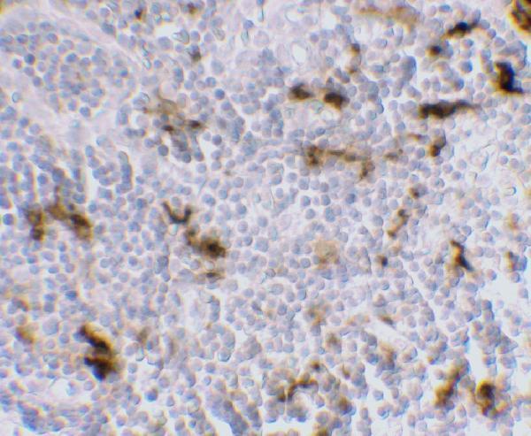
The anti-CD209 catalog antibody has a human lymph node IHC figure (image alt: human lymph node IHC). No IF figure is supplied (catalog: IF image alts empty).

Real IHC data Immunohistochemistry of DC-SIGN in human lymph node tissue with DC-SIGN antibody at 5 μg/mL.
Anti-DC-SIGN CD209 Monoclonal Antibody [5D7]
Cat # M01025-1

M01025-1 is the rendered SKU and lists human IHC-P reactivity (cards: M01025-1; IHC-P; Human). Its figure shows human lymph node IHC at 5 μg/mL (image alt: human lymph node; 5 μg/mL).

Which to pick: For FFPE IHC, choose M01025-1, a mouse monoclonal clone 5D7 with human IHC-P validation (catalog: host Mouse; clone 5D7; applications IHC-P; reactivity Human). Neither M01025-1 nor A01025-2 lists IF/ICC validation (catalog: application lists). Neither SKU lists reactivity beyond human, so there is no catalog-supported cross-species choice (catalog: both reactivity lists Human).

Each figure is that product's own IHC / IF validation image from its datasheet.

References

  1. UniProt Consortium. UniProt entry Q9NNX6 (CD209_HUMAN, CD209 antigen).
  2. Human Protein Atlas. CD209 tissue IHC expression (reliability: Supported).
  3. Human Protein Atlas. CD209 subcellular location (ICC-IF): Membrane.
  4. Human Protein Atlas. CD209 antibody validation summary (2 antibodies).
  5. Emergence of dendritic cells in the myocardium after acute myocardial infarction - implications for inflammatory myocardial damage. International journal of biomedical science : IJBS 2010 — PMC3614732.
  6. Identification of Putative Biomarkers for the Early Stage of Porcine Spermatogonial Stem Cells Using Next-Generation Sequencing. PloS one 2016 — PMC4723225.
  7. Presence of increased inflammatory infiltrates accompanied by activated dendritic cells in the left atrium in rheumatic heart disease. PloS one 2018 — PMC6159861.
  8. Brain Immune Cell Composition in Multiple Sclerosis and Progressive Multifocal Leukoencephalopathy After Natalizumab: An Immunohistochemical Cohort Study. Neurology(R) neuroimmunology & neuroinflammation 2026 — PMC13262666.
  9. PubMed PMID:1518869 — UniProt-cited evidence.
  10. PubMed PMID:10975799 — UniProt-cited evidence.
  11. PubMed PMID:11257134 — UniProt-cited evidence.