This website uses cookies to ensure you get the best experience on our website.


- Table of Contents
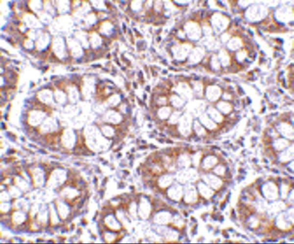
Plan CD248 IHC-P using the catalog antibody at 2.5 μg/mL in human colon sections (datasheet A04735). Compare the low cytoplasmic tissue staining (HPA tissue IHC) with the expected membrane location (UniProt).

Expected localisation, validated protocols, controls and antibodies — the at-a-glance facts below, then the full design guide.
| Expected localisation | Low cytoplasmic tissue staining (HPA tissue IHC); membrane expected (UniProt) | |
| Staining pattern | Low cytoplasmic staining in several tissues (HPA tissue IHC) | |
| Antigen retrieval | Citrate pH 6 HIER, heat-mediated (datasheet A04735) | |
| Positive control | Duodenum+4 more · see all | |
| Negative control | Adipose tissue+4 more · see all |
| Fixation | Formaldehyde-fixed colon shown (selected-SKU IHC image A04735); keep fixation consistent (standard IHC practice; not target-specific) | |
| Caveat | Staining and RNA show medium consistency (HPA tissue IHC) | |
| Regulation | Seen in angiogenesis and wound healing (UniProt) | |
| Isoform / epitope | 2 isoforms; map extracellular vs cytoplasmic epitope (UniProt) |
The catalog antibody’s IHC-P protocol is followed by published CD248 staining methods for fibrotic lung, renal carcinoma, and murine ankle joints (PMC4832513; PMC8006336; PMC3672796).
| Sample | Formaldehyde-fixed, paraffin-embedded Human Colon tissue (datasheet A04735) |
| Fixation | Image formalin-fixed; duration unreported (datasheet A04735); verify before use. |
| Sectioning | 4–5 µm sections on charged slides (standard) |
| Deparaffinisation | Xylene, graded ethanol series to water (standard) |
| Antigen retrieval | Heat retrieval: Citrate pH 6 (datasheet A04735); 20 min, 95–100 °C (standard) |
| Peroxidase block | 3% H2O2, 10 min, room temperature (standard) |
| Blocking | 10% serum (datasheet A04735) |
| Primary antibody | Rabbit anti-CD248, 2.5 μg/mL (datasheet A04735) |
| Primary incubation | Overnight at 4 °C (standard) |
| Detection | HRP-polymer secondary, DAB chromogen 5–10 min (standard) |
| Counterstain | Hematoxylin, blue, dehydrate and mount (standard) |
| Expected result | CD248-positive staining in paneth cells of duodenum (HPA tissue IHC: High). HPA tissue profile: Low cytoplasmic expression in several tissues. No signal in the no-primary control. |
CD248 is a membrane protein with an extracellular region at residues 18–687 and a transmembrane segment at 688–708 (UniProt Q9HCU0 topology). Expect staining in pericytes and stromal fibroblasts (UniProt Q9HCU0 tissue specificity). HPA also reports high staining in intestinal Paneth cells, while describing overall tissue IHC as low cytoplasmic expression in several tissues with Enhanced reliability and medium staining–RNA consistency (HPA tissue IHC).
| Distinct staining outlines perivascular or stromal cells, with little signal in nearby normal vessel endothelium. | This fits the membrane location and reported pericyte and stromal fibroblast expression (UniProt Q9HCU0 topology and tissue specificity; HPA subcellular). Assess the stained cell's position and morphology before assigning it to a vessel wall: UniProt also reports CD248 in tumor endothelial cells. |
| Paneth cells stain strongly in a duodenum or small intestine section. | This matches the High Paneth cell staining recorded in both tissues (HPA tissue IHC). It offers a tissue IHC comparison, though HPA's overall profile includes low cytoplasmic staining and its Enhanced rating has medium consistency with RNA expression (HPA tissue IHC). |
| A broad nuclear signal dominates, or stain fills unrelated tissue structures without a clear cellular pattern. | Predominant nuclear staining conflicts with the plasma membrane location (HPA subcellular; UniProt Q9HCU0 topology). Treat the pattern as suspect and compare controls; misplaced stain alone cannot identify whether antibody binding or chromogenic detection caused it (general IHC practice). |
| Adipocytes or other cells listed as undetected show strong staining across the section. | HPA lists adipocytes in adipose tissue and breast as Not detected (HPA tissue IHC). Strong staining in those specific cells warrants a cross-reactivity or endogenous detection check. Do not classify the entire tissue as negative: HPA's entries identify cell types, not every cell in a section. |
| No staining appears in Paneth cells in a technically valid intestinal section. | That differs from HPA's High Paneth cell result (HPA tissue IHC). Check that Paneth cells are present, then review the antibody's IHC validation and the staining run. A blank section alone cannot distinguish a failed assay from a sampled area lacking the expected cells (general IHC practice). |
| Compartment and epitope location | CD248 has a large extracellular region, one transmembrane segment, and a short cytoplasmic tail (UniProt Q9HCU0 topology). Its reported plasma membrane location guides compartment scoring (HPA subcellular). An antibody's exact epitope is not supplied, so topology cannot establish its retrieval requirements. |
| Tissue and cell-type context | HPA reports High Paneth cell staining, Medium endometrial stromal cell and glomerular cell staining, and Not detected adipocytes (HPA tissue IHC). These are cell-specific observations; score identified cells within each section rather than using a tissue name as a whole-section positive or negative label. |
| Antibody evidence and interpretation limit | HPA051856 has Enhanced IHC validation (HPA antibodies). HPA describes the tissue IHC result as Enhanced but notes medium consistency between antibody staining and RNA expression, plus stained cells or structures that are not annotated (HPA tissue IHC). Review morphology and controls before assigning an unexpected cell identity. |
| Antigen retrieval | Target-specific fixation effects are not established by the supplied assay evidence. Verify with a matched IHC source before attributing a result to fixation. |
| IF/ICC Q&A: where should signal appear? | At the plasma membrane: HPA lists this location as approved and lists ICC-IF images for ASC52telo and BJ [Human fibroblast] (HPA subcellular). HPA051856 has Approved ICC validation (HPA antibodies). This location evidence does not supply an IF/ICC protocol or establish staining intensity for either cell line. |
| Situation | Likely cause | Next action |
|---|---|---|
| No stain in an expected positive section | The examined area may lack identifiable positive cells, or the IHC run may have failed (general IHC practice). | Confirm Paneth cells are present when using duodenum or small intestine, where HPA reports High staining (HPA tissue IHC). Review the IHC-validated antibody's instructions and run controls before changing the interpretation (general IHC practice). |
| Positive-looking stain in normal vessel endothelium | Cell assignment may be uncertain in a crowded vessel wall; UniProt reports CD248 in pericytes and stromal fibroblasts and says normal endothelial cells are absent or barely detectable (UniProt Q9HCU0 tissue specificity). | Inspect morphology and the position of stained cells around the vessel. If the signal is truly endothelial, compare a suitable negative control and assess nonspecific staining before accepting it as CD248 (general IHC practice). |
| Strong signal in adipocytes | This conflicts with HPA's Not detected adipocyte entries for adipose tissue and breast; nonspecific binding or detection background is possible (HPA tissue IHC; general IHC practice). | Check the no-primary control and blocking and wash steps (general IHC practice). Score adipocytes separately from stromal or vascular cells, since the HPA negative designation applies to adipocytes (HPA tissue IHC). |
| Color develops in the no-primary control | The signal can arise from the detection system or endogenous tissue activity rather than primary-antibody binding (general chromogenic IHC practice). | Check the detection-only control, reagent blocking, and washes. Where a peroxidase-based system is used, verify the endogenous peroxidase block (general chromogenic IHC practice). Reassess the CD248 pattern only after control color is resolved. |
| Diffuse haze obscures cell borders | Excess background can reflect insufficient blocking or washing, or excessive reagent concentration (general IHC practice). It prevents a reliable comparison with the plasma membrane location (HPA subcellular). | Review blocking, washes, primary-antibody concentration, and detection exposure using controls (general IHC practice). Re-score only when cell outlines and tissue structures can be distinguished. |
| Cytoplasmic staining appears without a clear membrane outline | HPA's tissue overview reports low cytoplasmic expression in several tissues, although its ICC-IF location is plasma membrane (HPA tissue IHC; HPA subcellular). Compartment alone therefore does not settle whether a faint IHC signal is genuine. | Record intensity, cell identity, and any membrane accentuation; compare a matched negative control and HPA's cell-specific tissue observations (general IHC practice; HPA tissue IHC). Avoid calling widespread cytoplasmic color a strong positive solely from the tissue name. |


Comprehensive Human Protein Atlas IHC scoring per tissue (reliability: Enhanced — Medium consistency between antibody staining and RNA expression data. Antibody staining in cells/structures not annotated, view images.). Rows are taken directly from the HPA tissue chart — click any row's HPA link to view the source.
| Tissue | Cell type | Level | Evidence | Source |
|---|---|---|---|---|
| Duodenum | Paneth cells | High | Protein (IHC) | HPA → |
| Small intestine | Paneth cells | High | Protein (IHC) | HPA → |
| Appendix | Germinal center cells | Medium | Protein (IHC) | HPA → |
| Colon | Goblet cells | Medium | Protein (IHC) | HPA → |
| Endometrium | Cells in endometrial stroma | Medium | Protein (IHC) | HPA → |
| Tissue | Cell type | Level | Evidence | Source |
|---|---|---|---|---|
| Adipose tissue | Adipocytes | Not detected | Protein (IHC) | HPA → |
| Adrenal gland | Glandular cells | Not detected | Protein (IHC) | HPA → |
| Bone marrow | Hematopoietic cells | Not detected | Protein (IHC) | HPA → |
| Breast | Adipocytes | Not detected | Protein (IHC) | HPA → |
| Bronchus | Respiratory epithelial cells | Not detected | Protein (IHC) | HPA → |
Troubleshoot CD248 staining in paraffin sections by checking retrieval, compartment, cell type and controls before scoring chromogenic signal.
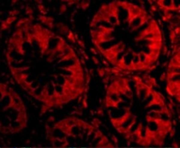
A04735 has real IHC and IF images from human colon tissue (A04735 IHC and IF image captions).
A04735 was validated for IHC-P on paraffin-embedded human colon tissue at 2.5 μg/mL (A04735 IHC image caption). A04735 was also validated for IF on human colon tissue at 20 μg/mL (A04735 IF image caption).
Which to pick: Choose A04735 for human tissue IHC-P; its own image caption documents paraffin sections and formaldehyde fixation (A04735 IHC image caption; catalog: Human reactivity). For IF, A04735 has human colon tissue data, while ICC validation is unreported (A04735 IF image caption; catalog: applications). No cross-species IHC/IF choice is supported: A04735 lists Human reactivity, and its clone and clonality are unreported (catalog: reactivity and clone).