Click image to see more details
-

-
-
-
-
+1
Product Info Summary
| SKU: | A04735 |
|---|---|
| Size: | 0.1 mg |
| Reactive Species: | Human |
| Host: | Rabbit |
| Application: | ELISA, IF, IHC-P, WB |
Customers Who Bought This Also Bought

Product info
Product Name
Anti-TEM1 CD248 Antibody
SKU/Catalog Number
A04735
Size
0.1 mg
Form
Liquid
Description
Boster Bio Anti-TEM1 CD248 Antibody (Catalog # A04735). Tested in ELISA, WB, IHC-P, IF applications. This antibody reacts with Human.
Storage & Handling
TEM1 antibody can be stored at 4°C up to one year. Antibodies should not be exposed to prolonged high temperatures.
Cite This Product
Anti-TEM1 CD248 Antibody (Boster Biological Technology, Pleasanton CA, USA, Catalog # A04735)
Host
Rabbit
Contents
TEM1 Antibody is supplied in PBS containing 0.02% sodium azide.
Clonality
Polyclonal
Isotype
IgG
Immunogen
Anti-TEM-1 antibody was raised against a peptide corresponding to 14 amino acids near the carboxy terminus of human TEM-1. The immunogen is located within the last 50 amino acids of TEM-1.
Cross-reactivity
At least two isoforms of TEM1 are known to exist; this antibody recognizes both isoforms.
Reactive Species
A04735 is reactive to CD248 in Human
Observed Molecular Weight
68 kDa
Calculated molecular weight
80.9 kDa
Background of CD248
Tumor endothelial marker (TEM) 1 was originally identified as a human embryonic fibroblast-specific antigen and was later determined to be endosialin, a single-pass transmembrane glycoprotein that has multiple extracellular domains, including three EGF-like domains, a sushi-like domain, and a C lectin-like domain. TEM proteins are significantly up-regulated during angiogenesis and neoangiogenesis that are crucial for the growth of solid tumors. While TEM1 is not required for angiogenesis during fetal development, postnatal growth or wound healing, it plays a role in tumor growth, invasion, and metastasis. Fibronectin and collagen types I and IV act as specific ligands of TEM1, leading to suggestions that these molecules may cause changes in the extracellular matrix, cell adhesion and migration during tumor invasion.
Antibody Validation
Boster validates all antibodies on WB, IHC, ICC, Immunofluorescence, and ELISA with known positive control and negative samples to ensure specificity and high affinity, including thorough antibody incubations.
Application & Images
Applications
A04735 is guaranteed for ELISA, IF, IHC-P, WB Boster Guarantee
Recommend Dilution
WB: 0.5-2 μg/mL; IHC-P: 2.5 μg/mL; IF: 20 μg/mL.
Antibody validated: Western Blot in human and mouse samples; Immunohistochemistry in human samples and Immunofluorescence in human samples. All other applications and species not yet tested. Optimal dilutions for each application should be determined by the researcher.
Validation Images & Assay Conditions
Click image to see more details
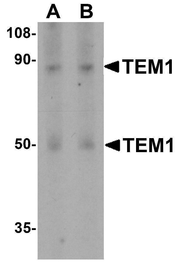
Western Blot Validation in Human Colon Tissue
Loading: 15 μg of lysates per lane.
Antibodies: TEM-1 A04735, (A, 0.5 μg/mL; B, 1 μg/mL), 1h incubation at RT in 5% NFDM/TBST.
Secondary: Goat anti-rabbit IgG HRP conjugate at 1:10000 dilution.

Click image to see more details
Independent Antibody Validation (IAV) via Protein Expression Profile in Cell Lines
Loading: 15 μg of lysates per lane.
Antibodies: TEM-1 A04735, (2 μg/mL), TEM-1 4359, ( 1μg/mL), and GAPDH (0.02 μg/mL), 1h incubation at RT in 5% NFDM/TBST.
Secondary: Goat anti-rabbit IgG HRP conjugate at 1:10000 dilution.

Click image to see more details
Western Blot Validation in Human Cell Lines
Loading: 15 μg of lysates per lane.
Antibodies: TEM-1 A04735, (2 μg/mL), 1h incubation at RT in 5% NFDM/TBST.
Secondary: Goat anti-rabbit IgG HRP conjugate at 1:10000 dilution.

Click image to see more details
Immunohistochemistry Validation of TEM-1 in Human Colon Tissue
Immunohistochemical analysis of paraffin-embedded Human Colon tissue using anti-TEM-1 antibody (A04735) at 2.5 μg/ml. Tissue was fixed with formaldehyde and blocked with 10% serum for 1 h at RT; antigen retrieval was by heat mediation with a citrate buffer (pH6). Samples were incubated with primary antibody overnight at 4˚C. A goat anti-rabbit IgG H&L (HRP) at 1/250 was used as secondary. Counter stained with Hematoxylin.

Click image to see more details
Immunofluorescence Validation of TEM-1 in Human Colon Tissue
Immunofluorescent analysis of 4% paraformaldehyde-fixed Human Colon Tissue labeling TEM-1 with A04735 at 20 μg/mL, followed by goat anti-rabbit IgG secondary antibody at 1/500 dilution (red).
Specific Publications For Anti-TEM1 CD248 Antibody (A04735)
Loading publications
Recommended Resources
Here are featured tools and databases that you might find useful.
- Boster's Pathways Library
- Protein Databases
- Bioscience Research Protocol Resources
- Data Processing & Analysis Software
- Photo Editing Software
- Scientific Literature Resources
- Research Paper Management Tools
- Molecular Biology Software
- Primer Design Tools
- Bioinformatics Tools
- Phylogenetic Tree Analysis
Customer Reviews
Have you used Anti-TEM1 CD248 Antibody?
Share your experimental results or join a short interview to earn up to $1,000 in product credits or other rewards.
0 Reviews For Anti-TEM1 CD248 Antibody
Customer Q&As
Have a question?
Find answers in Q&As, reviews.
Can't find your answer?
Submit your question
