This website uses cookies to ensure you get the best experience on our website.


- Table of Contents
Real validated CD248 Western blot protocols, expected-band and isoform facts, troubleshooting for weak or shifted signal, and recommended anti-CD248 WB antibodies. Everything you need to plan the experiment before you commit precious samples.
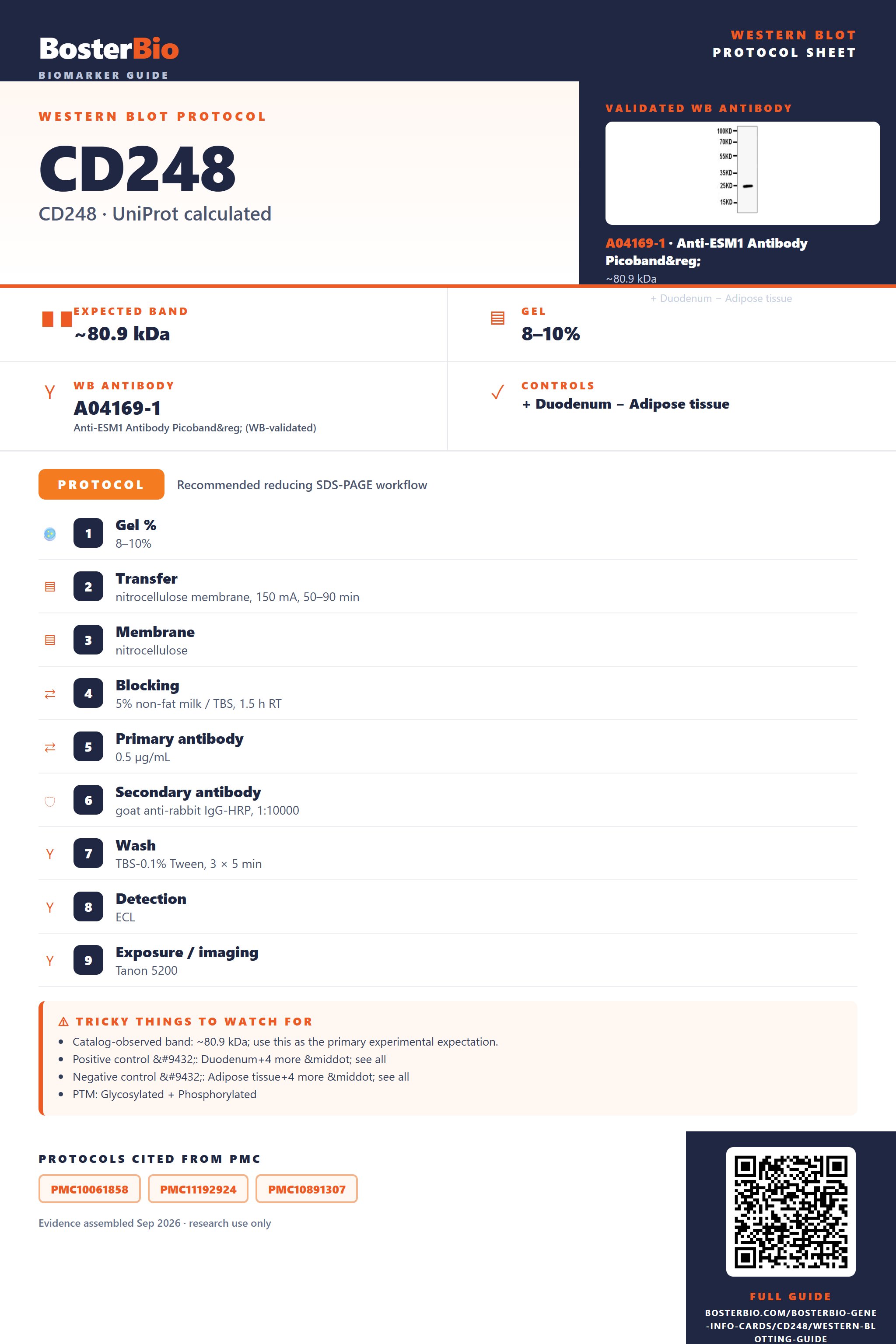
Expected bands, validated protocols, controls and antibodies — the at-a-glance facts below, then the full design guide.
| Expected band | ~80.9 kDa | |
| Gel | 8–10% | |
| Positive control | Duodenum+4 more · see all | |
| Negative control | Adipose tissue+4 more · see all |
| PTM | Glycosylated + Phosphorylated | |
| Caveat | Heavy O-glycosylation | |
| Regulation | LPS-induced | |
| Isoform | 2 isoform(s) |
Literature-validated Western blot parameters for CD248 — gel percentage, transfer, blocking, antibody incubation and detection, extracted from published methods.
| Gel % | 8–10% |
| Transfer | nitrocellulose membrane, 150 mA, 50–90 min |
| Membrane | nitrocellulose |
| Blocking | 5% non-fat milk / TBS, 1.5 h RT |
| Primary antibody | 0.5 µg/mL |
| Secondary antibody | goat anti-rabbit IgG-HRP, 1:10000 |
| Wash | TBS-0.1% Tween, 3 × 5 min |
| Detection | ECL |
| Exposure / imaging | Tanon 5200 |
| Observed band | 25 kDa |
CD248/Endosialin has an 80.9 kDa predicted backbone but runs higher and broader on blots mainly due to extensive O-linked glycosylation across 27 sites.
| band above the 80.9 kDa predicted mass | reflects mass added by extensive O-linked glycosylation at up to 27 sites |
| broad or smeared band rather than a sharp line | heterogeneous glycoform occupancy across the 27 O-glycosylation sites |
| band slightly below the unprocessed precursor size | N-terminal signal peptide (residues 1-17) has been cleaved to generate the mature membrane protein |
| two bands at different molecular weights | co-expression of isoform 1 and isoform 2 arising from alternative splicing |
| faint or absent band in a standard soluble lysate prep | single-pass type I membrane protein that is not efficiently solubilized without adequate detergent |
| slight mobility shift between reduced and non-reduced samples | intrachain disulfide bonds stabilize a compact fold that migrates differently without reduction |
| Predicted mass (UniProt) | 80.9 kDa backbone before any modification; the observed band typically runs above this |
| O-linked glycosylation (27 sites, e.g. Thr60, Thr401, Thr428) | adds substantial mass and heterogeneity, shifting the band well above 80.9 kDa and broadening it into a smear |
| Signal peptide cleavage (residues 1-17) | removes the N-terminal leader from the precursor, so the mature membrane-bound form runs slightly lower than the unprocessed translation product |
| Alternative splicing (isoforms 1 and 2) | can generate an additional band at a different apparent size distinct from the canonical isoform |
| Intrachain disulfide bonds (4 pairs in EGF-like/lectin domains) | stabilize a compact folded structure, so non-reduced samples may migrate slightly faster than fully reduced samples despite no true increase in mass |
| Situation | Likely cause | Next action |
|---|---|---|
| Band higher than expected | extensive O-linked glycosylation across 27 sites adds substantial mass to the mature protein | treat lysate with a deglycosylation enzyme and compare mobility shift toward the 80.9 kDa predicted mass |
| Broad smear instead of sharp band | heterogeneous occupancy of the 27 O-glycosylation sites produces a ladder of glycoforms | run a lower-percentage or gradient gel for longer to better resolve the smear, or deglycosylate before loading |
| Multiple bands | isoform 1 and isoform 2 arise from alternative splicing and can co-migrate as separate species | check which isoform the antibody epitope targets and confirm band assignment against isoform sequence differences |
| Band lower than expected | cleavage of the residues 1-17 signal peptide removes mass relative to the unprocessed precursor | confirm whether the predicted mass used for comparison is for the full-length precursor or the cleaved mature protein |
| Weak or no signal | single-pass type I membrane protein is poorly extracted by lysis buffers that do not adequately solubilize membrane proteins | use a detergent-based lysis buffer suited to membrane protein extraction and avoid excessive sample heating before loading |
Comprehensive Human Protein Atlas IHC scoring per tissue. Rows are taken directly from the HPA tissue chart — click any row's HPA link to view the source.
| Tissue | Cell type | Level | Evidence | Source |
|---|---|---|---|---|
| Duodenum | paneth cells | High | Protein (IHC) | HPA → |
| Rectum | fibroblasts | High | Protein (IHC) | HPA → |
| Skin | extracellular matrix | High | Protein (IHC) | HPA → |
| Small intestine | paneth cells | High | Protein (IHC) | HPA → |
| Appendix | germinal center cells | Medium | Protein (IHC) | HPA → |
| Tissue | Cell type | Level | Evidence | Source |
|---|---|---|---|---|
| Adipose tissue | adipocytes | Not detected | Protein (IHC) | HPA → |
| Adrenal gland | glandular cells | Not detected | Protein (IHC) | HPA → |
| Bone marrow | hematopoietic cells | Not detected | Protein (IHC) | HPA → |
| Breast | adipocytes | Not detected | Protein (IHC) | HPA → |
| Bronchus | respiratory epithelial cells | Not detected | Protein (IHC) | HPA → |
Deeper troubleshooting and optimisation questions for CD248, answered from its protein features.
BosterBio's CD248 antibodies are among the best-performing WB antibodies on the market — well cited, thoroughly validated, and orthogonally cross-validated against negative tissues and complementary methods.
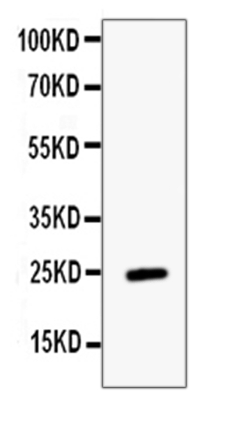
Our recommended CD248 Western blot antibodies are top-performing, extensively cited in the literature, and rigorously validated, with specificity confirmed through negative-tissue controls and orthogonal, complementary detection methods, giving you dependable, reproducible results for confident CD248 protein detection and expression analysis.
Which to pick: Only one anti-CD248 antibody is catalogued here, A04169-1, which includes an actual Western blot validation image, so it's the clear and only choice from this list for your CD248 blotting experiments.