CD28 / T-cell-specific surface glycoprotein CD28 · IHC design guide

Design Immunohistochemistry for CD28

Plan CD28 chromogenic IHC using lymph node or tonsil, where non-germinal center cells show medium staining (HPA tissue IHC). Start the catalog antibody at 2 μg/mL (datasheet) and assess the selective cytoplasmic pattern in immune-cell subsets (HPA tissue IHC).

Evidence assembled Oct 2026 · For research use; verify linked source records and product datasheet before use
Immunohistochemistry protocol sheet for CD28 (IHC for CD28): expected localisation Selective cytoplasmic staining in immune-cell subsets (HPA tissue IHC), antibody A00065, validated IHC image, and IHC protocol steps
Printable CD28 IHC protocol sheet — expected localisation Selective cytoplasmic staining in immune-cell subsets (HPA tissue IHC), antibody A00065, controls and protocol steps. Open the full CD28 IHC guide →

CD28 Immunohistochemistry Experimental Design Guide

Expected localisation, validated protocols, controls and antibodies — the at-a-glance facts below, then the full design guide.

Must know before staining
Expected localisation Selective cytoplasmic staining in immune-cell subsets (HPA tissue IHC)
Staining pattern Subset of immune cells shows cytoplasmic staining (HPA tissue IHC)
Antigen retrieval Citrate pH 6.0 HIER, 95–98 °C, 20 min (rule: cytoplasmic / membrane antigen)
Positive control ⓘ Lymph node+1 more · see all
Negative control ⓘ Adipose tissue+4 more · see all
Important caveats
Reasons your staining may differ from the expected pattern.
Fixation Keep fixation consistent across sections (standard IHC practice; not target-specific)
Caveat Immune-cell subsets stain selectively (HPA tissue IHC)
Regulation No expression regulator specified (UniProt)
Isoform / epitope 7 isoforms; check extracellular versus cytoplasmic epitope (UniProt)
Section 1

Recommended CD28 IHC & IF Protocols

The catalog antibody’s IHC-P protocol accompanies published CD28 IHC workflows on ATLL tissue and paraffin-embedded tissue sections (PMC8748252; PMC4914341).

Recommended immunohistochemistry (IHC-P) protocol parameters
SampleTissue sections; selected-image fixative not specified (standard IHC workflow)
FixationImage fixative and duration unreported (datasheet A00065); verify before use.
Sectioning4–5 µm sections on charged slides (standard)
DeparaffinisationXylene, graded ethanol series to water (standard)
Antigen retrievalHeat-induced epitope retrieval in citrate buffer, pH 6.0, 20 min at 95–98 °C (standard rule: cytoplasmic / membrane antigen)
Peroxidase block3% H2O2, 10 min, room temperature (standard)
Blocking10% normal serum of the secondary host, 30 min, room temperature (standard)
Primary antibodyRabbit anti-CD28, 2 μg/mL (datasheet A00065)
Primary incubationOvernight at 4 °C (standard)
DetectionHRP-polymer secondary, DAB chromogen 5–10 min (standard)
CounterstainHematoxylin, blue, dehydrate and mount (standard)
Expected resultCD28-positive staining in non-germinal center cells of lymph node (HPA tissue IHC: Medium). HPA tissue profile: Selective cytoplasmic expression in subsets of immune cells. No signal in the no-primary control.
💡Decision noteStart with citrate pH 6.0 retrieval at 95–98 °C for 20 min (page antigen-retrieval rule); follow each article’s stated conditions when reproducing its workflow (PMC8748252; PMC4914341).
Section 2

What Is the Expected CD28 Staining Pattern?

CD28 is a cell-surface receptor with an extracellular region, one transmembrane segment and a cytoplasmic tail (UniProt P10747 topology). Expect staining in subsets of immune cells, consistent with expression in T cells and plasma cells (UniProt P10747 tissue specificity). HPA tissue IHC describes selective cytoplasmic staining and rates its tissue pattern Enhanced for consistency with RNA data, pending external verification (HPA: tissue IHC).

What am I looking at on my slide?
Discrete staining in subsets of non-germinal-center cells in lymph node or tonsil.This matches HPA's Medium IHC staining in those cells (HPA: lymph node and tonsil). A membrane outline is biologically plausible from CD28 topology, but HPA describes its tissue IHC pattern as selective cytoplasmic staining (UniProt P10747 topology; HPA: tissue IHC). Do not require a crisp membrane rim to accept a chromogenic IHC result.
Predominantly nuclear staining, with little signal in the expected immune-cell subsets.Nuclear localization is unsupported by the supplied localization records (UniProt P10747: cell surface; HPA: tissue IHC and subcellular). Treat it as suspect and review counterstain, nonspecific antibody binding and detection controls using general IHC practice. Cytoplasmic staining alone is a different case: HPA reports that appearance in tissue IHC (HPA: tissue IHC).
Strong staining of adipocytes or glandular cells rather than scattered immune cells.HPA reports no detection in adipocytes of adipose tissue or breast, and in adrenal or cervical glandular cells (HPA: tissue IHC). Check whether the signal follows those cells or nearby immune cells. If it follows the reported negative cell types, investigate cross-reactivity or endogenous detection activity using general IHC controls.
Diffuse color across many cell types, extracellular spaces or a broad area of the section.That distribution does not resemble HPA's selective staining of immune-cell subsets (HPA: tissue IHC). Review background in a no-primary control, blocking, washing and chromogen development using general IHC practice. A low-level signal in every cell should not be scored as widespread CD28 expression without cell-level evidence.
No convincing staining in lymph node or tonsil, including the expected non-germinal-center cells.These are useful positive-reference tissues because HPA reports Medium staining in their non-germinal-center cells (HPA: tissue IHC). First confirm that the relevant cells are present on the section; then review the antibody's IHC validation and routine retrieval, detection and control performance. One negative run does not establish CD28 absence.
💡Expected CD28 appearanceA convincing positive is selective staining of non-germinal-center immune-cell subsets at approximately HPA's Medium level in lymph node or tonsil, allowing the reported cytoplasmic IHC appearance; broad staining of reported negative adipocytes or glandular cells is suspect (HPA: tissue IHC; UniProt P10747 topology).
How each factor affects the staining
Localization and assay readoutUniProt places CD28 at the cell surface, and HPA ICC-IF supports plasma-membrane localization; HPA tissue IHC nevertheless describes selective cytoplasmic expression (UniProt P10747 topology; HPA: subcellular and tissue IHC). Interpret each assay against its own reported appearance.
Reference tissue and cell selectionHPA records Medium staining in lymph-node and tonsil non-germinal-center cells, but Low staining in appendix lymphoid tissue, splenic red-pulp cells and marrow hematopoietic cells (HPA: tissue IHC). A weak result in a Low category is less decisive than loss of signal in a Medium category.
Antibody validation scopeHPA lists Enhanced IHC validation for HPA070003 and Supported ICC validation for HPA041233; those labels apply to different antibodies and assays (HPA: antibodies). HPA also says its tissue-pattern reliability awaits external verification (HPA: tissue IHC).
Protein features and epitope choiceCD28 has an extracellular region at residues 19–152, a transmembrane region at 153–179, five listed glycosylation sites and seven isoforms (UniProt P10747 topology, glycosylation and isoforms). These features make the antibody's stated epitope relevant to interpretation; the supplied record does not establish their effect on retrieval or staining.
Why is my staining missing, weak or wrong?
SituationLikely causeNext action
Lymph node or tonsil has no signal in the expected cell population.The HPA Medium reference pattern is missing; the record alone cannot distinguish biology from an assay failure (HPA: tissue IHC).Confirm the expected cells on the section, then review the IHC-validated antibody's instructions and routine retrieval, primary-antibody, detection and positive-control steps (general IHC practice).
Only the counterstain or a nuclear-looking signal is visible.A nuclear CD28 pattern lacks support from the supplied localization records (UniProt P10747: cell surface; HPA: subcellular).Compare the stained section with a no-primary control and inspect chromogen and counterstain balance before assigning cellular localization (general IHC practice).
Adipocytes or glandular cells stain strongly.Those cell types include HPA Not detected examples; cross-reactivity or endogenous detection activity is possible, but the image alone cannot identify which (HPA: tissue IHC; general IHC practice).Check the cell identity at higher magnification and compare no-primary and detection controls; score the expected immune cells separately (general IHC practice).
The whole section has diffuse color or poor contrast.Diffuse staining departs from the selective HPA tissue pattern and can arise from routine IHC background sources (HPA: tissue IHC; general IHC practice).Inspect the no-primary control, blocking and washes, and chromogen development; reassess only cell-resolved signal after background is controlled (general IHC practice).
The stain looks cytoplasmic rather than sharply membranous.HPA reports selective cytoplasmic tissue IHC, while UniProt and HPA ICC-IF support surface or plasma-membrane localization (HPA: tissue IHC and subcellular; UniProt P10747 topology).Judge tissue IHC by selective cell distribution and controls; do not reject it solely for lacking an IF-like membrane outline (HPA: tissue IHC; general IHC practice).
IF/ICC question: should its membrane pattern be required for IHC?The supported plasma-membrane ICC-IF result and the Enhanced tissue IHC result come from distinct HPA assay records and antibodies (HPA: subcellular, antibodies and tissue IHC).No. Use the plasma-membrane result to interpret IF/ICC on its own guide page; use HPA's selective tissue IHC pattern for chromogenic sections (HPA: subcellular and tissue IHC).

Sample controls for CD28 IHC & IF

🧪Run lymph node first: non-germinal center cells should stain (HPA: Medium). Use adipose tissue adipocytes as the negative tissue (HPA: Not detected); cells without CD28 staining on the lymph node slide should lack specific membrane signal, but the supplied HPA row does not identify an internal negative cell type.
Positive control tissue: Lymph node (Non-germinal center cells, HPA Medium)
Negative control tissue: Adipose tissue (HPA Not detected)
ICC-IF cell lines (HPA subcellular resource): HPA ICC-IF images show CD28 in JURKAT, U2OS, with annotated localisation: Plasma membrane (supported) (HPA subcellular).
Technical controls: Include no-primary (secondary-only) and host-species-matched isotype controls appropriate to the antibody’s clonality, plus CD28-knockout material if available. For chromogenic lymph node IHC, block endogenous peroxidase; check endogenous biotin if using avidin–biotin detection.
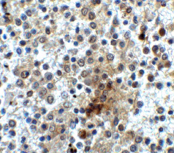
⚠️Feasibility: A target-specific fixation window and retrieval effect are unreported in the supplied evidence; the selected A00065 spleen IHC caption reports 2 µg/ml but does not report a fixative (selected-SKU tissue-IHC caption). IF/ICC may make membrane localization easier to assess (HPA: supported plasma membrane), but the evidence does not establish whether frozen sections are easier than paraffin sections. Endogenous peroxidase in lymph node leukocytes can complicate chromogenic scoring.

HPA tissue IHC evidence for CD28

Comprehensive Human Protein Atlas IHC scoring per tissue (reliability: Enhanced — High consistency between antibody staining and RNA expression data. Pending external verification.). Rows are taken directly from the HPA tissue chart — click any row's HPA link to view the source.

Positive expression · recommended positive controls

TissueCell typeLevelEvidenceSource
Lymph node Non-germinal center cells Medium Protein (IHC) HPA →
Tonsil Non-germinal center cells Medium Protein (IHC) HPA →

Undetected expression · recommended negative controls

TissueCell typeLevelEvidenceSource
Adipose tissue Adipocytes Not detected Protein (IHC) HPA →
Adrenal gland Glandular cells Not detected Protein (IHC) HPA →
Breast Adipocytes Not detected Protein (IHC) HPA →
Bronchus Respiratory epithelial cells Not detected Protein (IHC) HPA →
Caudate Glial cells Not detected Protein (IHC) HPA →
Section 3

Advanced CD28 IHC Tips

Troubleshoot CD28 chromogenic IHC in paraffin sections by checking retrieval, cellular distribution, controls, and scoring before interpreting immune cell staining.

What retrieval should I start with if CD28 staining is weak?
Start with citrate buffer at pH 6.0, heated to 95–98 °C for 20 min (page retrieval rule). Allow sections to cool in the buffer, then compare the staining with a matched section processed without retrieval; excessive heat can damage morphology and increase nonspecific staining (standard IHC practice). Assess signal in lymph node or tonsil non-germinal center cells, where medium staining has been reported (HPA: tissue IHC). If signal remains weak, test a second retrieval condition on adjacent sections while keeping antibody concentration and detection identical (standard IHC practice). Record whether any gain preserves cell boundaries and tissue morphology before adopting it (standard IHC practice).
How should I troubleshoot fixation when CD28 staining varies between blocks?
CD28-specific fixation sensitivity is unknown from the supplied evidence; the human spleen IHC caption reports 2 µg/ml antibody but does not state a fixative (catalog antibody: A00065 caption). Compare sections from blocks with documented processing histories using the same retrieval, antibody incubation, and chromogenic development conditions (standard IHC practice). Include a shared positive control section in each run so variation between runs can be separated from variation between blocks (standard IHC practice). If a block shows weak signal, inspect morphology and compare nearby immune cell areas before changing retrieval or antibody concentration (standard IHC practice). Record any improvement as specific to the tested processing conditions, pending a controlled fixation comparison (standard IHC practice).
Where should convincing CD28 staining appear in a tissue section?
Expect CD28 at the cell surface: it has an extracellular region at residues 19–152, a transmembrane segment at 153–179, and a cytoplasmic tail at 180–220 (UniProt P10747 topology). Plasma membrane localisation is independently supported (HPA: subcellular). In chromogenic sections, check whether staining follows the outlines of individual immune cells rather than spreading uniformly through the tissue (standard IHC interpretation). Some cytoplasmic staining in subsets of immune cells has been reported, so compartment alone should not determine whether a cell is positive (HPA: tissue IHC). Compare the pattern with matched controls and morphology, particularly where closely packed cells make membrane boundaries difficult to resolve (standard IHC practice).
Could CD28 isoforms or epitope position explain inconsistent staining?
CD28 has 7 listed isoforms, but the supplied catalog caption does not identify the antibody epitope or establish which isoforms it detects (UniProt P10747: isoforms; catalog antibody: A00065 caption). Check the antibody documentation for its immunogen or mapped epitope before interpreting differences between samples as isoform changes (standard IHC practice). An extracellular epitope lies within residues 19–152, whereas a tail epitope would lie within residues 180–220 (UniProt P10747 topology). The extracellular region includes 5 listed glycosylation sites, and the tail includes phosphorylation sites (UniProt P10747: modifications). Test a documented second epitope with matched controls if epitope dependence is central to the interpretation (standard IHC practice).
How can IF help assess an ambiguous CD28 IHC pattern?
On matched material, pair CD28 IF with a T-cell marker to assess whether the signal occurs in the expected cell population (UniProt P10747: T-cell expression; standard IF practice). Choose a fluorophore channel with low tissue autofluorescence and inspect an unstained section before assigning faint signal as positive (standard IF practice). CD28 has an extracellular region at residues 19–152 and a cytoplasmic tail at 180–220; choose and document permeabilisation according to the antibody’s known epitope (UniProt P10747 topology; standard IF practice). If the epitope is unknown, compare permeabilised and nonpermeabilised preparations rather than assuming surface access (standard IF practice). Interpret IF as a separate assay with its own controls, alongside the chromogenic IHC result (standard IF practice).
How do I reduce diffuse brown staining around CD28-positive immune cells?
First compare a no-primary control and a matched positive section to determine whether brown signal depends on the primary antibody (standard chromogenic IHC practice). Block endogenous peroxidase before horseradish peroxidase detection, and check whether changing chromogen development time reduces background while retaining cellular signal (standard chromogenic IHC practice). Inspect tissue edges, folds, and damaged regions separately because they can stain disproportionately (standard IHC practice). CD28 is reported in T cells and plasma cells, and HPA reports staining in subsets of immune cells; diffuse staining across unrelated cell types deserves further control testing (UniProt P10747: tissue specificity; HPA: tissue IHC). Titrate the antibody around a documented starting concentration rather than treating stronger brown colour as greater specificity (standard IHC practice).
How should I score CD28 when immune cell abundance differs between samples? ⚠ ANSWER MARKED FOR VERIFICATION
Define the region of interest and the cell type before scoring, then report CD28-positive cells as a percentage of eligible immune cells or as density per mm² (standard IHC quantification practice). If using an H-score, record staining intensity and the percentage of cells at each intensity under one consistent scoring rule (standard IHC quantification practice). Normalise to the assessed immune cell population when comparing expression per cell; report area-based density separately when immune infiltration itself is relevant (standard IHC quantification practice). CD28 expression is reported in T cells and plasma cells, so total tissue area alone can obscure differences in cell composition (UniProt P10747: tissue specificity). Apply the same positivity threshold, controls, and region selection across cases (standard IHC quantification practice).
When is a CD28-positive cell likely to be an artefact?
A convincing result identifies discrete immune cells with a pattern compatible with CD28 cell surface localisation, interpreted alongside tissue morphology and controls (UniProt P10747: localisation and tissue specificity; standard IHC interpretation). HPA reports medium staining in lymph node and tonsil non-germinal center cells, providing reference locations for comparison rather than proof for every stained cell (HPA: tissue IHC). Question staining confined to section edges or necrotic areas, and investigate widespread colour in unexpected cell types (standard IHC interpretation). A no-primary control helps identify detection-related colour, including residual endogenous enzyme activity (standard chromogenic IHC practice). Treat diffuse cytoplasmic colour cautiously, while recognising that HPA also reports selective cytoplasmic staining in immune cell subsets (HPA: tissue IHC).
Boster reagents

Best CD28 / T-cell-specific surface glycoprotein CD28 IHC Antibodies

The catalog lists one human-reactive anti-CD28 antibody with IHC and IF images from human spleen tissue (catalog: reactivity; IHC and IF image captions).

Real IHC data Immunohistochemistry of CD28 in human spleen tissue with CD28 antibody at 2 μg/ml.
Anti-CD28 Antibody
Cat # A00065

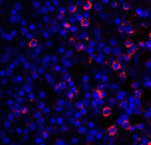
A00065 is listed for human IHC and IF (catalog: applications, reactivity). Its IHC image shows human spleen tissue at 2 μg/mL, and its IF image shows human spleen tissue at 20 μg/mL (catalog: IHC and IF image captions).

Which to pick: Choose A00065 for human tissue IHC; its IHC caption shows human spleen tissue, but does not report section processing or fixative (catalog: IHC image caption). For IF, A00065 has a human spleen image; ICC is not listed, and clonality is unreported (catalog: IF image caption, applications, clone). No cross-species option is documented because A00065 lists human reactivity only (catalog: reactivity).

Each figure is that product's own IHC / IF validation image from its datasheet.

References

  1. UniProt Consortium. UniProt entry P10747 (CD28_HUMAN, T-cell-specific surface glycoprotein CD28).
  2. Human Protein Atlas. CD28 tissue IHC expression (reliability: Enhanced).
  3. Human Protein Atlas. CD28 subcellular location (ICC-IF): Localized to the plasma membrane..
  4. Human Protein Atlas. CD28 antibody validation summary (2 antibodies).
  5. Clinicopathological significance of CD28 overexpression in adult T-cell leukemia/lymphoma. Cancer science 2022 — PMC8748252.
  6. Immunohistochemical biomarkers and distribution of telocytes in ApoE(-/-) mice. Cell biology international 2019 — PMC6851738.
  7. GLUT5-Mediated Disruption of the Gut-Testis Axis Deteriorates Olanzapine-Induced Testicular Fibrosis and is Ameliorated by Dapagliflozin. Drug design, development and therapy 2026 — PMC13600974.
  8. Tumor CTLA-4 overexpression predicts poor survival in patients with nasopharyngeal carcinoma. Oncotarget 2016 — PMC4914341.
  9. PubMed PMID:2825196 — UniProt-cited evidence.
  10. PubMed PMID:2162892 — UniProt-cited evidence.
  11. PubMed PMID:11916166 — UniProt-cited evidence.