This website uses cookies to ensure you get the best experience on our website.


- Table of Contents
Plan CD3D paraffin-section IHC using tonsil non-germinal center cells as a positive control (HPA tissue IHC) and an IHC-validated antibody at 1:50 (datasheet: M04405). Interpret cytoplasmic staining in a subset of immune cells (HPA tissue IHC) alongside the expected membrane location (UniProt) and presumed off-target staining (HPA tissue IHC).

Expected localisation, validated protocols, controls and antibodies — the at-a-glance facts below, then the full design guide.
| Expected localisation | Cytoplasmic in immune cells (HPA tissue IHC); membrane expected (UniProt) | |
| Staining pattern | Cytoplasmic staining in a subset of immune cells (HPA tissue IHC) | |
| Antigen retrieval | Citrate pH 6.0 HIER, 95–98 °C, 20 min (rule: cytoplasmic / membrane antigen) | |
| Positive control | Kidney+2 more · see all | |
| Negative control | Adipose tissue+4 more · see all |
| Fixation | Keep formalin fixation consistent across paraffin sections (standard IHC practice; not target-specific) | |
| Caveat | Presumed off-target staining can complicate interpretation (HPA tissue IHC) | |
| Regulation | Staining may vary with T-cell abundance (UniProt) | |
| Isoform / epitope | 2 isoforms; extracellular vs cytoplasmic epitope coverage unknown (UniProt) |
Compare the catalog antibody’s IHC-P protocol with published CD3D protocols for gastric, liver, and cervical cancer tissues (PMC9198637; PMC8192185; PMC11375541).
| Sample | Paraffin-embedded human spleen tissue; fixative not specified (datasheet M04405) |
| Fixation | Image fixative and duration unreported (datasheet M04405); verify before use. |
| Sectioning | 4–5 µm sections on charged slides (standard) |
| Deparaffinisation | Xylene, graded ethanol series to water (standard) |
| Antigen retrieval | Heat-induced epitope retrieval in citrate buffer, pH 6.0, 20 min at 95–98 °C (standard rule: cytoplasmic / membrane antigen) |
| Peroxidase block | 3% H2O2, 10 min, room temperature (standard) |
| Blocking | 10% normal serum of the secondary host, 30 min, room temperature (standard) |
| Primary antibody | Rabbit monoclonal (clone IDE-3) anti-CD3D, 1:50 (datasheet M04405) |
| Primary incubation | Overnight at 4 °C (standard) |
| Detection | HRP-polymer secondary, DAB chromogen 5–10 min (standard) |
| Counterstain | Hematoxylin, blue, dehydrate and mount (standard) |
| Expected result | CD3D-positive staining in distal tubules of kidney (HPA tissue IHC: Medium). HPA tissue profile: Cytoplasmic expression in a subset of immune cells. No signal in the no-primary control. |
CD3D is a cell-membrane component of the TCR–CD3 complex, with extracellular residues 22–105, a transmembrane segment at 106–126, and a cytoplasmic tail at 127–171 (UniProt P04234 topology). Expect staining chiefly in T lymphocytes (UniProt P04234 tissue specificity). HPA reports cytoplasmic staining in a subset of immune cells; its tissue IHC reliability is Enhanced, with presumed off-target binding observed and disregarded (HPA tissue IHC).
| Discrete staining in immune cells of tonsil or thymus, with a membranous outline or cytoplasmic signal. | This fits CD3D’s T-cell membrane location (UniProt P04234 subcellular location) and HPA’s observed cytoplasmic staining in a subset of immune cells (HPA tissue IHC). HPA reports Medium staining in tonsil non-germinal center cells and thymus cortical cells (HPA tissue IHC). Judge the stained cells and their location together, rather than requiring a perfect membrane ring in every section. |
| Strong nuclear staining dominates cells that otherwise lack a plausible immune-cell pattern. | Nuclear localization is unsupported by the supplied CD3D record: UniProt places CD3D at the cell membrane, while HPA describes cytoplasmic immune-cell staining in tissue IHC (UniProt P04234 subcellular location; HPA tissue IHC). Treat the nuclear pattern as suspect and review controls and detection conditions before scoring it as CD3D (general IHC practice). |
| Staining is concentrated in glandular or tubular cells instead of recognizable immune cells. | This does not, by itself, establish a T-cell result (UniProt P04234 tissue specificity). HPA reports Medium staining in kidney distal tubules and Low staining in stomach and small-intestine glandular cells, despite noting presumed off-target binding (HPA tissue IHC). Record the cell type and assess possible cross-reactivity or endogenous detection activity with appropriate controls (general IHC practice). |
| Broad, diffuse color covers tissue structures and obscures individual positive cells. | A field-wide haze cannot be reconciled confidently with HPA’s subset-of-immune-cells profile (HPA tissue IHC). Background from detection reagents, incomplete blocking, or overly strong assay conditions is possible (general IHC practice). Compare a negative reagent control and reduce background before interpreting faint cells; diffuse staining alone is not evidence of CD3D. |
| No convincing signal appears in tonsil or thymus where positive cells were expected. | HPA reports Medium staining in tonsil non-germinal center cells and thymus cortical cells (HPA tissue IHC). A blank result in suitable tissue calls for an assay check, including antibody dilution, retrieval, detection reagents, and section quality (general IHC practice). First verify that the expected cellular area is present; a single blank section cannot establish absent CD3D expression. |
| Cell compartment and epitope position | CD3D spans the membrane, with extracellular residues 22–105 and cytoplasmic residues 127–171 (UniProt P04234 topology). The supplied record does not map the antibody epitope, so an exclusively extracellular or intracellular antibody-binding claim is unsupported. |
| Tissue pattern and validation | HPA rates tissue IHC reliability Enhanced and reports cytoplasmic staining in a subset of immune cells, while noting presumed off-target binding (HPA tissue IHC). HPA071778 and CAB013055 both have Enhanced IHC validation; this supports using the cellular pattern, while leaving anomalous staining open to review (HPA antibodies; HPA tissue IHC). |
| Protein processing and variants | The UniProt precursor has a signal peptide at residues 1–21 and a mature chain spanning 22–171; two isoforms are listed (UniProt P04234 processing; isoforms). No epitope sequence or isoform-specific IHC performance is supplied, so these features do not predict a distinct staining pattern for this antibody. |
| Antigen retrieval | Target-specific fixation effects are not established by the supplied assay evidence. Verify with a matched IHC source before attributing a result to fixation. |
| IF/ICC Q&A: what localization is reported? | HPA reports uncertain endoplasmic-reticulum and vesicle localization in ICC-IF, and rates HPA071778 ICC validation Uncertain (HPA subcellular ICC-IF; HPA antibodies). Interpret that observation within an IF/ICC experiment; it does not replace the tissue-IHC immune-cell pattern (HPA tissue IHC). |
| Situation | Likely cause | Next action |
|---|---|---|
| Tonsil or thymus has no discrete positive cells. | The sampled area may lack the HPA-listed cell population, or the IHC assay may have failed (HPA tissue IHC; general IHC practice). | Confirm the relevant region on the section, then check the positive control, antibody dilution, retrieval, and detection steps (general IHC practice). HPA lists Medium staining in tonsil non-germinal center and thymus cortical cells (HPA tissue IHC). |
| A nuclear pattern is stronger than immune-cell staining. | This location conflicts with UniProt’s cell-membrane assignment and HPA’s cytoplasmic immune-cell profile (UniProt P04234 subcellular location; HPA tissue IHC). | Compare reagent controls and a known-positive immune-cell area; review primary-antibody and detection conditions before accepting the signal (general IHC practice). |
| Kidney tubules or glandular cells appear positive. | HPA records Medium distal-tubule staining and Low stomach or small-intestine glandular staining, while noting presumed off-target binding (HPA tissue IHC). | Document the exact cell type and compare appropriate controls and immune-cell staining. Do not score epithelial color as T-cell localization without cellular evidence (UniProt P04234 tissue specificity; general IHC practice). |
| Diffuse chromogen masks individual cells. | The result cannot be matched reliably to HPA’s subset-of-immune-cells profile; assay background is possible (HPA tissue IHC; general IHC practice). | Inspect negative reagent controls and review blocking, antibody dilution, washes, and chromogen development (general IHC practice). Reassess whether discrete immune cells remain visible. |
| A faint cytoplasmic signal appears without a crisp membrane rim. | HPA describes cytoplasmic staining in a subset of immune cells, although UniProt assigns CD3D to the cell membrane (HPA tissue IHC; UniProt P04234 subcellular location). | Use cell identity, distribution, and controls to judge the signal; do not reject it solely for lacking a continuous rim (HPA tissue IHC; general IHC practice). |
| ICC-IF shows punctate intracellular staining that differs from the tissue slide. | HPA’s ICC-IF summary lists uncertain endoplasmic-reticulum and vesicle localization, whereas tissue IHC reports cytoplasmic immune-cell staining (HPA subcellular ICC-IF; HPA tissue IHC). | Assess the ICC-IF result within its own controls and validation context. HPA071778 is ICC Uncertain but IHC Enhanced (HPA antibodies); use the tissue-IHC pattern to interpret the paraffin section. |
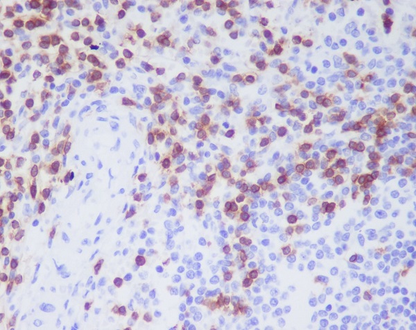
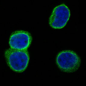
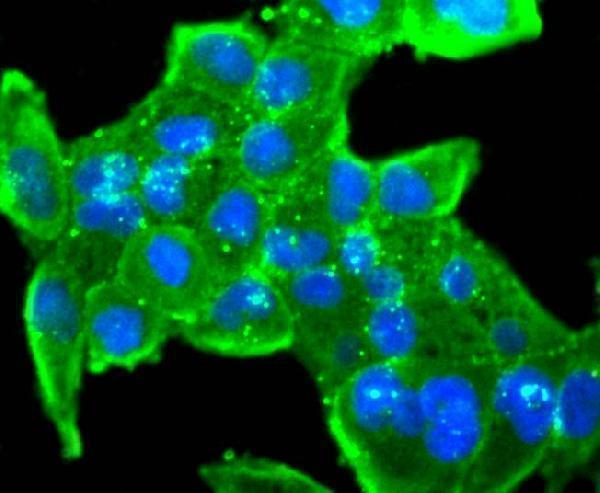


Comprehensive Human Protein Atlas IHC scoring per tissue (reliability: Enhanced — Medium consistency between antibody staining and RNA expression data. Presumed off target binding observed and disregarded.). Rows are taken directly from the HPA tissue chart — click any row's HPA link to view the source.
| Tissue | Cell type | Level | Evidence | Source |
|---|---|---|---|---|
| Kidney | Distal tubules | Medium | Protein (IHC) | HPA → |
| Thymus | Cortical cells | Medium | Protein (IHC) | HPA → |
| Tonsil | Non-germinal center cells | Medium | Protein (IHC) | HPA → |
| Tissue | Cell type | Level | Evidence | Source |
|---|---|---|---|---|
| Adipose tissue | Adipocytes | Not detected | Protein (IHC) | HPA → |
| Adrenal gland | Glandular cells | Not detected | Protein (IHC) | HPA → |
| Appendix | Glandular cells | Not detected | Protein (IHC) | HPA → |
| Bone marrow | Hematopoietic cells | Not detected | Protein (IHC) | HPA → |
| Breast | Glandular cells | Not detected | Protein (IHC) | HPA → |
Troubleshoot CD3D chromogenic staining in paraffin sections using the stated retrieval method, membrane topology, tissue evidence, and appropriate controls.
Human-reactive anti-CD3D antibodies have IHC images from paraffin-embedded spleen and fluorescence images from HeLa and Jurkat cells (catalog reactivity; image captions).
M04405 is listed for human IHC and IF, with paraffin-section spleen IHC and Jurkat IF images (M04405 applications, reactivity, image captions). A04405-1 is listed for human IHC and ICC, with a paraformaldehyde-fixed HeLa ICC image (A04405-1 applications, reactivity, image caption).
Which to pick: Choose M04405 for tissue IHC: this rabbit monoclonal has a human spleen paraffin-section IHC image; its caption does not report the fixative (M04405 catalog clone, reactivity, IHC caption). For cellular fluorescence, choose A04405-1 for ICC in paraformaldehyde-fixed, Triton-permeabilised HeLa cells, or M04405 for IF in Jurkat cells (A04405-1 ICC caption; M04405 applications, IF caption). Neither SKU has listed nonhuman reactivity to support cross-species use (catalog reactivity: Human for both).