This website uses cookies to ensure you get the best experience on our website.


- Table of Contents
Real validated CD58 Western blot protocols, expected-band and isoform facts, troubleshooting for weak or shifted signal, and recommended anti-CD58 WB antibodies. Everything you need to plan the experiment before you commit precious samples.
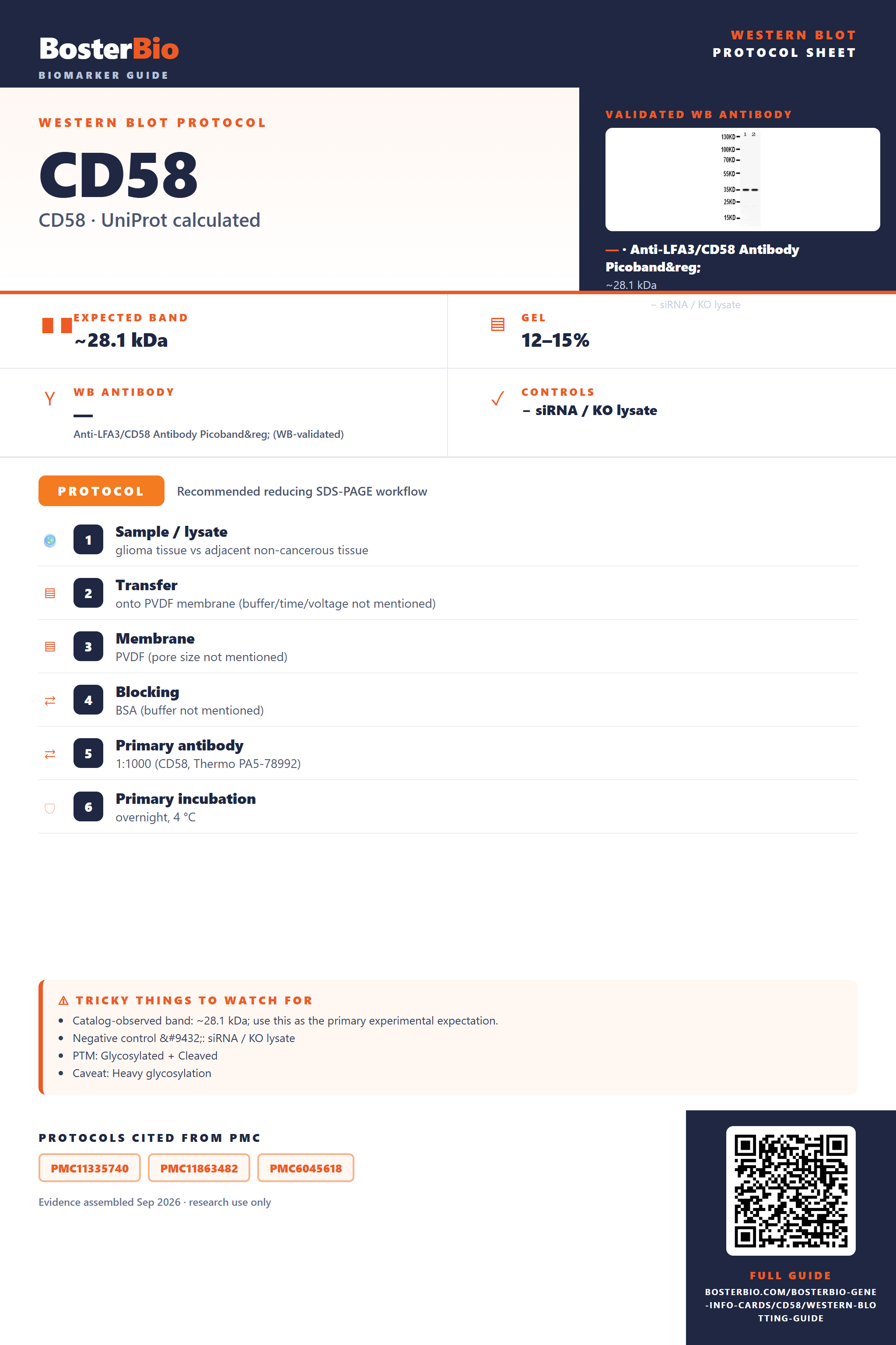
Expected bands, validated protocols, controls and antibodies — the at-a-glance facts below, then the full design guide.
| Expected band | ~28.1 kDa | |
| Observed band | 35 kDa | |
| Gel | 12–15% | |
| Negative control | siRNA / KO lysate |
| PTM | Glycosylated + Cleaved | |
| Caveat | Heavy glycosylation | |
| Regulation | LPS-induced | |
| Isoform | 3 isoform(s) |
Literature-validated Western blot parameters for CD58 — gel percentage, transfer, blocking, antibody incubation and detection, extracted from published methods.
| Sample / lysate | glioma tissue vs adjacent non-cancerous tissue |
| Transfer | onto PVDF membrane (buffer/time/voltage not mentioned) |
| Membrane | PVDF (pore size not mentioned) |
| Blocking | BSA (buffer not mentioned) |
| Primary antibody | 1:1000 (CD58, Thermo PA5-78992) |
| Primary incubation | overnight, 4 °C |
CD58 has a 28.1 kDa predicted core but runs at an observed ~35 kDa on Western blot due to extensive N-glycosylation at its six sites.
| prominent band at approximately 35 kDa | mature, fully N-glycosylated CD58 monomer running well above its 28.1 kDa unmodified core mass |
| diffuse or smeared band spanning roughly 30-40 kDa | heterogeneous occupancy and branching across the six N-linked glycosylation sites produces a mixture of glycoforms |
| band smaller than the calculated 28.1 kDa full-length translation product | cleavage of the 28-residue N-terminal signal peptide during membrane insertion removes several kDa from the precursor to yield the mature protein |
| two or more discrete bands at different apparent molecular weights | CD58 has three annotated splice isoforms of differing length, each capable of contributing a separate band |
| single clean band near 28 kDa | unglycosylated or fully deglycosylated CD58 core protein, seen after glycosidase treatment or in non-glycosylated expression systems |
| Predicted core mass (28.1 kDa, 250 aa) | unmodified polypeptide baseline; rarely observed directly since the native protein is glycosylated |
| Six N-linked glycosylation sites (Asn40, Asn94, Asn109, Asn135, Asn169, Asn195) | cumulative glycan attachment raises the apparent mass to the observed ~35 kDa and can broaden the band |
| N-terminal signal peptide (residues 1-28) | cleaved upon membrane insertion, so the mature protein runs smaller than the full-length translated precursor |
| Alternative splicing (isoforms 1, 2, 3) | different isoforms vary in length, so lysates can show additional bands relatively larger or smaller than the canonical isoform |
| Non-glycosylated recombinant or bacterially expressed standard | lacking the six N-glycans, this form runs closer to the 28 kDa core mass, lower than native cell-derived CD58 |
| Situation | Likely cause | Next action |
|---|---|---|
| Band higher than expected | heavy N-glycosylation across six sites raises apparent mass well above the 28.1 kDa core | treat lysate with PNGase F and confirm the band collapses toward the core mass to verify glycan-dependent shift |
| Broad smear instead of sharp band | heterogeneous occupancy and branching of the six N-glycosylation sites generates a range of glycoforms | run a lower-percentage or longer gel for better resolution, or deglycosylate with PNGase F/Endo H to sharpen the band |
| Multiple bands | three alternatively spliced isoforms of differing length can be co-expressed | confirm which isoform is detected using isoform-specific epitope mapping and check isoform expression in the cell type used |
| Band lower than expected | signal peptide cleavage removes the 28-residue leader from the precursor, or the sample is a non-glycosylated recombinant construct | compare against the mature membrane-bound mass and verify the glycosylation state of the construct or standard used |
| No band in lysate | as a single-pass type I membrane glycoprotein, CD58 requires adequate detergent solubilization from the plasma membrane, and expression is low in some cell lines | use a membrane-compatible lysis buffer, confirm surface expression by flow cytometry, and include a known CD58-positive line such as HeLa as a positive control |
| Weak or no signal | low endogenous CD58 expression in certain cell types or antibody binding hindered by glycan masking of the epitope | use higher-expressing lines, increase protein loading, or select an antibody clone raised against a non-glycosylated region |

Comprehensive Human Protein Atlas IHC scoring per tissue. Rows are taken directly from the HPA tissue chart — click any row's HPA link to view the source.
| Tissue | Cell type | Level | Evidence | Source |
|---|
| Tissue | Cell type | Level | Evidence | Source |
|---|
Deeper troubleshooting and optimisation questions for CD58, answered from its protein features.
BosterBio's CD58 antibodies are among the best-performing WB antibodies on the market — well cited, thoroughly validated, and orthogonally cross-validated against negative tissues and complementary methods.
For CD58 (LFA-3) Western blotting, Boster's rabbit polyclonal RP1106 is the recommended choice, with a validated blot showing specific detection at 35 kDa in HeLa and K562 whole-cell lysates — clear, reproducible band-size confirmation for confident use.
Which to pick: Only one Boster CD58 antibody is catalogued: RP1106, a rabbit anti-LFA-3 affinity-purified polyclonal with an actual WB image (HeLa and K562 lysates, band at 35 kDa). Since it's the sole option listed, it's the default pick for WB.