This website uses cookies to ensure you get the best experience on our website.


- Table of Contents
Plan chromogenic CD79B IHC in paraffin sections using lymphoid tissue controls and the observed cytoplasmic pattern in lymphoid cells (HPA tissue IHC). Compare staining with CD79B's membrane topology (UniProt) and consider the HPA antibody specificity caution (HPA tissue IHC).

Expected localisation, validated protocols, controls and antibodies — the at-a-glance facts below, then the full design guide.
| Expected localisation | Lymphoid-cell cytoplasm (HPA tissue IHC); cell membrane (UniProt) | |
| Staining pattern | Strong, selective cytoplasmic staining in lymphoid cells (HPA tissue IHC) | |
| Antigen retrieval | EDTA pH 8.0 HIER, heat-mediated (datasheet M01399-1) | |
| Positive control | Appendix+3 more · see all | |
| Negative control | Adipose tissue+4 more · see all |
| Fixation | Keep paraffin-section fixation consistent (standard IHC practice; not target-specific) | |
| Caveat | HPA antibody may detect proteins from multiple genes (HPA tissue IHC) | |
| Regulation | B-cell-specific expression (UniProt) | |
| Isoform / epitope | 3 isoforms; map epitopes to extracellular or cytoplasmic regions (UniProt) |
The catalog antibody protocol is paired with one published CD79B IHC method (PMC7032881).
| Sample | Paraffin-embedded human tonsil tissue; fixative not specified (datasheet M01399-1) |
| Fixation | Image fixative and duration unreported (datasheet M01399-1); verify before use. |
| Sectioning | 4–5 µm sections on charged slides (standard) |
| Deparaffinisation | Xylene, graded ethanol series to water (standard) |
| Antigen retrieval | Heat retrieval: EDTA pH 8.0 (datasheet M01399-1); 20 min, 95–100 °C (standard) |
| Peroxidase block | 3% H2O2, 10 min, room temperature (standard) |
| Blocking | 10% goat serum (datasheet M01399-1) |
| Primary antibody | Mouse monoclonal (clone 6H11) anti-CD79B, 1μg/ml (datasheet M01399-1) |
| Primary incubation | Overnight at 4 °C (datasheet M01399-1) |
| Detection | Streptavidin-biotin complex (SABC), DAB chromogen (datasheet M01399-1) |
| Counterstain | Hematoxylin, blue, dehydrate and mount (standard) |
| Expected result | CD79B-positive staining in non-germinal center cells of appendix (HPA tissue IHC: High). HPA tissue profile: High and selective cytoplasmic expression in lymphoid cells. No signal in the no-primary control. |
CD79B is a B-cell membrane protein with an extracellular region, one transmembrane segment and a cytoplasmic tail (UniProt P40259 topology; UniProt P40259 tissue specificity). In paraffin-section IHC, expect selective staining of lymphoid cells; HPA describes a high, mainly cytoplasmic tissue pattern (HPA: tissue IHC). Interpret that pattern with caution: HPA rates tissue reliability Enhanced but reports only medium consistency with RNA and warns that the antibodies may target proteins from multiple genes (HPA: tissue IHC reliability).
| Strong, selective staining in lymphoid areas of appendix, lymph node, spleen or tonsil. | This agrees with HPA's High staining in appendix non-germinal center cells, lymph-node germinal center cells, splenic white-pulp cells and tonsil non-germinal center cells (HPA: tissue IHC). Compare the stained cell population with the relevant tissue, since the listed positive populations differ by organ (HPA: tissue IHC). |
| Membrane-associated staining, with apparent cytoplasmic color in lymphoid cells. | A membrane component fits CD79B topology (UniProt P40259 topology). Apparent cytoplasmic staining is also compatible with HPA's reported high, selective cytoplasmic IHC pattern in lymphoid cells (HPA: tissue IHC). A crisp membrane rim is therefore not required to call the paraffin-section result plausible. |
| Predominantly nuclear staining, with little signal in the expected lymphoid population. | Treat this as an uncertain IHC result: UniProt places CD79B at the cell membrane, while HPA's tissue IHC profile is cytoplasmic in lymphoid cells (UniProt P40259 subcellular location; HPA: tissue IHC). HPA calls nucleoplasmic localization uncertain in ICC-IF, so nuclear color alone does not establish a specific IHC signal (HPA: subcellular ICC-IF). |
| Strong color in adipocytes, glandular cells or respiratory epithelial cells. | Those cell types are Not detected in the listed adipose, adrenal or bronchus IHC observations (HPA: tissue IHC). Check whether the color persists in a no-primary control; cross-reactivity or endogenous chromogenic activity are possible general IHC explanations. HPA's multiple-gene antibody caution makes cell identity especially important (HPA: tissue IHC reliability). |
| Diffuse color across the section, or no staining in a listed positive lymphoid tissue. | Diffuse color is difficult to score as selective lymphoid staining against the HPA pattern (HPA: tissue IHC). A blank positive tissue calls for checking the IHC run before interpreting the sample as negative: the listed lymphoid populations have High staining in HPA, but that evidence does not establish CD79B's sensitivity to fixation or retrieval (HPA: tissue IHC). |
| Tissue and cell population | Use a listed positive population when assessing the stain: appendix and tonsil non-germinal center cells, lymph-node germinal center cells, or splenic white-pulp cells are High in HPA (HPA: tissue IHC). Bone-marrow hematopoietic cells are Not detected in the supplied HPA record, so do not assume every hematopoietic compartment is a positive control (HPA: tissue IHC). |
| Epitope position and processing | CD79B has a signal peptide at residues 1–28, an extracellular region at 29–159, a transmembrane segment at 160–180, and a cytoplasmic tail at 181–229 (UniProt P40259 topology and processing). Antibody epitope location is unspecified here; these coordinates cannot predict that antibody's retrieval requirement or staining intensity. |
| Molecular variants and modifications | UniProt lists 3 isoforms, glycosylation sites at residues 73, 101, 127 and 128, and phosphorylated tyrosines at 196 and 207 (UniProt P40259 isoforms and modifications). The supplied records do not show whether these features alter recognition by the IHC antibody, so do not infer an isoform-specific or modification-specific slide pattern. |
| Antibody evidence | HPA marks HPA044107 IHC Enhanced and HPA009178 and CAB009751 IHC Supported (HPA: antibody validation). The tissue profile's Enhanced reliability comes with medium RNA–staining consistency and a multiple-gene targeting caution; agreement with a tissue pattern supports interpretation but cannot alone prove antibody specificity (HPA: tissue IHC reliability). |
| IF/ICC: where should signal appear? | Mainly at the plasma membrane (supported); HPA also reports nucleoplasm and cytosol as uncertain additional locations (HPA: subcellular ICC-IF). This IF/ICC observation provides context for localization; it does not replace the separate IF/ICC guide or define a paraffin-section protocol. |
| Situation | Likely cause | Next action |
|---|---|---|
| No color in a listed positive lymphoid tissue. | The run may have failed, or staining may be below detection; HPA reports High staining in specified lymphoid populations (HPA: tissue IHC). | Confirm that the chosen section contains the relevant cells, then review the general IHC steps: retrieval, primary-antibody application, detection reagents and chromogen. Use the catalog antibody's IHC-P instructions for its specified conditions; no target-specific retrieval or fixation effect is established here. |
| Diffuse brown color obscures cell boundaries. | Excess background from the staining workflow can hide HPA's selective lymphoid pattern (general IHC practice; HPA: tissue IHC). | Inspect a no-primary control and review general IHC blocking, washes, antibody concentration and chromogen development. Score CD79B only where stained cells remain identifiable against neighboring tissue (general IHC practice; HPA: tissue IHC). |
| Color appears in a listed negative cell population. | Adipocytes, adrenal glandular cells and bronchial respiratory epithelial cells are Not detected in the supplied HPA observations (HPA: tissue IHC). | Check cell identity and a no-primary control, then review endogenous detection activity and primary-antibody specificity (general IHC practice). The HPA multiple-gene targeting caution means this discrepancy needs verification before a CD79B call (HPA: tissue IHC reliability). |
| Only nuclei appear strongly positive. | This does not match the principal membrane localization or selective cytoplasmic tissue IHC description; HPA labels nucleoplasmic ICC-IF localization uncertain (UniProt P40259 subcellular location; HPA: tissue IHC; HPA: subcellular ICC-IF). | Recheck counterstain versus chromogen and compare with no-primary and known-positive sections (general IHC practice). Do not score isolated nuclear color as confirmed CD79B on the basis of the uncertain ICC-IF observation. |
| A membrane rim is weak but lymphoid-cell cytoplasmic color is clear. | HPA describes the tissue IHC profile as cytoplasmic, despite UniProt membrane localization (HPA: tissue IHC; UniProt P40259 subcellular location). | Assess whether staining is selective for the expected lymphoid cells and agrees with the positive control (HPA: tissue IHC). Do not reject an otherwise matching paraffin-section result solely because a sharp membrane outline is absent. |
| Positive and negative controls both show substantial color. | The contrast needed to recognize selective HPA lymphoid staining is lost; background or detection activity is possible (HPA: tissue IHC; general IHC practice). | Review no-primary staining, blocking, washing and chromogen development, then repeat the comparison with an HPA-listed positive population and an appropriate listed negative cell population (general IHC practice; HPA: tissue IHC). |


Comprehensive Human Protein Atlas IHC scoring per tissue (reliability: Enhanced — Medium consistency between antibody staining and RNA expression data. Caution, targets protein from more than one gene.). Rows are taken directly from the HPA tissue chart — click any row's HPA link to view the source.
| Tissue | Cell type | Level | Evidence | Source |
|---|---|---|---|---|
| Appendix | Non-germinal center cells | High | Protein (IHC) | HPA → |
| Lymph node | Germinal center cells | High | Protein (IHC) | HPA → |
| Spleen | Cells in white pulp | High | Protein (IHC) | HPA → |
| Tonsil | Non-germinal center cells | High | Protein (IHC) | HPA → |
| Tissue | Cell type | Level | Evidence | Source |
|---|---|---|---|---|
| Adipose tissue | Adipocytes | Not detected | Protein (IHC) | HPA → |
| Adrenal gland | Glandular cells | Not detected | Protein (IHC) | HPA → |
| Bone marrow | Hematopoietic cells | Not detected | Protein (IHC) | HPA → |
| Breast | Adipocytes | Not detected | Protein (IHC) | HPA → |
| Bronchus | Respiratory epithelial cells | Not detected | Protein (IHC) | HPA → |
Troubleshoot CD79B staining in paraffin sections by checking retrieval, cellular context and controls before interpreting signal intensity.
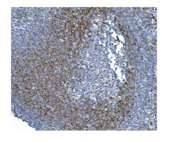
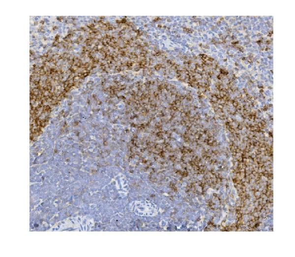
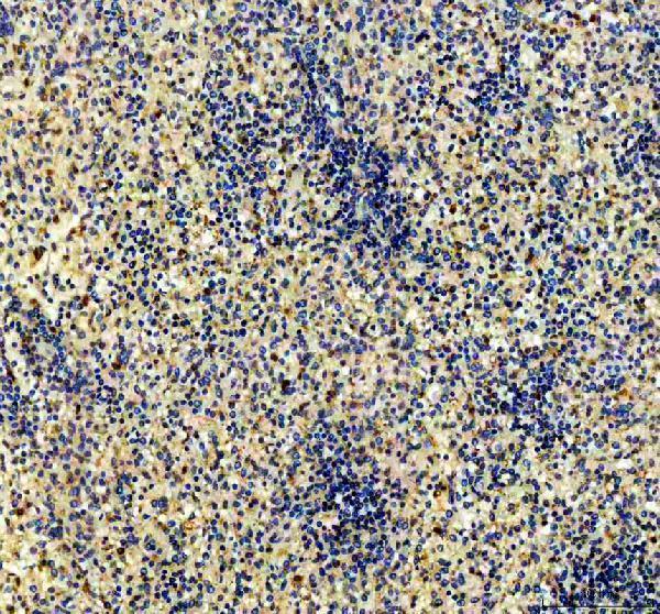
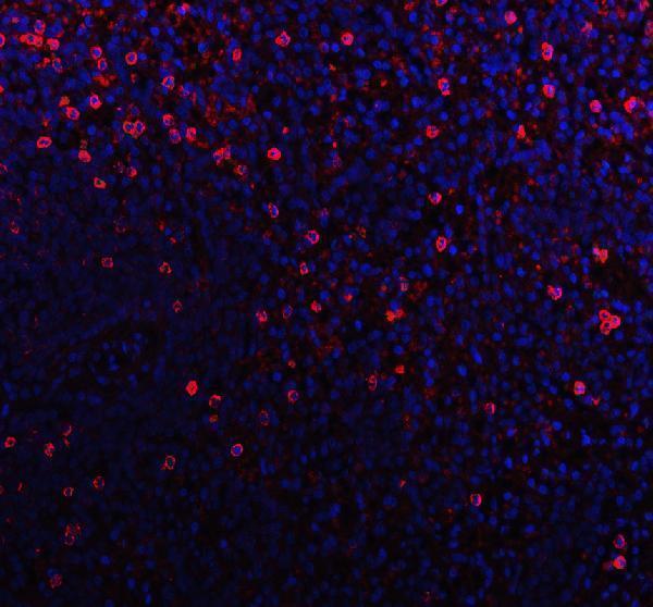
IHC images show CD79B staining in human tonsil, human spleen and rat spleen (M01399-1 and PB9169 IHC captions); an IF image shows human spleen (PB9169 IF caption).
M01399-1 has paraffin-section IHC images from human tonsil and rat spleen (M01399-1 IHC captions). PB9169 has paraffin-section IHC and IF images from human spleen (PB9169 IHC and IF captions).
Which to pick: For paraffin-section IHC, choose mouse monoclonal M01399-1 for human tonsil or rat spleen, or rabbit PB9169 for human spleen (catalog hosts; M01399-1 and PB9169 IHC captions). For IF, PB9169 has a human spleen image; for ICC, rabbit monoclonals M01399 and M01399-3 list that application (PB9169 IF caption; catalog hosts and applications). For cross-species tissue IHC, M01399-1 has human and rat images; the fixative is unreported in its captions (M01399-1 IHC captions).