This website uses cookies to ensure you get the best experience on our website.


- Table of Contents
Plan chromogenic IHC on paraffin sections with catalog antibody PA1363 at 0.5–1 μg/mL (datasheet). Assess membranous staining in squamous epithelia (HPA tissue IHC), using high-staining placental cytotrophoblasts or skin basal-layer cells as positive controls (HPA tissue IHC).

Expected localisation, validated protocols, controls and antibodies — the at-a-glance facts below, then the full design guide.
| Expected localisation | Cell membrane (UniProt) | |
| Staining pattern | Membranous staining in squamous epithelial cells (HPA tissue IHC) | |
| Antigen retrieval | Citrate pH 6 HIER, heat-mediated (datasheet PA1363) | |
| Positive control | Bronchus+4 more · see all | |
| Negative control | Adipose tissue+4 more · see all |
| Fixation | Matched tissue-IHC evidence does not establish the fixation claim. Validate the specimen-specific method before use. (selected-SKU IHC image PA1363) | |
| Caveat | Skin staining may favor basal-layer cells (HPA tissue IHC) | |
| Regulation | Expression varies across tissues (HPA tissue IHC) | |
| Isoform / epitope | 2 isoforms; extracellular vs cytoplasmic epitope matters (UniProt) |
Use the catalog antibody’s IHC-P protocol (datasheet: PA1363) alongside these 4 published CDH3 protocols (PMC10670999; PMC5058855; PMC11951682; PMC5325383).
| Sample | Paraffin-embedded human placenta tissues; fixative not specified (datasheet PA1363) |
| Fixation | Image fixative and duration unreported (datasheet PA1363); verify before use. |
| Sectioning | 4–5 µm sections on charged slides (standard) |
| Deparaffinisation | Xylene, graded ethanol series to water (standard) |
| Antigen retrieval | Heat retrieval: Citrate pH 6, 20 min (datasheet PA1363) |
| Peroxidase block | 3% H2O2, 10 min, room temperature (standard) |
| Blocking | 10% goat serum (datasheet PA1363) |
| Primary antibody | Rabbit anti-CDH3, 0.5-1μg/ml (datasheet PA1363) |
| Primary incubation | Overnight at 4 °C (datasheet PA1363) |
| Detection | Streptavidin-biotin complex (SABC), DAB chromogen (datasheet PA1363) |
| Counterstain | Hematoxylin, blue, dehydrate and mount (standard) |
| Expected result | CDH3-positive staining in respiratory epithelial cells of bronchus (HPA tissue IHC: High). HPA tissue profile: Membranous expression in squamous epithelia. No signal in the no-primary control. |
CDH3 is a cell membrane cadherin with an extracellular region at residues 108–654 and a cytoplasmic region at 678–829 (UniProt P22223 topology). Expect membranous staining in selected epithelial cells, especially squamous epithelia, and in placental cytotrophoblasts (HPA tissue IHC). HPA rates the tissue IHC profile Enhanced, while reporting medium consistency between antibody staining and RNA expression (HPA tissue IHC).
| Distinct staining outlines squamous epithelial cells, especially in oral mucosa or tonsil (HPA tissue IHC). | This fits CDH3 at the cell membrane (UniProt P22223 topology; HPA tissue IHC). HPA reports High staining in those cell populations; judge the outlined cells rather than the section's overall color. |
| Strong nuclear staining, or cytoplasmic staining that dominates over cell borders, appears in an otherwise positive epithelium. | That is discordant with the reported cell membrane pattern (UniProt P22223 topology; HPA tissue IHC). Treat it as a possible artefact and review localization and staining controls before scoring it as CDH3. |
| Adipocytes or bone marrow hematopoietic cells stain, despite their Not detected HPA entries (HPA tissue IHC). | Consider cross-reactivity or endogenous detection activity (standard IHC practice). The HPA entries describe those specific cell populations; they do not establish that every cell in either tissue must be negative. |
| Diffuse chromogen covers cells and spaces without reproducible cell border definition. | Diffuse background cannot establish the membrane localization expected for CDH3 (UniProt P22223 topology; HPA tissue IHC). Check blocking, detection background, washing, and the negative control (standard IHC practice). |
| No signal appears in a correctly identified oral mucosa squamous epithelium or placental cytotrophoblast control. | Both populations have High HPA staining (HPA tissue IHC). Check control integrity and the IHC detection workflow before interpreting an unstained test section as CDH3 negative (standard IHC practice). |
| Cell population and tissue choice | HPA reports High staining in bronchial and nasopharyngeal respiratory epithelium, oral mucosa and tonsil squamous epithelium, skin basal layer cells, and placental cytotrophoblasts (HPA tissue IHC). Breast glandular and cervical squamous cells are Medium (HPA tissue IHC). |
| Reported negative cells | HPA lists adipocytes, bone marrow hematopoietic cells, and duodenal glandular cells as Not detected (HPA tissue IHC). Interpret these as cell specific reference patterns, since an unstained named population cannot validate every cell in the same section. |
| Topology and antigen location | The mature CDH3 chain spans residues 108–829; residues 108–654 are extracellular and 678–829 cytoplasmic (UniProt P22223 topology and processing). Antibody epitope location is unspecified here, so this record cannot predict an epitope specific retrieval response. |
| Processing, glycosylation, and isoforms | UniProt records a signal peptide at 1–24, propeptide at 25–107, glycosylation sites at 200 and 566, and two isoforms (UniProt P22223). These facts alone do not establish shedding, differential IHC staining, or fixation sensitivity. |
| How much confidence to place in a pattern | HPA labels the tissue IHC profile Enhanced but describes antibody staining versus RNA expression as medium consistency (HPA tissue IHC). Its antibody entries also give Enhanced IHC status to HPA001767 and CAB002487 (HPA antibodies). |
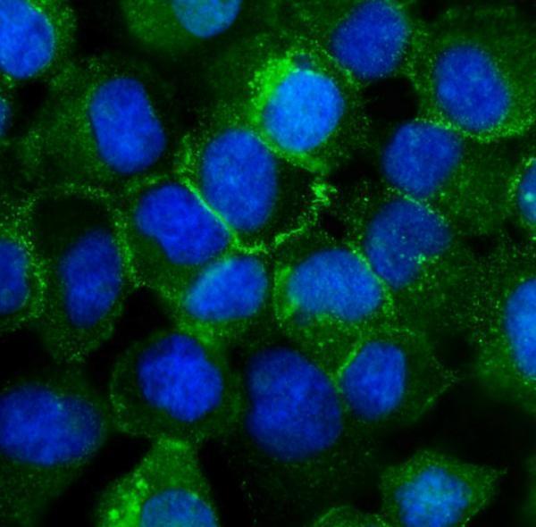
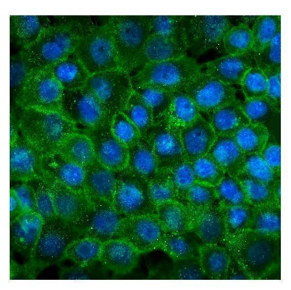
| IF/ICC Q: Where should CDH3 fluorescence appear? | A: At the plasma membrane and cell junctions, both supported HPA locations (HPA subcellular ICC-IF). This localization supports the compartment check; use the separate IF/ICC guide for its workflow. |
| Situation | Likely cause | Next action |
|---|---|---|
| A known High control has no visible membrane signal. | The run may have failed at tissue identification, antigen retrieval, primary antibody application, or detection (standard IHC practice). | Confirm the named positive cells, then check each IHC step and a run control (standard IHC practice). HPA High is an observed pattern, not proof of a fixation specific failure (HPA tissue IHC). |
| Expected borders are faint in breast glandular or cervical squamous cells. | Both populations are reported at Medium rather than High staining (HPA tissue IHC). | Compare with a High control such as oral mucosa squamous epithelium before changing interpretation (HPA tissue IHC). Score visible borders in the named cells, not overall tissue darkness (standard IHC practice). |
| The whole section has diffuse brown haze. | Nonspecific background or endogenous detection activity may obscure cell borders (standard IHC practice). | Review blocking, washes, chromogen exposure, and a negative control (standard IHC practice). Require membrane localization before assigning CDH3 positivity (UniProt P22223 topology; HPA tissue IHC). |
| Nuclei or cytoplasm stain more strongly than junctional borders. | The compartment conflicts with reported membrane localization (UniProt P22223 topology; HPA tissue IHC); the observation alone does not identify the technical cause. | Check a positive control for border staining and examine detection background (standard IHC practice). Do not score nuclear only color as the expected CDH3 pattern. |
| Adipocytes or marrow hematopoietic cells appear positive. | Those named populations are Not detected in HPA tissue IHC; cross-reactivity or endogenous detection activity is possible (HPA tissue IHC; standard IHC practice). | Compare a negative control and a membrane positive epithelial control (standard IHC practice; HPA tissue IHC). Resolve the unexpected signal before calling these cells CDH3 positive. |
| A low level glandular population is called negative after comparison with a High epithelial control. | HPA reports Low staining in thyroid, salivary, stomach, gallbladder, seminal vesicle, and prostate glandular cells (HPA tissue IHC). | Assess the named glandular cells at appropriate magnification and record whether cell border signal is detectable (standard IHC practice). Avoid applying a High control's intensity threshold to an HPA Low population. |




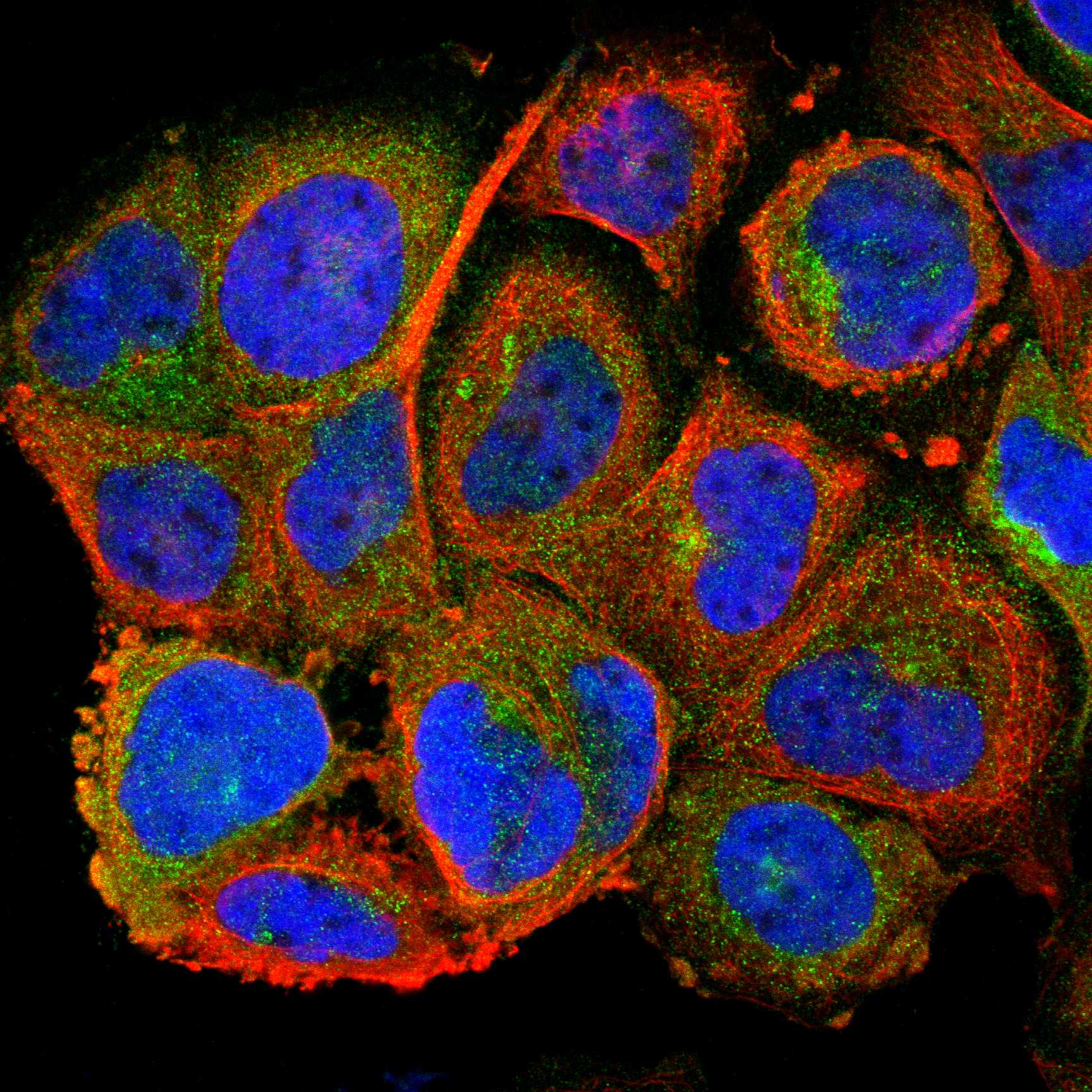

Comprehensive Human Protein Atlas IHC scoring per tissue (reliability: Enhanced — Medium consistency between antibody staining and RNA expression data.). Rows are taken directly from the HPA tissue chart — click any row's HPA link to view the source.
| Tissue | Cell type | Level | Evidence | Source |
|---|---|---|---|---|
| Bronchus | Respiratory epithelial cells | High | Protein (IHC) | HPA → |
| Nasopharynx | Respiratory epithelial cells | High | Protein (IHC) | HPA → |
| Oral mucosa | Squamous epithelial cells | High | Protein (IHC) | HPA → |
| Placenta | Cytotrophoblasts | High | Protein (IHC) | HPA → |
| Skin | Cells in basal layer | High | Protein (IHC) | HPA → |
| Tissue | Cell type | Level | Evidence | Source |
|---|---|---|---|---|
| Adipose tissue | Adipocytes | Not detected | Protein (IHC) | HPA → |
| Adrenal gland | Glandular cells | Not detected | Protein (IHC) | HPA → |
| Bone marrow | Hematopoietic cells | Not detected | Protein (IHC) | HPA → |
| Caudate | Glial cells | Not detected | Protein (IHC) | HPA → |
| Cerebellum | Cells in granular layer | Not detected | Protein (IHC) | HPA → |
Troubleshoot CDH3 chromogenic IHC in paraffin sections by checking retrieval, compartment, cell identity and assay controls before scoring.
The catalog includes CDH3 antibodies with IHC data from human placenta sections and IF data from human A431 cells (PA1363, A03353-1 and M03353 image captions).
PA1363 has IHC data from paraffin-embedded human placenta (PA1363 IHC image caption). A03353-1 and M03353 have IF/ICC data from A431 cells (A03353-1 and M03353 IF image captions).
Which to pick: For tissue IHC, choose PA1363: its own caption documents paraffin-embedded human placenta, citrate retrieval at pH 6 for 20 minutes, and DAB detection; the fixative is unreported (PA1363 IHC image caption). For IF/ICC, choose A03353-1 for a rabbit polyclonal antibody or M03353 for mouse monoclonal clone 3C9 (A03353-1 dilution_raw; M03353 catalog entry). No listed SKU has documented cross-species reactivity beyond human (catalog reactivity fields).