CDIP1 / Cell death-inducing p53-target protein 1 · IHC design guide

Design Immunohistochemistry for CDIP1

Plan CDIP1 paraffin IHC using the general cytoplasmic and nuclear tissue profile and high staining in fallopian tube glandular cells and pachytene spermatocytes (HPA tissue IHC). Use spleen red pulp, reported as not detected (HPA tissue IHC), as a comparison and start the catalog antibody at 2.5 μg/mL (datasheet).

Evidence assembled Oct 2026 · For research use; verify linked source records and product datasheet before use
Immunohistochemistry protocol sheet for CDIP1 (IHC for CDIP1): expected localisation Cytoplasmic/nuclear (HPA tissue IHC); late endosome/lysosome membrane (UniProt), antibody A10988, validated IHC image, and IHC protocol steps
Printable CDIP1 IHC protocol sheet — expected localisation Cytoplasmic/nuclear (HPA tissue IHC); late endosome/lysosome membrane (UniProt), antibody A10988, controls and protocol steps. Open the full CDIP1 IHC guide →

CDIP1 Immunohistochemistry Experimental Design Guide

Expected localisation, validated protocols, controls and antibodies — the at-a-glance facts below, then the full design guide.

Must know before staining
Expected localisation Cytoplasmic/nuclear (HPA tissue IHC); late endosome/lysosome membrane (UniProt)
Staining pattern High in fallopian tube glandular cells; general cytoplasmic/nuclear staining (HPA tissue IHC)
Antigen retrieval Citrate pH 6.0 HIER, 95–98 °C, 20 min (rule: cytoplasmic / membrane antigen)
Positive control ⓘ Fallopian tube+4 more · see all
Negative control ⓘ Spleen
Important caveats
Reasons your staining may differ from the expected pattern.
Fixation Keep fixation consistent across paraffin sections. (standard IHC practice; not target-specific)
Caveat IHC staining and RNA show medium consistency; verify specificity (HPA tissue IHC)
Regulation p53 target linked to TNF-mediated apoptosis (UniProt)
Isoform / epitope 3 isoforms; no annotated processing; epitope impact unknown (UniProt)
Section 1

Recommended CDIP1 IHC & IF Protocols

Compare the catalog antibody’s IHC-P protocol with one published CDIP1 IHC method (PMC7965692).

Recommended immunohistochemistry (IHC-P) protocol parameters
SampleTissue sections; selected-image fixative not specified (standard IHC workflow)
FixationImage fixative and duration unreported (datasheet A10988); verify before use.
Sectioning4–5 µm sections on charged slides (standard)
DeparaffinisationXylene, graded ethanol series to water (standard)
Antigen retrievalHeat-induced epitope retrieval in citrate buffer, pH 6.0, 20 min at 95–98 °C (standard rule: cytoplasmic / membrane antigen)
Peroxidase block3% H2O2, 10 min, room temperature (standard)
Blocking10% normal serum of the secondary host, 30 min, room temperature (standard)
Primary antibodyRabbit anti-CDIP1, 2.5 μg/mL (datasheet A10988)
Primary incubationOvernight at 4 °C (standard)
DetectionHRP-polymer secondary, DAB chromogen 5–10 min (standard)
CounterstainHematoxylin, blue, dehydrate and mount (standard)
Expected resultCDIP1-positive staining in glandular cells of fallopian tube (HPA tissue IHC: High). HPA tissue profile: General cytoplasmic and nuclear expression. No signal in the no-primary control.
💡Decision noteStart with citrate pH 6.0 HIER at 95–98 °C for 20 min (page antigen-retrieval setting); the published method also uses heated citrate (PMC7965692).
Section 2

What Is the Expected CDIP1 Staining Pattern?

In paraffin-section IHC, expect general cytoplasmic and nuclear CDIP1 staining, with high signal reported in fallopian tube glandular cells and testis pachytene spermatocytes (HPA tissue IHC). CDIP1 is annotated at late endosome and lysosome membranes, with no transmembrane segment (UniProt Q9H305 topology). The tissue IHC profile is Approved, with medium consistency between staining and RNA data and external verification pending (HPA tissue IHC).

What am I looking at on my slide?
Clear cytoplasmic and nuclear stain in fallopian tube glandular cells or testis pachytene spermatocytes.This matches the general IHC distribution and High staining reported for those cell types (HPA tissue IHC). Judge the signal in its tissue context; the HPA profile is Approved but awaits external verification (HPA tissue IHC).
Stain appears only along cell borders or outside cells, without the reported cytoplasmic or nuclear pattern.Treat an exclusive border or extracellular pattern as suspect: HPA reports general cytoplasmic and nuclear IHC staining, while UniProt places CDIP1 at late endosome and lysosome membranes (HPA tissue IHC; UniProt Q9H305). Review morphology and controls before assigning it to CDIP1 (standard IHC practice).
Strong stain appears in spleen red-pulp cells, especially if expected positive cells stain weakly.HPA reports CDIP1 as not detected in spleen red-pulp cells (HPA tissue IHC). Unexpected signal warrants checks for antibody cross-reactivity or endogenous detection activity; that tissue observation alone cannot identify the cause (standard IHC practice).
Color coats many structures evenly, obscuring cell boundaries and compartment differences.Interpret this as background until a localized signal can be separated from it (standard IHC practice). Uniform color does not establish the general cytoplasmic and nuclear pattern reported by HPA, or its differences between listed cell types (HPA tissue IHC).
No stain appears in fallopian tube glandular cells or testis pachytene spermatocytes.These are useful positive reference cells because HPA reports High staining in each (HPA tissue IHC). An absent result calls for review of the IHC run and tissue identification; it does not, by itself, establish that the specimen lacks CDIP1 (standard IHC practice).
💡Expected CDIP1 appearanceCall a result positive when identifiable fallopian tube glandular cells or testis pachytene spermatocytes show strong cytoplasmic and/or nuclear staining (HPA tissue IHC); isolated border or extracellular color is suspect against the reported IHC pattern and endolysosomal annotation (HPA tissue IHC; UniProt Q9H305).
How each factor affects the staining
Tissue and cell selectionHPA reports High staining in fallopian tube glandular cells and testis pachytene spermatocytes, Medium staining in several other listed cells, and no detected stain in spleen red-pulp cells (HPA tissue IHC). Compare the specified cells, not whole-organ averages.
Evidence strengthThe HPA tissue IHC profile is Approved, with medium staining-to-RNA consistency and external verification pending; the listed antibody's IHC status is Approved (HPA tissue IHC; HPA antibody record). Treat a matching image as supportive, not definitive.
Compartment and resolutionUniProt annotates late endosome and lysosome membranes, with no transmembrane segment; HPA describes general cytoplasmic and nuclear tissue staining (UniProt Q9H305; HPA tissue IHC). Routine chromogenic IHC cannot resolve that organelle assignment from pattern alone (standard IHC practice).
Isoforms and processingUniProt lists 3 isoforms and a chain spanning residues 1–208, with no annotated signal peptide, propeptide, glycosylation sites or modified residues (UniProt Q9H305). The supplied records do not locate the antibody epitope or establish isoform-specific staining.
IF/ICC Q&A: should I expect the same pattern?HPA reports mainly centrosomal IF/ICC localization, with additional nucleoplasmic signal, whereas its tissue IHC profile says general cytoplasmic and nuclear expression (HPA subcellular; HPA tissue IHC). Interpret each application against its own reported pattern.
Why is my staining missing, weak or wrong?
SituationLikely causeNext action
No signal in a selected positive tissue.Tissue selection, staining-run failure or low detectable signal are possibilities (standard IHC practice). HPA reports High stain only for the specified fallopian tube glandular cells and testis pachytene spermatocytes (HPA tissue IHC).Confirm the cell population on the section; review reagent, retrieval and detection run controls before interpreting the specimen (standard IHC practice).
Widespread diffuse chromogen masks cells.Nonspecific binding or detection background may obscure a localized result (standard IHC practice). The HPA tissue profile describes cytoplasmic and nuclear expression, not uniform slide-wide color (HPA tissue IHC).Review blocking, antibody concentration, washing and a detection control; score only cellular signal distinguishable from background (standard IHC practice).
Spleen red-pulp cells stain strongly.This conflicts with the not detected red-pulp observation (HPA tissue IHC); cross-reactivity or endogenous detection activity is possible (standard IHC practice).Check a detection-only control and inspect whether color follows cells, pigment or deposits before attributing it to CDIP1 (standard IHC practice).
Stain is confined to cell borders or extracellular deposits.That distribution differs from the reported general cytoplasmic and nuclear IHC profile and the late endosome/lysosome annotation (HPA tissue IHC; UniProt Q9H305).Recheck morphology and negative controls, then compare the same run with an HPA-listed positive cell population (standard IHC practice; HPA tissue IHC).
Only nuclear stain is visible.Nuclear signal is within HPA's general IHC profile, but its relative intensity versus cytoplasm is not specified for every cell type (HPA tissue IHC).Record the compartment and cell type; compare a positive reference and detection control before calling the nuclear-only result erroneous (standard IHC practice).
IF/ICC shows a small centrosomal focus that seems unlike the tissue section.HPA reports mainly centrosomal IF/ICC signal, plus nucleoplasmic signal, while its tissue IHC summary is general cytoplasmic and nuclear (HPA subcellular; HPA tissue IHC).Use the HPA IF/ICC localization as the comparison for that application; score paraffin-section chromogenic IHC against the tissue pattern (HPA subcellular; HPA tissue IHC).

Sample controls for CDIP1 IHC & IF

🧪Run fallopian tube first; its glandular cells should stain (HPA: High in fallopian tube glandular cells). Use spleen red-pulp cells as the biological comparison (HPA: Not detected in spleen red-pulp cells); any unstained cells within the fallopian tube section should show only background chromogen, without assigning an unsupported cell identity (standard IHC practice).
Positive control tissue: Fallopian tube (Glandular cells, HPA High)
Negative control tissue: Spleen (HPA Not detected)
ICC-IF cell lines (HPA subcellular resource): HPA ICC-IF images show CDIP1 in HeLa, Hep-G2, U2OS, NIH 3T3, with annotated localisation: Centrosome (supported) (HPA subcellular).
Technical controls: Include no-primary/secondary-only and host-species-matched controls: an isotype-matched antibody for a monoclonal primary or nonimmune IgG for a polyclonal primary; confirm specificity with CDIP1 knockout tissue where available (standard IHC practice). Check endogenous peroxidase and, if using avidin–biotin detection, endogenous biotin on the fallopian tube section (standard IHC practice).
⚠️Feasibility: A CDIP1-specific fixation window and antigen-retrieval dependency are unreported in the supplied evidence; optimize retrieval empirically for paraffin IHC (standard IHC practice). The selected A10988 tissue-IHC caption names FNIP2 in mouse brain and leaves its fixative unreported (selected caption), so it does not establish CDIP1 processing conditions. The evidence does not establish whether frozen sections or IF are easier, or identify a fallopian tube-specific artefact; check tissue background during optimization (standard IHC practice).

HPA tissue IHC evidence for CDIP1

Comprehensive Human Protein Atlas IHC scoring per tissue (reliability: Approved — Medium consistency between antibody staining and RNA expression data. Pending external verification.). Rows are taken directly from the HPA tissue chart — click any row's HPA link to view the source.

Positive expression · recommended positive controls

TissueCell typeLevelEvidenceSource
Fallopian tube Glandular cells High Protein (IHC) HPA →
Testis Pachytene spermatocytes High Protein (IHC) HPA →
Adrenal gland Glandular cells Medium Protein (IHC) HPA →
Appendix Glandular cells Medium Protein (IHC) HPA →
Breast Glandular cells Medium Protein (IHC) HPA →

Undetected expression · recommended negative controls

TissueCell typeLevelEvidenceSource
Spleen Cells in red pulp Not detected Protein (IHC) HPA →
Section 3

Advanced CDIP1 IHC Tips

Troubleshoot CDIP1 staining in paraffin sections by checking retrieval, controls and cellular distribution before comparing signal across samples.

What retrieval should I try first when CDIP1 staining is weak?
Start with citrate pH 6.0 heat-induced antigen retrieval at 95–98 °C for 20 min (page retrieval rule). Keep section thickness, cooling, antibody incubation and chromogenic development consistent while comparing retrieval conditions (standard IHC practice). If staining remains weak, test a different retrieval buffer or heating time on adjacent sections, alongside a no-primary control (standard IHC practice). Evaluate whether signal improves in the expected cells without increasing diffuse background or tissue damage (HPA: tissue IHC profile; standard IHC practice). Do not use the supplied A10988 image to optimize CDIP1 retrieval, because its caption identifies FNIP2 (selected hero caption: A10988).
Could fixation explain inconsistent CDIP1 staining between paraffin blocks?
CDIP1-specific sensitivity to fixative type or fixation duration is unknown from the supplied evidence; the selected image caption concerns FNIP2 and does not state a fixative (selected hero caption: A10988). Record fixative, time to fixation, fixation duration and processing history for each block before comparing staining (standard IHC practice). When practical, stain similarly processed sections together with the same retrieval, detection and development conditions (standard IHC practice; page retrieval rule). Compare morphology and signal in matched cellular compartments, and include a no-primary control to reveal processing-related background (standard IHC practice). Do not infer a CDIP1 fixation effect from tissue expression or protein topology alone (HPA: tissue IHC profile; UniProt Q9H305 topology).
How should I assess cytoplasmic, nuclear and punctate CDIP1 staining?
Score cytoplasmic, nuclear and punctate staining separately because the reported observations differ by method (HPA: general cytoplasmic and nuclear tissue expression; HPA: centrosome and nucleoplasm ICC/IF; UniProt Q9H305: late endosome and lysosome membranes). In paraffin sections, examine whether puncta remain associated with intact cells and recur in comparable cells across fields (standard IHC practice). Use a nuclear counterstain and inspect serial sections to distinguish intracellular signal from overlapping cells or debris (standard IHC practice). Do not require every positive cell to show one universal compartment pattern, since tissue IHC and cultured-cell ICC/IF provide different observations (HPA: tissue IHC and subcellular profiles). Record compartment, cell type and intensity rather than collapsing them into a single positive call (standard IHC practice).
Can an unknown antibody epitope complicate CDIP1 IHC interpretation?
CDIP1 has 3 annotated isoforms and a LITAF domain spanning residues 122–206, but the supplied evidence does not map this antibody’s epitope (UniProt Q9H305). Check the antibody documentation for its immunogen or epitope before claiming that staining represents every isoform (standard IHC practice). If that information is unavailable, report the result as antibody-detected CDIP1 staining without assigning an isoform (standard IHC practice; UniProt Q9H305: alternative splicing). CDIP1 has no annotated transmembrane segment or glycosylation sites; neither fact establishes where an undocumented epitope is exposed in a fixed section (UniProt Q9H305 topology and PTMs). Compare staining across appropriately controlled sections before interpreting a negative result as isoform absence (standard IHC practice).
How can IF help assess a puzzling chromogenic CDIP1 pattern?
Use the separate IF/ICC guide for fluorescence conditions; this page’s curated workflow concerns paraffin-section chromogenic IHC (application scope). For an IF comparison, multiplex CDIP1 with a marker for the cell type being scored, such as an appropriate glial or glandular-cell marker where those cells are under study (HPA: caudate glial cells and fallopian-tube glandular cells; standard IF practice). Choose spectrally separated fluorophores and place the weaker signal in a channel with low tissue autofluorescence (standard IF practice). Set permeabilisation according to the documented epitope: intracellular access may require detergent, but the epitope’s membrane-facing side is not supplied here (standard IF practice; UniProt Q9H305: endosome and lysosome membranes). Interpret agreement by cell type and compartment, allowing for the distinct IHC and ICC/IF observations (HPA: tissue IHC and subcellular profiles).
What should I check when CDIP1 chromogenic staining looks diffuse?
Run no-primary and detection-only controls to identify signal from secondary reagents or endogenous tissue activity (standard chromogenic IHC practice). Check peroxidase blocking, washing and DAB development on the same staining run before increasing confidence in weak diffuse signal (standard chromogenic IHC practice). Inspect section edges, folds, necrotic areas and pigment under the counterstain, since these can mimic cellular staining (standard IHC practice). Compare intact cells with the expected tissue and compartment observations, while recognizing that HPA rates tissue IHC consistency as medium and external verification as pending (HPA: tissue IHC reliability and profile). If background rises across controls, adjust the general workflow before assigning biological meaning to intensity differences (standard IHC practice).
How should I quantify CDIP1 staining across sections? ⚠ ANSWER MARKED FOR VERIFICATION
Define the cell population and compartment before scoring, then apply identical thresholds to all sections in the comparison (standard IHC quantification practice). For cellular chromogenic signal, report the percentage of positive cells and an H-score based on intensity and positive-cell fraction; for discrete positive cells, density per mm² can be useful (standard IHC quantification practice). Normalize counts to evaluable tissue area or the number of eligible cells, excluding folds, necrosis and damaged edges (standard IHC quantification practice). Keep cytoplasmic and nuclear scores separate because both occur in the reported tissue profile (HPA: general cytoplasmic and nuclear expression). Record staining batch and tissue region so differences in sampling or development do not masquerade as expression changes (standard IHC practice).
How can I distinguish credible CDIP1 positivity from artefact?
A credible positive has reproducible cellular staining in an anatomically identified population, supported by clean negative controls and intact morphology (standard IHC practice). Compare candidate cells with reported high staining in fallopian-tube glandular cells and pachytene spermatocytes, while treating spleen red-pulp cells as a reported nondetected population rather than an absolute negative standard (HPA: tissue IHC profile). Examine nuclear and cytoplasmic distribution alongside puncta, since tissue IHC and cellular localization reports are not identical (HPA: tissue IHC and subcellular profiles; UniProt Q9H305: endosome and lysosome membranes). Treat signal restricted to edges, necrosis or endogenous-enzyme control staining as suspect, especially when the relevant cells lack consistent signal (standard chromogenic IHC practice). Interpret apparent positivity cautiously because the HPA tissue IHC rating remains pending external verification (HPA: reliability description).
Boster reagents

Best CDIP1 / Cell death-inducing p53-target protein 1 IHC Antibodies

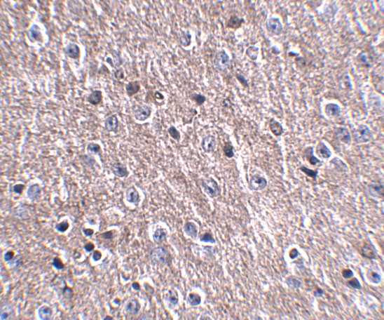
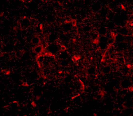
A10988 lists IHC-P and IF for human and mouse reactivity (catalog: applications and reactivity). Its IF image shows CDIP1 in mouse brain; the IHC caption names FNIP2, so it does not establish CDIP1 staining (catalog: image captions).

Real IHC data Immunohistochemistry of FNIP2 in mouse brain tissue with FNIP2 antibody at 2.5 μg/mL.
Anti-CDIP CDIP1 Antibody
Cat # A10988

A10988 is listed for IHC-P and IF with human and mouse reactivity (catalog: applications and reactivity). Its IF caption shows CDIP1 in mouse brain at 20 μg/mL, while its IHC caption names FNIP2 in mouse brain at 2.5 μg/mL (catalog: IF and IHC image captions).

Which to pick: For paraffin-section IHC, A10988 is the listed option at a starting concentration of 2.5 μg/mL, but its IHC image caption names FNIP2, and the fixative is unreported (catalog: IHC-P application, dilution and IHC image caption). For IF, A10988 has a CDIP1 mouse-brain image at 20 μg/mL; ICC validation is unreported (catalog: IF image caption and applications). A10988 lists human and mouse reactivity, although both image captions describe mouse tissue; clonality is unreported (catalog: reactivity, image captions and clone field).

Each figure is that product's own IHC / IF validation image from its datasheet.

References

  1. UniProt Consortium. UniProt entry Q9H305 (CDIP1_HUMAN, Cell death-inducing p53-target protein 1).
  2. Human Protein Atlas. CDIP1 tissue IHC expression (reliability: Approved).
  3. Human Protein Atlas. CDIP1 subcellular location (ICC-IF): Mainly localized to the centrosome. In addition localized to the nucleoplasm..
  4. Human Protein Atlas. CDIP1 antibody validation summary (1 antibodies).
  5. Cardiac telocytes inhibit cardiac microvascular endothelial cell apoptosis through exosomal miRNA-21-5p-targeted cdip1 silencing to improve angiogenesis following myocardial infarction. Theranostics 2021 — PMC7681094.
  6. IL-33 Promotes the Growth of Non-Small Cell Lung Cancer Cells Through Regulating miR-128-3p/CDIP1 Signalling Pathway. Cancer management and research 2021 — PMC7965692.
  7. PubMed PMID:10570909 — UniProt-cited evidence.
  8. PubMed PMID:17599062 — UniProt-cited evidence.
  9. PubMed PMID:11230166 — UniProt-cited evidence.