CELSR3 / Cadherin EGF LAG seven-pass G-type receptor 3 · IHC design guide

Design Immunohistochemistry for CELSR3

Plan CELSR3 chromogenic IHC on paraffin sections using the observed cytoplasmic tissue pattern (HPA tissue IHC) and the catalog antibody’s 1:100–1:300 IHC range (datasheet). Compare positive and negative tissues while accounting for presumed off-target staining (HPA tissue IHC).

Evidence assembled Oct 2026 · For research use; verify linked source records and product datasheet before use
Immunohistochemistry protocol sheet for CELSR3 (IHC for CELSR3): expected localisation Cytoplasmic staining (HPA tissue IHC); cell membrane (UniProt), antibody A07204-2, validated IHC image, and IHC protocol steps
Printable CELSR3 IHC protocol sheet — expected localisation Cytoplasmic staining (HPA tissue IHC); cell membrane (UniProt), antibody A07204-2, controls and protocol steps. Open the full CELSR3 IHC guide →

CELSR3 Immunohistochemistry Experimental Design Guide

Expected localisation, validated protocols, controls and antibodies — the at-a-glance facts below, then the full design guide.

Must know before staining
Expected localisation Cytoplasmic staining (HPA tissue IHC); cell membrane (UniProt)
Staining pattern Cytoplasmic staining in Purkinje, Leydig and adrenal glandular cells (HPA tissue IHC)
Antigen retrieval Citrate pH 6.0 HIER, 95–98 °C, 20 min (rule: cytoplasmic / membrane antigen)
Positive control ⓘ Cerebellum+1 more · see all
Negative control ⓘ Adipose tissue+4 more · see all
Important caveats
Reasons your staining may differ from the expected pattern.
Fixation Keep fixation consistent across sections (standard IHC practice; not target-specific); Matched tissue-IHC evidence does not establish the fixation claim. Validate the specimen-specific method before use. (selected-SKU IHC image A07204-2)
Caveat Presumed off-target binding; external verification pending (HPA tissue IHC)
Regulation No expression regulator annotated (UniProt)
Isoform / epitope 2 isoforms; check whether the epitope is extracellular or cytoplasmic (UniProt)
Section 1

Recommended CELSR3 IHC & IF Protocols

The catalog antibody IHC-P protocol is paired with a published protocol for human embryonic kidney and urinary tract (PMC10907620).

Recommended immunohistochemistry (IHC-P) protocol parameters
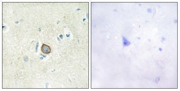
SampleParaffin-embedded human brain tissue; fixative not specified (datasheet A07204-2)
FixationImage fixative and duration unreported (datasheet A07204-2); verify before use.
Sectioning4–5 µm sections on charged slides (standard)
DeparaffinisationXylene, graded ethanol series to water (standard)
Antigen retrievalHeat-induced epitope retrieval in citrate buffer, pH 6.0, 20 min at 95–98 °C (standard rule: cytoplasmic / membrane antigen)
Peroxidase block3% H2O2, 10 min, room temperature (standard)
Blocking10% normal serum of the secondary host, 30 min, room temperature (standard)
Primary antibodyRabbit anti-CELSR3, 1:100-1:300 (datasheet A07204-2)
Primary incubationOvernight at 4 °C (standard)
DetectionHRP-polymer secondary, DAB chromogen 5–10 min (standard)
CounterstainHematoxylin, blue, dehydrate and mount (standard)
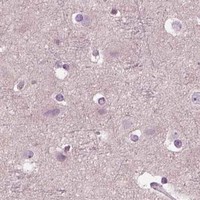
Expected resultCELSR3-positive staining in purkinje cells of cerebellum (HPA tissue IHC: Medium). HPA tissue profile: Cytoplasmic expression in cerebellum, testis and adrenal gland. No signal in the no-primary control.
💡Decision noteStart with citrate pH 6.0 heat retrieval at 95–98 °C for 20 min (page retrieval rule).
Section 2

What Is the Expected CELSR3 Staining Pattern?

CELSR3 is a seven-pass cell-membrane receptor (UniProt Q9NYQ7 topology). In tissue IHC, expect cytoplasmic staining in cerebellar Purkinje cells and testicular Leydig cells, with lower staining in adrenal glandular cells (HPA tissue IHC). HPA rates its tissue staining “Approved” but notes presumed off-target binding was disregarded and external verification is pending (HPA tissue IHC). Interpret the observed cytoplasmic pattern alongside the membrane annotation rather than requiring a crisp membrane outline.

What am I looking at on my slide?
Moderate chromogenic staining in Purkinje cells or Leydig cells (HPA tissue IHC).This matches HPA's reported positive cell types and staining level. Assess which cells stain and where the signal sits; a membrane outline is compatible with UniProt topology, while HPA describes the tissue IHC profile as cytoplasmic (HPA tissue IHC; UniProt Q9NYQ7 topology).
Predominantly nuclear staining in the expected positive cells.Nuclear localisation is unsupported by the supplied localisation records (UniProt Q9NYQ7; HPA tissue IHC; HPA subcellular). Treat a nuclear-only result as a possible artefact and review the controls and detection conditions before scoring it as CELSR3 (general IHC practice).
Strong signal in a cell type HPA lists as not detected, such as adipocytes in adipose tissue (HPA tissue IHC).This conflicts with that cell-specific HPA observation. Consider antibody cross-reactivity or endogenous detection activity, check a reagent-omission control, and avoid extending the HPA negative finding to every cell in the tissue (HPA tissue IHC; general IHC practice).
Diffuse colour across tissue structures, including spaces or cells outside the expected pattern.Poorly localised colour makes cell-specific scoring unreliable (general IHC practice). Compare the section with a primary-antibody omission control to assess background from the detection system; diffuse colour alone does not establish CELSR3 localisation.
No signal in Purkinje cells or Leydig cells on the test section (HPA tissue IHC).HPA reports medium staining in these cells, so a blank result calls for a check of section quality, retrieval, antibody application and chromogen development (HPA tissue IHC; general IHC practice). It does not, by itself, establish that CELSR3 is absent.
💡Expected CELSR3 appearanceCall the IHC result consistent with CELSR3 when Purkinje or Leydig cells show cell-associated, approximately medium staining, including the cytoplasmic pattern reported by HPA; isolated nuclear signal or strong staining in an HPA-listed negative cell type warrants review (HPA tissue IHC; UniProt Q9NYQ7 topology).
How each factor affects the staining
Membrane topology versus tissue IHC appearanceUniProt places CELSR3 at the cell membrane with seven transmembrane segments; HPA describes cytoplasmic tissue staining (UniProt Q9NYQ7 topology; HPA tissue IHC). Use the observed cell-specific pattern when reading chromogenic sections, and do not infer an epitope from either record.
Antibody validationThe listed antibody, HPA062866, has “Approved” IHC status; HPA also reports presumed off-target binding that was disregarded and says external verification is pending (HPA antibodies; HPA tissue IHC). The status supports a reference pattern, not certainty for every stained cell.
Antigen retrievalRetrieval is a standard variable to check when paraffin-section IHC fails (general IHC practice). The supplied records give no CELSR3-specific retrieval condition or fixation-sensitivity result, so establish conditions using controls rather than treating one setting as validated.
Epitope and isoform coverageUniProt lists two CELSR3 isoforms and a long extracellular region plus a cytoplasmic tail (UniProt Q9NYQ7). The supplied antibody record gives no epitope, so its recognition of individual isoforms or regions cannot be inferred (HPA antibodies).
IF/ICC localisation?HPA's subcellular summary says “Membrane,” but supplies no cell-line ICC-IF images or main-location assignment (HPA subcellular). This can guide interpretation of an IF image; it does not establish an IF staining pattern or an IF protocol.
Why is my staining missing, weak or wrong?
SituationLikely causeNext action
Purkinje or Leydig cells are blank.These are medium-staining reference cells in HPA tissue IHC; a blank section may reflect a workflow failure (HPA tissue IHC; general IHC practice).Check tissue integrity and a working positive control, then review retrieval, antibody application and detection steps one at a time (general IHC practice). Do not assign a CELSR3-specific retrieval requirement from this result.
Colour is widespread and obscures cell boundaries.Background from antibody or detection reagents can obscure localised staining (general IHC practice).Inspect a primary-antibody omission control; review blocking, washing and detection development, then score only signal that remains distinguishable in the expected cells (general IHC practice; HPA tissue IHC).
The dominant signal is nuclear.A nuclear pattern lacks support in the supplied CELSR3 localisation records (UniProt Q9NYQ7; HPA tissue IHC; HPA subcellular).Check the omission control and compare with positive-tissue staining. Record nuclear-only signal as unresolved unless independent evidence establishes its identity (general IHC practice).
Adipocytes in adipose tissue stain strongly.HPA lists those cells as not detected; cross-reactivity or endogenous detection activity is possible (HPA tissue IHC; general IHC practice).Compare matched sections with and without primary antibody, and verify whether the expected positive cells stain in the same run (general IHC practice; HPA tissue IHC).
Cytoplasmic staining appears without a crisp membrane rim.HPA reports a cytoplasmic tissue IHC profile even though UniProt places CELSR3 at the membrane (HPA tissue IHC; UniProt Q9NYQ7 topology).Check whether the signal is cell-associated and occurs in HPA's positive cell types. Do not reject an otherwise matching IHC pattern solely because membrane edges are indistinct (HPA tissue IHC).
Weak adrenal staining is difficult to score.HPA reports low staining in adrenal glandular cells, below its medium observations in Purkinje and Leydig cells (HPA tissue IHC).Use the medium-staining tissues to assess whether the run worked, compare adrenal signal with background controls, and report uncertain low-level staining as uncertain (HPA tissue IHC; general IHC practice).

Sample controls for CELSR3 IHC & IF

🧪Run cerebellum first and require staining in Purkinje cells (HPA: Medium in Purkinje cells); run adipose tissue as the negative comparator (HPA: Not detected in adipocytes). On the cerebellum slide, assess cells outside the Purkinje cell population for background staining, without assuming they are CELSR3-negative (HPA: Purkinje cells are the reported positive population).
Positive control tissue: Cerebellum (Purkinje cells, HPA Medium)
Negative control tissue: Adipose tissue (HPA Not detected)
ICC-IF cell lines (HPA subcellular resource): HPA carries no ICC-IF cell line for CELSR3; derive a cell-line control from the positive tissue's cell type (Purkinje cells) and confirm it by RNA or western blot first.
Technical controls: Include a no-primary, secondary-only control and an isotype control matched to the primary antibody’s host species and clonality (standard IHC practice); use a CELSR3 knockout sample or the peptide-block control shown for the selected antibody as a biological specificity check (catalog caption: peptide block). For cerebellum, quench endogenous peroxidase before chromogenic detection and distinguish tissue pigment from specific staining (standard IHC practice).
⚠️Feasibility: No target-specific fixation window or antigen-retrieval dependency is reported in the supplied evidence, so optimize retrieval empirically (supplied target/application evidence). The selected A07204-2 paraffin-section caption does not report a fixative (catalog caption: fixative unreported). The supplied evidence does not establish that frozen sections or IF are easier; in cerebellum, tissue pigment can complicate chromogenic scoring, and autofluorescence can complicate IF (standard IHC/IF practice).

HPA tissue IHC evidence for CELSR3

Comprehensive Human Protein Atlas IHC scoring per tissue (reliability: Approved — Presumed off target binding observed and disregarded. Pending external verification.). Rows are taken directly from the HPA tissue chart — click any row's HPA link to view the source.

Positive expression · recommended positive controls

TissueCell typeLevelEvidenceSource
Cerebellum Purkinje cells Medium Protein (IHC) HPA →
Testis Leydig cells Medium Protein (IHC) HPA →

Undetected expression · recommended negative controls

TissueCell typeLevelEvidenceSource
Adipose tissue Adipocytes Not detected Protein (IHC) HPA →
Appendix Glandular cells Not detected Protein (IHC) HPA →
Bone marrow Hematopoietic cells Not detected Protein (IHC) HPA →
Breast Adipocytes Not detected Protein (IHC) HPA →
Bronchus Respiratory epithelial cells Not detected Protein (IHC) HPA →
Section 3

Advanced CELSR3 IHC Tips

Troubleshoot CELSR3 chromogenic IHC in paraffin sections using the selected antibody image, annotated topology and tissue expression as distinct evidence sources.

What retrieval should I start with for CELSR3 in paraffin sections?
Start with citrate buffer at pH 6.0, heated to 95–98 °C for 20 min (page retrieval rule). Keep section thickness, cooling time and detection conditions constant while comparing retrieved and unretrieved sections, so a staining change can be assigned to retrieval (standard IHC practice). CELSR3 has a large extracellular region and 7 transmembrane segments, but the antibody epitope is unspecified here, so topology alone cannot predict retrieval sensitivity (UniProt Q9NYQ7 topology; supplied antibody evidence). If signal remains weak, test a more alkaline retrieval buffer as a documented fallback, watching for tissue damage and increased background (standard IHC practice). Score the same cell population across conditions, including cerebellar Purkinje cells where medium staining is reported (HPA tissue IHC).
How should I assess fixation effects without a CELSR3-specific fixation study?
Record fixative, fixation duration, tissue size and processing history for every section before comparing CELSR3 staining (standard IHC practice). The selected A07204-2 image shows paraffin-embedded human brain tissue, but its caption does not report the fixative; target-specific fixation sensitivity therefore remains unknown (selected A07204-2 caption). Compare sections processed under defined fixation conditions using the same citrate pH 6.0 retrieval and chromogenic detection settings (page retrieval rule; standard IHC practice). Check morphology and staining together, since poor preservation can obscure cell boundaries needed to assess a membrane receptor (UniProt Q9NYQ7 subcellular location; standard IHC practice). Do not treat HPA cytoplasmic staining or CELSR3 glycosylation sites as evidence for a particular fixation response (HPA tissue IHC; UniProt Q9NYQ7 glycosylation).
How should membrane and cytoplasmic CELSR3 staining be reconciled?
Evaluate membrane-associated staining against the predicted cell-membrane location and 7-segment topology of CELSR3 (UniProt Q9NYQ7 subcellular location and topology). Also record cytoplasmic signal separately: the HPA tissue IHC profile describes cytoplasmic expression in cerebellum, testis and adrenal gland (HPA tissue IHC). Its reported medium staining in cerebellar Purkinje cells and testicular Leydig cells provides cell-specific comparators, not proof that every cytoplasmic deposit is specific (HPA tissue IHC; standard IHC interpretation). Review matched negative controls and adjacent morphology before assigning diffuse cytoplasmic DAB to CELSR3 (standard IHC practice). State the compartment and cell type in results, because disagreement between predicted location and observed pattern needs independent confirmation (UniProt Q9NYQ7 subcellular location; HPA tissue IHC; standard IHC practice).
Can this antibody distinguish CELSR3 isoforms or extracellular from cytoplasmic epitopes?
Do not assign an isoform or membrane side from staining alone: CELSR3 has 2 listed isoforms, while the selected caption does not map this antibody’s epitope (UniProt Q9NYQ7 isoforms; selected A07204-2 caption). Its major extracellular region spans residues 33–2540, whereas the terminal cytoplasmic region spans 2775–3312 (UniProt Q9NYQ7 topology). Obtain the immunogen coordinates and align them with both isoform sequences before claiming isoform coverage or epitope orientation (standard antibody validation practice). Fifteen annotated glycosylation sites occur in CELSR3, but their presence does not establish whether this antibody binds a glycosylated epitope (UniProt Q9NYQ7 glycosylation; supplied antibody evidence). The caption’s peptide-blocked image supports a competition comparison for this SKU, not isoform specificity (selected A07204-2 caption; standard antibody validation practice).
How can I assess CELSR3 by IF alongside a cell-type marker?
For a separate IF experiment, pair CELSR3 with a marker that identifies the expected cell population, such as a validated Purkinje-cell marker when examining cerebellum (HPA tissue IHC; standard IF practice). Choose spectrally separated fluorophores after inspecting unstained tissue, and place the weaker channel away from prominent tissue autofluorescence (standard IF practice). Determine whether the antibody recognizes the extracellular region 33–2540 or a cytoplasmic region before choosing permeabilisation: compare intact-cell access with gentle permeabilisation when appropriate (UniProt Q9NYQ7 topology; standard IF practice). Include single-channel and secondary-only controls to assess bleed-through and background before interpreting overlap (standard IF practice). The HPA subcellular payload lists no ICC/IF image-bearing cell lines, so an IF localisation claim needs validation in the chosen preparation (HPA subcellular).
What should I check when CELSR3 DAB staining is widespread?
Compare the stained section with a no-primary control and inspect endogenous pigment, tissue edges and damaged regions before adjusting antibody concentration (standard IHC practice). Apply a peroxidase block and assess its effectiveness with a detection control, since endogenous enzyme activity can also produce chromogenic signal (standard IHC practice). The selected A07204-2 caption describes a peptide-blocked image; compare the paired images under equivalent imaging conditions, while recognizing that peptide competition alone does not establish tissue specificity (selected A07204-2 caption; standard antibody validation practice). HPA reports presumed off-target binding that was disregarded and external verification pending, so interpret diffuse deposits cautiously (HPA tissue IHC). Reassess blocking, washes and DAB development time together, then retain settings that preserve morphology and cell-specific contrast (standard IHC practice).
How should I quantify CELSR3 staining across paraffin sections? ⚠ ANSWER MARKED FOR VERIFICATION
Define the anatomical region and eligible cell type before scoring, then apply the same segmentation and exclusion rules to every section (standard IHC quantification practice). Report the percentage of positive cells with intensity categories or an H-score, and record membrane-associated and cytoplasmic staining separately (standard IHC quantification practice; UniProt Q9NYQ7 subcellular location; HPA tissue IHC). Normalize positive-cell counts to eligible cells, or express counted positive cells as density per mm² of evaluated tissue (standard IHC quantification practice). Cerebellar Purkinje cells and testicular Leydig cells have medium HPA staining, which can guide region selection without setting a universal positivity threshold (HPA tissue IHC). Keep retrieval, chromogen development and image acquisition consistent across the comparison set (page retrieval rule; standard IHC quantification practice).
What evidence separates convincing CELSR3 staining from artefact?
Prioritize a reproducible cell-specific pattern with preserved morphology, and compare membrane-associated signal with CELSR3’s annotated cell-membrane location (standard IHC interpretation; UniProt Q9NYQ7 subcellular location). HPA reports medium staining in Purkinje and Leydig cells and a cytoplasmic tissue profile, so describe any observed compartment explicitly rather than treating one pattern as definitive (HPA tissue IHC). Treat staining concentrated at section edges, in necrotic areas or in no-primary controls as potential artefact, including endogenous peroxidase signal (standard IHC interpretation). Compare the selected antibody’s peptide-blocked brain image with its paired unblocked image, but seek an independent specificity check for a disputed pattern (selected A07204-2 caption; standard antibody validation practice). HPA’s approved entry notes presumed off-target binding and pending external verification, limiting certainty from that reference alone (HPA tissue IHC).
Boster reagents

Best CELSR3 / Cadherin EGF LAG seven-pass G-type receptor 3 IHC Antibodies

Anti-CELSR3 antibodies have IHC data from paraffin-embedded human brain (A07204-2 image caption) and IF data from A549 cells (A07204-1 image caption); both list human, mouse, and rat reactivity (catalog reactivity).

Real IHC data Immunohistochemistry analysis of paraffin-embedded human brain tissue, using CELSR3 Antibody. The picture on the right is blocked with the synthesized peptide.
Anti-CDHF11 CELSR3 Antibody
Cat # A07204-2
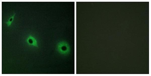
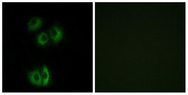
Real IF data Immunofluorescence analysis of A549 cells, using CELSR3 Antibody. The picture on the right is blocked with the synthesized peptide.
Anti-CELSR3/Flamingo Homolog 1 Antibody
Cat # A07204-1

A07204-2 lists IHC and IF (catalog applications), with IHC shown in paraffin-embedded human brain and IF shown in HepG2 cells (A07204-2 image captions). A07204-1 lists IF and ICC (catalog applications), with IF shown in A549 cells (A07204-1 image caption).

Which to pick: For tissue IHC, choose A07204-2 at a starting dilution within 1:100–1:300 (catalog applications and dilution); its IHC image shows paraffin-embedded human brain, but the fixative is unreported (A07204-2 IHC image caption). For IF/ICC, choose the polyclonal A07204-1, which lists both applications and shows IF in A549 cells (A07204-1 dilution_raw, applications, and IF image caption). Both list mouse and rat reactivity (catalog reactivity), but their supplied images show human samples only (A07204-1 and A07204-2 image captions).

Each figure is that product's own IHC / IF validation image from its datasheet.

References

  1. UniProt Consortium. UniProt entry Q9NYQ7 (CELR3_HUMAN, Cadherin EGF LAG seven-pass G-type receptor 3).
  2. Human Protein Atlas. CELSR3 tissue IHC expression (reliability: Approved).
  3. Human Protein Atlas. CELSR3 subcellular location (ICC-IF): Membrane.
  4. Human Protein Atlas. CELSR3 antibody validation summary (1 antibodies).
  5. Increased expression of CELSR3 indicates a poor prognostic factor for Prostate Cancer. Journal of Cancer 2021 — PMC7797646.
  6. Effect of CELSR3 on the Cell Cycle and Apoptosis of Hepatocellular Carcinoma Cells. Journal of Cancer 2020 — PMC7086248.
  7. Bi-allelic variants in CELSR3 are implicated in central nervous system and urinary tract anomalies. NPJ genomic medicine 2024 — PMC10907620.
  8. Celsr3 is required for normal development of GABA circuits in the inner retina. PLoS genetics 2011 — PMC3154962.
  9. PubMed PMID:10716726 — UniProt-cited evidence.
  10. PubMed PMID:16641997 — UniProt-cited evidence.
  11. PubMed PMID:9693030 — UniProt-cited evidence.