This website uses cookies to ensure you get the best experience on our website.


- Table of Contents
Plan CERS5 paraffin IHC using the IHC-validated A08612-1 antibody at a starting concentration of 2.5 μg/mL (datasheet). Compare high staining in testis Sertoli cells with undetected staining in heart cardiomyocytes, and assess the observed cytoplasmic and nucleolar pattern alongside the ER membrane annotation (HPA tissue IHC; UniProt).

Expected localisation, validated protocols, controls and antibodies — the at-a-glance facts below, then the full design guide.
| Expected localisation | ER membrane (UniProt); cytoplasmic/nucleolar tissue staining (HPA tissue IHC) | |
| Staining pattern | Sertoli cells: high; cytoplasmic/nucleolar tissue pattern (HPA tissue IHC) | |
| Antigen retrieval | Citrate pH 6.0 HIER, 95–98 °C, 20 min (rule: cytoplasmic / membrane antigen) | |
| Positive control | Testis+4 more · see all | |
| Negative control | Heart muscle+1 more · see all |
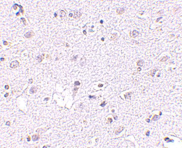
| Fixation | Keep fixation consistent across sections (standard IHC practice; not target-specific); Matched tissue-IHC evidence does not establish the fixation claim. Validate the specimen-specific method before use. (selected-SKU IHC image A08612-1) | |
| Caveat | Nucleolar staining warrants validation against the ER expectation (HPA tissue IHC; UniProt) | |
| Regulation | PAQR4 regulates CERS5 stability (UniProt) | |
| Isoform / epitope | 2 isoforms; check luminal versus cytoplasmic epitope coverage (UniProt) |
Compare the catalog antibody’s IHC-P protocol with published gastric and colorectal tissue protocols (PMC9552061; PMC4858121).
| Sample | Tissue sections; selected-image fixative not specified (standard IHC workflow) |
| Fixation | Image fixative and duration unreported (datasheet A08612-1); verify before use. |
| Sectioning | 4–5 µm sections on charged slides (standard) |
| Deparaffinisation | Xylene, graded ethanol series to water (standard) |
| Antigen retrieval | Heat-induced epitope retrieval in citrate buffer, pH 6.0, 20 min at 95–98 °C (standard rule: cytoplasmic / membrane antigen) |
| Peroxidase block | 3% H2O2, 10 min, room temperature (standard) |
| Blocking | 10% normal serum of the secondary host, 30 min, room temperature (standard) |
| Primary antibody | Rabbit anti-CERS5, 2.5 μg/mL (datasheet A08612-1) |
| Primary incubation | Overnight at 4 °C (standard) |
| Detection | HRP-polymer secondary, DAB chromogen 5–10 min (standard) |
| Counterstain | Hematoxylin, blue, dehydrate and mount (standard) |
| Expected result | CERS5-positive staining in sertoli cells of testis (HPA tissue IHC: High). HPA tissue profile: Cytoplasmic and nucleolar expression in several tissues. No signal in the no-primary control. |
CERS5 is an endoplasmic reticulum membrane protein with 6 transmembrane segments (UniProt Q8N5B7 topology). In tissue IHC, expect cytoplasmic staining, sometimes with nucleolar staining, across several tissues (HPA: tissue IHC profile). Sertoli cells show the strongest listed signal; several glandular and glial cell populations show medium signal (HPA: High in Sertoli cells; Medium in listed glandular and glial cells). HPA rates its tissue IHC profile Approved, with medium consistency between staining and RNA expression (HPA: reliability).
| Strong cytoplasmic signal in testis Sertoli cells, with possible nucleolar staining. | This matches the strongest listed tissue result and the reported tissue IHC compartments (HPA: High in Sertoli cells; cytoplasmic and nucleolar profile). Score the cells and compartments separately: a strong Sertoli cell result does not mean every cell in the section should stain equally (HPA: cell-specific tissue IHC data). |
| Moderate staining in glandular cells or cerebral cortex glial cells. | This is compatible with the listed medium signals in adrenal gland, appendix, breast, colon and duodenum glandular cells, and cerebral cortex glial cells (HPA: Medium in these cell populations). Compare like cell types when scoring; a field-wide intensity score can obscure the reported cell-specific pattern (general IHC practice). |
| Uniform nuclear staining without a discernible cytoplasmic or nucleolar pattern. | Treat this as questionable compartment assignment rather than confirmation of CERS5: tissue IHC reports cytoplasmic and nucleolar expression, while UniProt places CERS5 at the endoplasmic reticulum membrane (HPA: tissue IHC profile; UniProt Q8N5B7 subcellular location). Review morphology and controls before calling it an artefact; nucleolar signal itself is reported by HPA (HPA: tissue IHC profile). |
| Strong staining in cardiomyocytes or smooth muscle cells. | These cell types are listed as not detected in the sampled tissue IHC data (HPA: heart muscle cardiomyocytes; smooth muscle cells). Unexpected strong signal raises concern for cross-reactivity or endogenous detection activity (general IHC practice). Recheck controls and staining distribution; an HPA non-detection is a comparison point, not proof that all specimens must be negative (HPA: tissue IHC data). |
| Hazy, widespread color across cells and tissue spaces. | Diffuse background makes compartment and cell-type calls unreliable (general IHC practice). It does not reproduce the reported cytoplasmic and nucleolar, cell-dependent profile (HPA: tissue IHC profile). Inspect the negative detection control and tissue morphology before interpreting weak color as CERS5 (general IHC practice). |
| Which compartment should guide IHC scoring? | Use the observed cytoplasmic and nucleolar tissue pattern for IHC interpretation (HPA: tissue IHC profile). The endoplasmic reticulum membrane assignment and 6 transmembrane segments provide molecular context, but do not establish how every chromogenic section will resolve the protein (UniProt Q8N5B7 topology; general IHC practice). |
| How much should intensity vary by cell type? | The supplied IHC observations range from High in Sertoli cells to Medium in several glandular or glial populations, Low in listed respiratory, neural and lung populations, and Not detected in cardiomyocytes and smooth muscle cells (HPA: tissue IHC data). Choose a control and scoring threshold with its specific cell population in mind (general IHC practice). |
| What does antibody validation support? | The tissue IHC profile is Approved, with medium consistency between antibody staining and RNA expression (HPA: reliability). HPA026589 is IHC Approved, whereas HPA006780 has an ICC approval listed and no IHC status in the supplied antibody record (HPA: antibody validation). Do not transfer an ICC approval to IHC (HPA: antibody validation). |
| IF/ICC: should its location be read as the IHC pattern? | HPA ICC-IF reports mainly nuclear membrane localization, with additional endoplasmic reticulum and primary cilium localization (HPA: subcellular ICC-IF). Tissue IHC separately reports cytoplasmic and nucleolar staining (HPA: tissue IHC profile). Interpret each assay against its own reported pattern; this section does not establish an IF/ICC staining protocol (HPA: assay-specific observations). |
| Do topology or isoforms predict epitope access? | UniProt lists 2 isoforms, a lumenal N-terminus and a cytoplasmic C-terminus (UniProt Q8N5B7 isoforms; topology). The supplied record gives no antibody epitope, so it cannot predict which isoform is recognized or whether a particular retrieval condition improves staining (UniProt Q8N5B7 record; general IHC practice). |
| Situation | Likely cause | Next action |
|---|---|---|
| No signal in testis Sertoli cells. | A known high-staining comparison population is missing (HPA: High in Sertoli cells); a detection or tissue-processing problem is possible (general IHC practice). | Check that Sertoli cells are present and assess a matched positive control, primary-antibody incubation, retrieval condition and detection reagents (general IHC practice). Do not infer a CERS5-specific fixation sensitivity from this result (HPA: tissue IHC scope). |
| Only faint staining in a chosen control. | The chosen cells may have Low rather than High reported staining; lung macrophages and bronchial respiratory epithelial cells are listed Low (HPA: tissue IHC data). | Compare the same identified cell type across sections, or include the listed High Sertoli cell population to check assay performance (HPA: tissue IHC data; general IHC practice). Avoid treating every faint tissue as a failed run. |
| Color persists without primary antibody. | Signal can arise from the chromogenic detection workflow or endogenous activity (general IHC practice). | Review the negative detection control; apply appropriate endogenous-enzyme blocking and check detection reagents according to the chromogen system (general IHC practice). Reassess CERS5 only after background is controlled. |
| Strong signal appears in cardiomyocytes or smooth muscle cells. | This conflicts with the listed Not detected observations and may reflect nonspecific binding or detection background (HPA: heart muscle and smooth muscle IHC; general IHC practice). | Compare a primary-omission control, inspect the exact stained cell type and optimize blocking or antibody concentration as general IHC adjustments (general IHC practice). Treat the HPA observations as reference data, not absolute exclusions. |
| Nuclear staining dominates the section. | Nucleolar staining is reported, but uniform nuclear filling does not match the stated cytoplasmic and nucleolar tissue profile (HPA: tissue IHC profile). | Use morphology and counterstain to distinguish nucleoli from whole nuclei; compare a positive tissue and negative detection control before assigning specificity (general IHC practice). Keep the ICC-IF nuclear membrane result distinct from tissue IHC (HPA: subcellular ICC-IF). |
| Results differ after changing antigen retrieval. | Retrieval can alter general IHC signal and background, but target-specific retrieval or fixation sensitivity is unreported in the supplied CERS5 evidence (general IHC practice; UniProt Q8N5B7 and HPA scope). | Compare retrieval conditions on matched sections with the same positive and negative controls, then score the expected cells and compartments (general IHC practice; HPA: tissue IHC profile). Avoid claiming that one condition reveals a proven CERS5 epitope. |


Comprehensive Human Protein Atlas IHC scoring per tissue (reliability: Approved — Medium consistency between antibody staining and RNA expression data.). Rows are taken directly from the HPA tissue chart — click any row's HPA link to view the source.
| Tissue | Cell type | Level | Evidence | Source |
|---|---|---|---|---|
| Testis | Sertoli cells | High | Protein (IHC) | HPA → |
| Adrenal gland | Glandular cells | Medium | Protein (IHC) | HPA → |
| Appendix | Glandular cells | Medium | Protein (IHC) | HPA → |
| Breast | Glandular cells | Medium | Protein (IHC) | HPA → |
| Cerebral cortex | Glial cells | Medium | Protein (IHC) | HPA → |
Troubleshoot CERS5 staining in paraffin sections by checking retrieval, compartment pattern, cell identity and controls before comparing signal intensity.
The catalog includes CERS5 antibodies with human brain IHC or IF images and listed human, mouse, and rat reactivity (catalog applications, reactivity, and image captions).
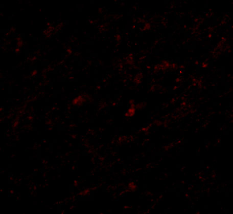
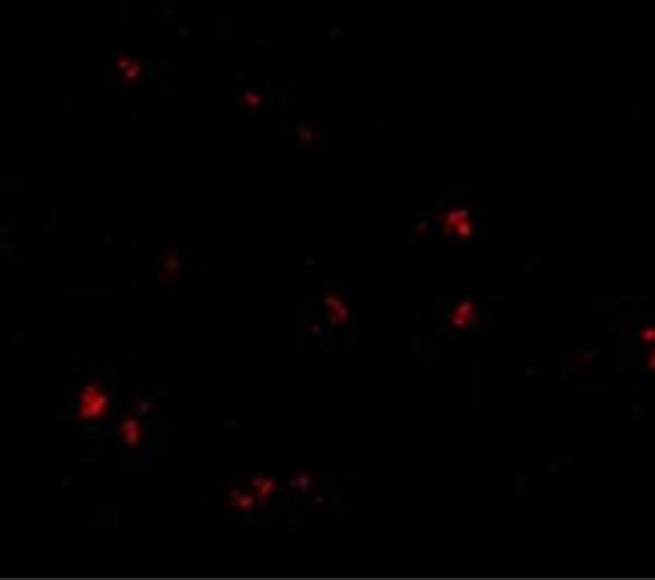
A08612-1 lists IHC-P and IF, with images of human brain tissue IHC at 2.5 μg/mL and human brain cells IF at 20 μg/mL (A08612-1 applications and image captions). A08612 lists IF and shows human brain tissue IF at 20 μg/mL (A08612 applications and image caption).
Which to pick: For paraffin-section tissue IHC, choose A08612-1: it lists IHC-P and shows human brain tissue IHC at 2.5 μg/mL; the fixative is unreported (A08612-1 applications and image caption). For IF, A08612 shows human brain tissue staining; A08612-1 also lists IF and shows human brain cells, but neither catalog entry separately lists ICC (A08612 and A08612-1 applications and image captions). Both list human, mouse, and rat reactivity, while their supplied images show human samples; clonality is unreported for both (catalog reactivity, image captions, and clone fields).