CHAF1B / Chromatin assembly factor 1 subunit B · IHC design guide

Design Immunohistochemistry for CHAF1B

Plan CHAF1B IHC in paraffin sections using nuclear staining in dividing cells as the expected pattern (HPA tissue IHC). Bone marrow hematopoietic cells and tonsil germinal center cells show high staining and can guide control selection (HPA tissue IHC).

Evidence assembled Oct 2026 · For research use; verify linked source records and product datasheet before use
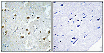
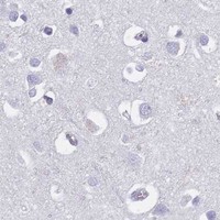
Immunohistochemistry protocol sheet for CHAF1B (IHC for CHAF1B): expected localisation Nuclear staining in dividing cells (HPA tissue IHC), antibody A30479, validated IHC image, and IHC protocol steps
Printable CHAF1B IHC protocol sheet — expected localisation Nuclear staining in dividing cells (HPA tissue IHC), antibody A30479, controls and protocol steps. Open the full CHAF1B IHC guide →

CHAF1B Immunohistochemistry Experimental Design Guide

Expected localisation, validated protocols, controls and antibodies — the at-a-glance facts below, then the full design guide.

Must know before staining
Expected localisation Nuclear staining in dividing cells (HPA tissue IHC)
Staining pattern Dividing cells show nuclear staining (HPA tissue IHC)
Antigen retrieval Tris-EDTA pH 8.0 HIER, heat-mediated (datasheet A30479)
Positive control ⓘ Bone marrow+4 more · see all
Negative control ⓘ Adipose tissue+4 more · see all
Important caveats
Reasons your staining may differ from the expected pattern.
Fixation Keep fixation consistent across samples. (standard IHC practice; not target-specific)
Caveat The fraction of dividing cells can alter apparent positivity (HPA tissue IHC)
Regulation Enriched in dividing cells (HPA tissue IHC)
Isoform / epitope No annotated isoforms; one 1–559 chain (UniProt)
Section 1

Recommended CHAF1B IHC & IF Protocols

The catalog antibody uses heat-mediated Tris-EDTA retrieval at pH 8.0 (datasheet A30479). Two published CHAF1B IHC methods provide paraffin-section protocols (PMC7249347; PMC12492380).

Recommended immunohistochemistry (IHC-P) protocol parameters
SampleParaffin-embedded Human brain tissue; fixative not specified (datasheet A30479)
FixationImage fixative and duration unreported (datasheet A30479); verify before use.
Sectioning4–5 µm sections on charged slides (standard)
DeparaffinisationXylene, graded ethanol series to water (standard)
Antigen retrievalHeat retrieval: Tris-EDTA pH 8.0 (datasheet A30479); 20 min, 95–100 °C (standard)
Peroxidase block3% H2O2, 10 min, room temperature (standard)
Blocking10% normal serum of the secondary host, 30 min, room temperature (standard)
Primary antibodyRabbit anti-CHAF1B, 1:100 - 1:300 (datasheet A30479)
Primary incubationOvernight at 4 °C (standard)
DetectionHRP-polymer secondary, DAB chromogen 5–10 min (standard)
CounterstainHematoxylin, blue, dehydrate and mount (standard)
Expected resultCHAF1B-positive staining in hematopoietic cells of bone marrow (HPA tissue IHC: High). HPA tissue profile: Nuclear expression mainly in dividing cells. No signal in the no-primary control.
💡Decision noteStart with Tris-EDTA pH 8.0 for the catalog antibody (datasheet A30479). Microwave retrieval in 0.01 mol/L sodium citrate for 20 min is a published alternative (PMC12492380).
Section 2

What Is the Expected CHAF1B Staining Pattern?

CHAF1B staining in paraffin sections should be predominantly nuclear, mainly in dividing cells. The HPA tissue profile is rated Enhanced, with medium consistency between antibody staining and RNA expression (HPA tissue IHC). CHAF1B is associated with DNA replication foci and can be cytoplasmic in M phase; it has no transmembrane segment (UniProt Q13112). Interpret staining by cell type and compartment, rather than expecting every cell in a positive tissue to stain.

What am I looking at on my slide?
Distinct nuclear staining in bone marrow hematopoietic cells or tonsil germinal center cells.These are strong positive reference populations: HPA reports High staining in both. Compare nuclei within the same section because expression is mainly seen in dividing cells (HPA tissue IHC).
Predominant membrane staining, or broad cytoplasmic staining across many cells with little nuclear signal.This does not match the principal tissue pattern, which is nuclear, or the supported ICC-IF nucleoplasmic location (HPA tissue IHC; HPA subcellular). UniProt notes cytoplasmic CHAF1B in M phase, so an isolated cytoplasmic cell is not conclusive evidence of an artefact (UniProt Q13112).
Strong staining in adipocytes or another cell population reported as Not detected.Adipocytes in adipose tissue are an HPA negative reference (HPA tissue IHC). Check whether the staining follows cell boundaries or persists in a no-primary control before attributing it to cross-reactivity or endogenous chromogenic activity (general IHC practice).
Brown haze across tissue and blank areas, obscuring nuclear boundaries.A diffuse deposit is difficult to score as CHAF1B because the expected tissue pattern is predominantly nuclear (HPA tissue IHC). Uneven background may reflect detection or wash conditions; inspect a no-primary control and the distribution of deposit (general IHC practice).
No convincing nuclear staining in a bone marrow or tonsil positive reference.HPA reports High staining in bone marrow hematopoietic cells and tonsil germinal center cells (HPA tissue IHC). Review whether the relevant cells are present, then assess staining controls and the IHC-validated antibody workflow before calling the sample CHAF1B-negative (general IHC practice).
💡Expected CHAF1B appearanceCall positive when a subset of expected cells shows clear, mainly nuclear chromogenic staining, strongest in bone marrow hematopoietic or tonsil germinal center cells (HPA tissue IHC); widespread membrane signal or diffuse brown haze without resolved nuclei is suspect (HPA tissue IHC; general IHC practice).
How each factor affects the staining
Cell population and tissue contextHPA reports High staining in bone marrow hematopoietic cells and tonsil germinal center cells; Medium staining occurs in several other listed populations, including duodenal glandular cells (HPA tissue IHC). Score the named cell population, not the entire tissue section (general IHC practice).
Cell cycle and compartmentThe predominantly nuclear tissue pattern is consistent with CHAF1B activity in replication-associated chromatin assembly; UniProt also notes DNA replication foci and cytoplasmic localization in M phase (HPA tissue IHC; UniProt Q13112). A single cytoplasmic cell needs morphologic context before classification.
Antibody evidenceThe tissue profile has Enhanced reliability but only medium consistency with RNA data (HPA tissue IHC). Of the listed antibodies, CAB033604 has Enhanced IHC status and HPA021679 has Approved IHC status (HPA antibodies); their status does not establish identical intensity in every specimen.
Topology and processingCHAF1B has no transmembrane segment, signal peptide or propeptide, and its annotated chain spans residues 1–559 (UniProt Q13112). This supports treating a predominantly membrane-bound pattern as unexpected; it does not identify the epitope recognized by the catalog antibody.
IF/ICC Q: Where should CHAF1B appear?A: The supported main location is the nucleoplasm in the HPA ICC-IF record (HPA subcellular). This is a localization cross-check for the IHC interpretation; the supplied IF evidence does not define an IF/ICC protocol.
Why is my staining missing, weak or wrong?
SituationLikely causeNext action
Positive-reference nuclei are blank.The expected cell population may be absent from the examined area, or the staining run may have failed (HPA tissue IHC; general IHC practice).Find marrow hematopoietic or tonsil germinal center cells on the section, then check control staining and the catalog antibody’s IHC-P instructions (HPA tissue IHC; general IHC practice).
Only weak, sparse nuclei stain in a tissue expected to be positive.CHAF1B is mainly expressed in dividing cells, so a positive tissue need not stain uniformly (HPA tissue IHC). Run conditions may also affect visible contrast (general IHC practice).Compare the relevant cell population with a positive reference on the same run; review the catalog antibody’s IHC-P conditions before interpreting weak staining (HPA tissue IHC; general IHC practice).
Cytoplasmic signal dominates the section.Broad cytoplasmic staining conflicts with the predominant nuclear tissue pattern, although cytoplasmic CHAF1B is noted in M phase (HPA tissue IHC; UniProt Q13112).Check whether signal is restricted to plausible mitotic cells. If it is widespread, compare with a no-primary control and reassess antibody-specific staining conditions (UniProt Q13112; general IHC practice).
Brown deposit appears in a reported negative cell population.The deposit may reflect nonspecific antibody binding or endogenous detection activity; HPA reports adipose-tissue adipocytes as Not detected (HPA tissue IHC; general IHC practice).Examine a no-primary control, review blocking and endogenous enzyme control appropriate to the chromogen, and score nuclei separately from diffuse deposit (general IHC practice).
Background obscures nuclear scoring.Diffuse chromogenic signal can reduce separation between positive nuclei and surrounding tissue (general IHC practice).Use the run’s negative control to locate background, then review washes, blocking, antibody concentration and chromogen development against the validated IHC-P workflow (general IHC practice).
Two antibodies give different staining distributions.The listed antibodies have different HPA IHC validation statuses: CAB033604 is Enhanced and HPA021679 is Approved (HPA antibodies). This record does not establish why a particular specimen differs.Compare nuclear staining in the same named positive and negative cell populations, document each antibody’s controls, and avoid resolving disagreement from intensity alone (HPA tissue IHC; general IHC practice).

Sample controls for CHAF1B IHC & IF

🧪Run bone marrow first; hematopoietic cells should show CHAF1B staining (HPA: High in hematopoietic cells). Use adipose tissue adipocytes as the negative tissue (HPA: Not detected in adipocytes); on the positive slide, assess nonhematopoietic cells for little or no nuclear signal as an internal comparator, without treating them as a validated negative population.
Positive control tissue: Bone marrow (Hematopoietic cells, HPA High)
Negative control tissue: Adipose tissue (HPA Not detected)
ICC-IF cell lines (HPA subcellular resource): HPA ICC-IF images show CHAF1B in A-431, U-251MG, U2OS, U2OS, siRNA 1 (10x), U2OS, siRNA 1 (40x), U2OS, scrambled (10x), U2OS, scrambled (40x), with annotated localisation: Nucleoplasm (supported) (HPA subcellular).
Technical controls: Include a no-primary (secondary-only) control and an isotype control matched to the primary antibody’s host species, immunoglobulin class, and concentration (standard IHC practice). Use a CHAF1B knockout specimen where available, or an immunogen-peptide block as shown for the catalog antibody (selected-SKU IHC caption: peptide pre-absorption); block endogenous peroxidase in bone marrow before chromogenic detection (standard IHC practice).
⚠️Feasibility: A target-specific fixation window or fixation effect is unreported, and the selected-SKU paraffin-section caption does not state the fixative (selected-SKU IHC caption: fixative unreported). The reported paraffin-section example used high-pressure, high-temperature Tris-EDTA retrieval at pH 8.0, with antibody at 1:100 overnight at 4°C; this is a starting condition, not proof that retrieval is required (selected-SKU IHC caption). HPA supports nucleoplasmic IF localisation, but the supplied evidence does not establish that frozen sections or IF are easier than paraffin IHC (HPA: Nucleoplasm supported); endogenous peroxidase in bone marrow can confound chromogenic signal (standard IHC practice).

HPA tissue IHC evidence for CHAF1B

Comprehensive Human Protein Atlas IHC scoring per tissue (reliability: Enhanced — Medium consistency between antibody staining and RNA expression data.). Rows are taken directly from the HPA tissue chart — click any row's HPA link to view the source.

Positive expression · recommended positive controls

TissueCell typeLevelEvidenceSource
Bone marrow Hematopoietic cells High Protein (IHC) HPA →
Tonsil Germinal center cells High Protein (IHC) HPA →
Appendix Lymphoid tissue Medium Protein (IHC) HPA →
Duodenum Glandular cells Medium Protein (IHC) HPA →
Esophagus Squamous epithelial cells Medium Protein (IHC) HPA →

Undetected expression · recommended negative controls

TissueCell typeLevelEvidenceSource
Adipose tissue Adipocytes Not detected Protein (IHC) HPA →
Adrenal gland Glandular cells Not detected Protein (IHC) HPA →
Breast Adipocytes Not detected Protein (IHC) HPA →
Caudate Glial cells Not detected Protein (IHC) HPA →
Cerebellum Cells in granular layer Not detected Protein (IHC) HPA →
Section 3

Advanced CHAF1B IHC Tips

Use the catalog antibody’s paraffin section evidence to plan CHAF1B chromogenic IHC, then assess nuclear staining against tissue controls and cell cycle context.

Which retrieval conditions should I try first for weak CHAF1B staining in paraffin sections?
Start with heat-mediated Tris-EDTA at pH 8.0, the retrieval specified for this IHC page (datasheet A30479). The catalog antibody’s paraffin-embedded human brain image used high-pressure, high-temperature retrieval in that buffer, followed by antibody at 1:100 overnight at 4°C (caption A30479). If staining is weak, compare heating duration on adjacent sections while keeping antibody concentration and detection constant (standard IHC practice). Include a known positive compartment, such as tonsil germinal center cells, to distinguish retrieval failure from low expression (HPA: High in tonsil germinal center cells). Watch for tissue detachment or widespread staining as heating increases (standard IHC practice).
How should I assess whether fixation is masking CHAF1B in my IHC sections?
Target-specific CHAF1B sensitivity to fixative type or duration is unknown from the supplied evidence (caption A30479: fixative unreported). The catalog image establishes staining in paraffin-embedded human brain after Tris-EDTA retrieval at pH 8.0; it does not identify the fixative (caption A30479). For a new specimen series, compare matched material processed under documented fixation conditions while holding section thickness, retrieval, and detection constant (standard IHC practice). Include a positive control with expected nuclear staining, such as bone marrow hematopoietic cells (HPA: High in bone marrow hematopoietic cells). Record nuclear intensity and tissue preservation separately so damaged morphology is not mistaken for antigen loss (standard IHC practice).
Should CHAF1B staining be nuclear, and when might cytoplasmic signal be credible?
Prioritize nuclear staining in dividing cells when reading chromogenic CHAF1B IHC (HPA: nuclear expression mainly in dividing cells). Supported subcellular imaging places CHAF1B in the nucleoplasm, while UniProt also describes DNA replication foci (HPA: supported nucleoplasm; UniProt Q13112: replication foci). Cytoplasmic localization is reported in M phase, so evaluate a cytoplasmic cell against its morphology and nearby nuclear staining before dismissing it (UniProt Q13112: cytoplasmic in M phase). A diffuse cytoplasmic pattern across unrelated cell types merits a no-primary control and review of detection background (standard IHC practice). Score nuclear and cytoplasmic compartments separately rather than combining their chromogen intensity (standard IHC practice).
Can an unknown antibody epitope explain variable CHAF1B staining after retrieval?
Treat epitope accessibility as an open question: the supplied record annotates 0 isoforms and one chain spanning residues 1–559, but gives no antibody epitope (UniProt Q13112: isoforms and processing). CHAF1B has no annotated transmembrane segment or glycosylation sites, so a membrane or glycan-specific explanation lacks support here (UniProt Q13112: topology and glycosylation). Several modified residues are annotated, including phosphothreonine 394 and phosphoserine 409; their effect on this antibody is unknown (UniProt Q13112: modified residues). If staining changes with retrieval, compare adjacent sections under controlled heating conditions (standard IHC practice). Use peptide pre-absorption as an available specificity comparison, as shown for the catalog image (caption A30479).
How can IF help check an unexpected CHAF1B IHC pattern?
Use IF as a separate localization check when chromogenic IHC shows unexpected cytoplasmic or widespread staining (standard IHC/IF practice). HPA IF images include A-431, U-251MG, and U2OS cells, and support nucleoplasmic CHAF1B localization (HPA: subcellular imaging). Multiplex with a proliferation marker such as Ki-67 and a nuclear counterstain to relate CHAF1B signal to dividing cells (HPA: nuclear expression mainly in dividing cells; standard IF practice). Choose a far-red fluorophore, such as a 647 nm channel, when tissue autofluorescence obscures shorter wavelengths (standard IF practice). Because the expected epitope is intracellular and the antibody epitope is unspecified, test permeabilization for nucleoplasmic access and check morphology (HPA: supported nucleoplasm; standard IF practice).
What should I check when CHAF1B chromogen appears throughout the section?
First compare a no-primary section with the stained section to identify signal from the detection system or tissue (standard IHC practice). Include a peroxidase block before DAB development, then inspect blood-rich areas for residual endogenous enzyme activity (standard chromogenic IHC practice). The catalog brain image includes a peptide pre-absorbed antibody negative control, which can help assess antibody-dependent staining under its reported conditions (caption A30479). Compare background at the documented 1:100 overnight antibody condition before increasing antibody concentration or detection time (caption A30479; standard IHC practice). Judge suspect staining against the expected nuclear pattern, while allowing for M-phase cytoplasmic localization (HPA: nuclear expression; UniProt Q13112: cytoplasmic in M phase).
How should I quantify CHAF1B IHC when cell populations vary across sections? ⚠ ANSWER MARKED FOR VERIFICATION
Define the cell population and count viable cells within the same anatomical compartment before comparing slides (standard IHC practice). For nuclear CHAF1B, report the percentage of positive nuclei and an H-score from 0–300 based on intensity-weighted nuclear percentages (HPA: nuclear expression mainly in dividing cells; standard IHC scoring). If abundance matters, report positive nuclei per mm² alongside the total viable nuclei counted per area (standard IHC practice). Normalize comparisons to the eligible cell population and sampled area, since a changing proportion of dividing cells can shift the section-wide result (HPA: nuclear expression mainly in dividing cells; standard IHC practice). Keep cytoplasmic staining in a separate field because M-phase localization can differ (UniProt Q13112).
How can I distinguish true CHAF1B staining from artefact in IHC?
A credible result favors nuclei in proliferating populations, including tonsil germinal center cells and bone marrow hematopoietic cells (HPA: nuclear expression mainly in dividing cells; High in both populations). Interpret broad staining in unrelated cell types cautiously, particularly when a no-primary section shows similar color (standard IHC practice). Exclude torn edges and necrotic areas from scoring, and assess brown deposits in blood-rich regions after a peroxidase block (standard chromogenic IHC practice). Cytoplasmic staining in a mitotic cell remains plausible, whereas a uniform cytoplasmic pattern requires further control work (UniProt Q13112: cytoplasmic in M phase; standard IHC practice). The catalog peptide pre-absorption control offers one antibody-dependent comparison for its paraffin brain image (caption A30479).
Boster reagents

Best CHAF1B / Chromatin assembly factor 1 subunit B IHC Antibodies

The catalog lists CHAF1B antibodies for IHC and IF in human samples, with mouse and rat reactivity for A30479 (catalog: applications and reactivity); only A30479 has a supplied IHC image (catalog: image captions).

Real IHC data Immunohistochemical analysis of paraffin-embedded Human brain. Antibody was diluted at 1:100(4° overnight). High-pressure and temperature Tris-EDTA,pH8.0 was used for antigen retrieval. Negetive contrl (right) obtaned from antibody was pre-absorbed by immunogen peptide.
Anti-CAF1B CHAF1B Antibody
Cat # A30479

A30479 is listed for IHC and IF in human, mouse and rat, with an IHC image of a paraffin-embedded human brain section (catalog: A30479 applications, reactivity and image caption). M03959-1 is listed for human IHC and IF, but has no supplied IHC or IF image (catalog: M03959-1 applications, reactivity and image captions).

Which to pick: For tissue IHC, choose A30479 because its own image documents staining in a paraffin-embedded human brain section; the caption does not report the fixative (A30479 image caption). For human IF/ICC, M03959-1 is a rabbit monoclonal listed for both applications, though no IF image is supplied (catalog: M03959-1 host, clone, applications and image captions). For mouse or rat work, choose A30479 based on its listed reactivity and IHC/IF applications (catalog: A30479 reactivity and applications).

Each figure is that product's own IHC / IF validation image from its datasheet.

References

  1. UniProt Consortium. UniProt entry Q13112 (CAF1B_HUMAN, Chromatin assembly factor 1 subunit B).
  2. Human Protein Atlas. CHAF1B tissue IHC expression (reliability: Enhanced).
  3. Human Protein Atlas. CHAF1B subcellular location (ICC-IF): Localized to the nucleoplasm..
  4. Human Protein Atlas. CHAF1B antibody validation summary (2 antibodies).
  5. USP4-Dependent CHAF1B Stabilization Regulates Distinct SETDB1 Ubiquitin States Linked to AKT T308 Signaling and Lipogenic Remodeling in HCC. Advanced science (Weinheim, Baden-Wurttemberg, Germany) 2026 — PMC13624420.
  6. Ubiquitin ligase CHAF1B induces cisplatin resistance in lung adenocarcinoma by promoting NCOR2 degradation. Cancer cell international 2020 — PMC7249347.
  7. CHAF1B: the hidden culprit behind sorafenib resistance in hepatocellular carcinoma. International journal of medical sciences 2025 — PMC12492380.
  8. The histone chaperone CAF-1 safeguards somatic cell identity. Nature 2015 — PMC4866648.
  9. PubMed PMID:7600578 — UniProt-cited evidence.
  10. PubMed PMID:10830953 — UniProt-cited evidence.
  11. PubMed PMID:15489334 — UniProt-cited evidence.