This website uses cookies to ensure you get the best experience on our website.


- Table of Contents
CHD2 shows variable nuclear staining across most tissue types (HPA tissue IHC). This IHC-P guide uses the catalog antibody and tissue evidence to help plan controls and interpret staining.

Expected localisation, validated protocols, controls and antibodies — the at-a-glance facts below, then the full design guide.
| Expected localisation | Nuclear staining in tissue sections (HPA tissue IHC) | |
| Staining pattern | Variable nuclear staining across most tissue types (HPA tissue IHC) | |
| Antigen retrieval | Citrate pH 6 HIER, heat-mediated (datasheet A04079) | |
| Positive control | Cerebellum+4 more · see all | |
| Negative control | Adipose tissue+1 more · see all |
| Fixation | Keep fixation conditions consistent across sections. (standard IHC practice; not target-specific) | |
| Caveat | Skeletal myocytes: not detected (HPA tissue IHC) | |
| Regulation | Staining intensity varies by tissue (HPA tissue IHC) | |
| Isoform / epitope | 3 isoforms; check epitope coverage (UniProt) |
The catalog antibody’s IHC-P protocol (datasheet: A04079) is paired with a published CHD2 staining protocol for mouse testis sections (PMC9700024).
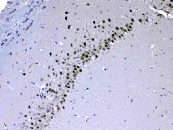
| Sample | Paraffin-embedded rat brain tissue; fixative not specified (datasheet A04079) |
| Fixation | Image fixative and duration unreported (datasheet A04079); verify before use. |
| Sectioning | 4–5 µm sections on charged slides (standard) |
| Deparaffinisation | Xylene, graded ethanol series to water (standard) |
| Antigen retrieval | Heat retrieval: Citrate pH 6, 20 min (datasheet A04079) |
| Peroxidase block | 3% H2O2, 10 min, room temperature (standard) |
| Blocking | 10% goat serum (datasheet A04079) |
| Primary antibody | Rabbit anti-CHD2, 0.5-1μg/ml (datasheet A04079) |
| Primary incubation | Overnight at 4 °C (datasheet A04079) |
| Detection | Streptavidin-biotin complex (SABC), DAB chromogen (datasheet A04079) |
| Counterstain | Hematoxylin, blue, dehydrate and mount (standard) |
| Expected result | CHD2-positive staining in cells in granular layer of cerebellum (HPA tissue IHC: High). HPA tissue profile: Nuclear expression at variable levels in most tissue types. No signal in the no-primary control. |
In paraffin-section IHC, expect CHD2 predominantly in nuclei, at variable levels across most tissues (HPA tissue IHC: Enhanced; medium consistency with RNA). Strong examples include cerebellar granular-layer cells, cortical and hippocampal neurons, and endometrial glandular cells (HPA tissue IHC: High). A nuclear pattern agrees with its annotated location and lack of a transmembrane segment (UniProt O14647: nucleus; topology).
| Distinct nuclear staining in cerebellar granular-layer cells or cortical neurons, with less staining in adjacent cells. | This fits the expected compartment and two reported High cell populations (UniProt O14647: nucleus; HPA tissue IHC: High). Compare nuclei within the same section: HPA describes variable expression, so uniform intensity across every cell is unnecessary (HPA tissue IHC: variable nuclear expression). |
| Strong, widespread cytoplasmic staining obscures nuclei in a paraffin section. | Treat this as a questionable IHC pattern because HPA reports nuclear tissue staining and UniProt places CHD2 in the nucleus (HPA tissue IHC: nuclear expression; UniProt O14647: nucleus). Check background and antibody controls before calling the cytoplasmic signal CHD2. ICC-IF has a separate localisation record (HPA subcellular ICC-IF: additional locations). |
| Adipocytes or skeletal-muscle myocytes stain strongly while nearby expected nuclei are weak. | Those cell types were Not detected in HPA tissue IHC; strong signal there raises concern for cross-reactivity or endogenous chromogenic activity (HPA tissue IHC: adipocytes and myocytes Not detected; general IHC practice). Judge the named cell types, since these records do not declare every cell in either tissue negative. |
| Colour spreads across stroma, extracellular spaces, or most cells without clear nuclear boundaries. | A diffuse deposit cannot establish CHD2-positive cells; assess nonspecific antibody binding, endogenous detection activity, and wash adequacy with suitable controls (general IHC practice). Compare the distribution with HPA's variable nuclear pattern rather than assigning a score to the entire field (HPA tissue IHC: nuclear expression). |
| No nuclear signal appears in an HPA High population, such as endometrial glandular cells. | A negative result there calls for a technical check of staining and tissue integrity; it does not by itself prove CHD2 absence (HPA tissue IHC: endometrial glandular cells High; general IHC practice). HPA's Enhanced tissue assessment still notes medium consistency with RNA, so avoid treating one reference image as an absolute threshold (HPA tissue IHC: reliability). |
| Tissue and cell selection | HPA reports variable nuclear expression across most tissues, with High signal in several neuronal, glandular, and Sertoli populations (HPA tissue IHC). Select a named cell population for scoring; a mixed section can contain cells with different staining levels. |
| Reference antibody validation | The HPA tissue profile is Enhanced, while its antibody entries differ: HPA060744 is IHC Enhanced and HPA060960 is IHC Supported (HPA antibodies; HPA tissue IHC). These labels support interpretation of the reference pattern; they do not validate every catalog antibody. |
| IF/ICC localisation Q&A | What should IF/ICC show? Mainly nucleoplasmic signal, with reported additional nucleolar, Golgi-apparatus, and vesicle localisation (HPA subcellular ICC-IF: approved locations). Those ICC-IF observations do not establish an equally prominent extranuclear pattern in paraffin-section IHC. |
| Isoforms and epitope coverage | UniProt lists 3 CHD2 isoforms (UniProt O14647: isoforms). If a tested antibody's epitope differs among them, staining could reflect its coverage; no epitope or isoform-specific IHC performance is supplied, so do not assign an observed pattern to one isoform. |
| Retrieval and detection context | Antigen retrieval and chromogenic detection are general paraffin-IHC workflow variables (general IHC practice). No supplied CHD2 source establishes fixation sensitivity or an optimal retrieval condition; use the IHC-validated antibody's stated conditions, then evaluate controls. |
| Situation | Likely cause | Next action |
|---|---|---|
| No stain in cerebellar granular-layer cells or another HPA High population. | The run may have insufficient detection or a tissue-quality problem; the image alone cannot identify which (HPA tissue IHC: High; general IHC practice). | Check the positive-control section, reagent sequence, and whether nuclei remain interpretable; review the IHC-validated antibody's stated conditions before adjusting retrieval or dilution (general IHC practice). |
| Nuclei are visible, but colour is mainly cytoplasmic throughout the section. | The distribution conflicts with the reported tissue-IHC pattern; nonspecific binding or detection background is possible (HPA tissue IHC: nuclear expression; general IHC practice). | Compare a matched negative control and a reported High cell population, then reassess blocking, washing, and detection conditions (HPA tissue IHC: High populations; general IHC practice). |
| Adipocytes or skeletal-muscle myocytes look strongly positive. | HPA reports CHD2 as Not detected in those named cell types; cross-reactivity or endogenous chromogenic activity may account for colour (HPA tissue IHC; general IHC practice). | Check controls for antibody-independent colour and compare nuclear staining in a reported High population; score each cell type separately (general IHC practice; HPA tissue IHC: High populations). |
| The whole section has diffuse colour and cell boundaries are hard to resolve. | Excess background can obscure a variable nuclear pattern; its source cannot be determined from the test section alone (HPA tissue IHC: variable nuclear expression; general IHC practice). | Inspect a matched negative control, confirm wash and blocking steps, and reassess chromogen development using the IHC-validated antibody's conditions (general IHC practice). |
| Signal differs sharply between cell types within one otherwise interpretable section. | Cell-dependent variation can be genuine: HPA reports High, Medium, Low, and Not detected populations (HPA tissue IHC). | Identify and score the named cell populations separately; compare their nuclear pattern with HPA tissue observations before changing the staining workflow (HPA tissue IHC: variable nuclear expression). |
| An IF/ICC image shows nucleolar or punctate extranuclear signal alongside nuclear staining. | HPA reports additional nucleolar, Golgi-apparatus, and vesicle locations in ICC-IF (HPA subcellular ICC-IF: approved locations). | Interpret that image using the separate IF/ICC guide and its controls; for paraffin IHC, retain the reported variable nuclear tissue pattern as the reference (HPA subcellular ICC-IF; HPA tissue IHC). |


Comprehensive Human Protein Atlas IHC scoring per tissue (reliability: Enhanced — Medium consistency between antibody staining and RNA expression data.). Rows are taken directly from the HPA tissue chart — click any row's HPA link to view the source.
| Tissue | Cell type | Level | Evidence | Source |
|---|---|---|---|---|
| Cerebellum | Cells in granular layer | High | Protein (IHC) | HPA → |
| Cerebral cortex | Neuronal cells | High | Protein (IHC) | HPA → |
| Endometrium | Glandular cells | High | Protein (IHC) | HPA → |
| Epididymis | Glandular cells | High | Protein (IHC) | HPA → |
| Hippocampus | Neuronal cells | High | Protein (IHC) | HPA → |
Troubleshoot CHD2 staining in paraffin sections by checking nuclear localisation, retrieval, cell context and controls before comparing chromogenic signal across samples.
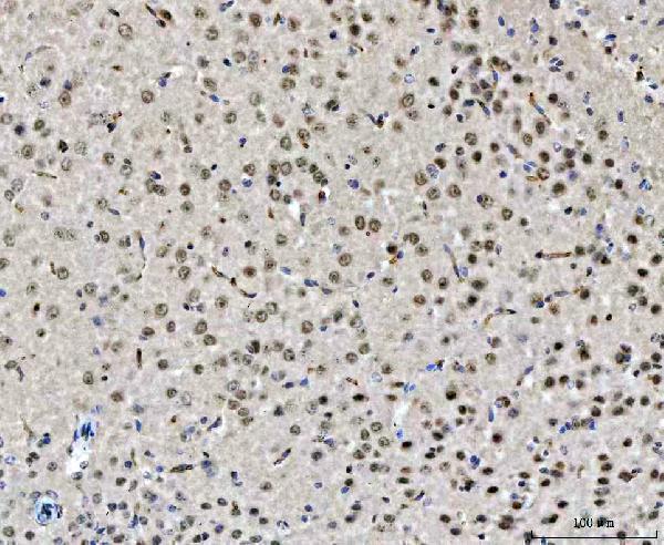
A04079 has IHC images of paraffin-embedded rat, mouse, and human brain sections (A04079 IHC captions). No IF/ICC application or image is listed (catalog applications and IF images).
A04079 will render with its rat brain IHC figure (A04079 figure tag and image caption). Its additional IHC captions show mouse and human brain sections, while its listed reactivity also includes Monkey (A04079 IHC captions; catalog reactivity).
Which to pick: Choose A04079 for paraffin-section tissue IHC: its captions document rat and mouse brain with citrate retrieval at pH 6, and human brain with EDTA retrieval at pH 8 (A04079 IHC captions). For cross-species work, A04079 lists Human, Monkey, Mouse, and Rat reactivity, though the supplied IHC images cover Human, Mouse, and Rat; the fixative is unreported (catalog reactivity; A04079 IHC captions). No supplied SKU supports an IF/ICC recommendation because A04079 has no listed IF/ICC application, dilution, or image (catalog applications, IF dilution, and IF images).