CHEK2 / Serine/threonine-protein kinase Chk2 · IHC design guide

Design Immunohistochemistry for CHEK2

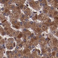
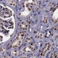
Plan chromogenic CHEK2 IHC in paraffin sections around the nuclear staining reported in most tissues (HPA tissue IHC). This guide covers fixation, antigen retrieval and antibody titration, with colon glandular cells as a high-staining reference (HPA tissue IHC).

Evidence assembled Oct 2026 · For research use; verify linked source records and product datasheet before use
Immunohistochemistry protocol sheet for CHEK2 (IHC for CHEK2): expected localisation Nuclear staining in most tissues (HPA tissue IHC), antibody PB9692, validated IHC image, and IHC protocol steps
Printable CHEK2 IHC protocol sheet — expected localisation Nuclear staining in most tissues (HPA tissue IHC), antibody PB9692, controls and protocol steps. Open the full CHEK2 IHC guide →

CHEK2 Immunohistochemistry Experimental Design Guide

Expected localisation, validated protocols, controls and antibodies — the at-a-glance facts below, then the full design guide.

Must know before staining
Expected localisation Nuclear staining in most tissues (HPA tissue IHC)
Staining pattern Nuclear staining across many tissue cell types (HPA tissue IHC)
Antigen retrieval EDTA pH 8.0 HIER, heat-mediated (datasheet PB9692)
Positive control ⓘ Adrenal gland+4 more · see all
Negative control ⓘ Adipose tissue+4 more · see all
Important caveats
Reasons your staining may differ from the expected pattern.
Fixation Keep formalin fixation consistent between sections (standard IHC practice; not target-specific)
Caveat Adipocytes may lack detectable staining (HPA tissue IHC)
Regulation DNA damage activates CHEK2; abundance unclear (UniProt)
Isoform / epitope 13 isoforms; isoform-specific staining is unestablished (UniProt)
Section 1

Recommended CHEK2 IHC & IF Protocols

The catalog antibody’s IHC-P protocol uses EDTA pH 8.0 heat retrieval (datasheet: PB9692). The published CHEK2 protocols below provide tissue-specific IHC conditions (PMC7967923; PMC8497153; PMC10886656; PMC6004653).

Recommended immunohistochemistry (IHC-P) protocol parameters
SampleParaffin-embedded human lung cancer tissue; fixative not specified (datasheet PB9692)
FixationImage fixative and duration unreported (datasheet PB9692); verify before use.
Sectioning4–5 µm sections on charged slides (standard)
DeparaffinisationXylene, graded ethanol series to water (standard)
Antigen retrievalHeat retrieval: EDTA pH 8.0 (datasheet PB9692); 20 min, 95–100 °C (standard)
Peroxidase block3% H2O2, 10 min, room temperature (standard)
Blocking10% goat serum (datasheet PB9692)
Primary antibodyRabbit anti-CHEK2, 0.5-1μg/ml (datasheet PB9692)
Primary incubationOvernight at 4 °C (datasheet PB9692)
DetectionStreptavidin-biotin complex (SABC), DAB chromogen (datasheet PB9692)
CounterstainHematoxylin, blue, dehydrate and mount (standard)
Expected resultCHEK2-positive staining in glandular cells of adrenal gland (HPA tissue IHC: High). HPA tissue profile: Nuclear expression in most tissues. No signal in the no-primary control.
💡Decision noteStart with EDTA pH 8.0 heat retrieval for the catalog antibody (datasheet: PB9692); compare the published retrieval conditions if optimizing staining (PMC7967923; PMC10886656; PMC6004653).
Section 2

What Is the Expected CHEK2 Staining Pattern?

CHEK2 staining should be predominantly nuclear in cells that express it, with nucleoplasmic localization and possible PML body association (UniProt O96017; HPA subcellular). HPA reports nuclear expression in most tissues, including high staining in colon glandular cells and bronchial basal cells (HPA tissue IHC). Its tissue IHC reliability is Enhanced, with medium agreement between antibody staining and RNA data (HPA tissue IHC). CHEK2 has no transmembrane segment or signal peptide (UniProt O96017 topology).

What am I looking at on my slide?
Distinct nuclear staining in colon glandular cells or bronchial basal cells, with relatively clear surrounding tissue (HPA tissue IHC).This matches the reported high staining in those cell types and CHEK2's predominantly nuclear location (HPA tissue IHC; UniProt O96017). Assess the named cell population rather than assigning one intensity to the whole section (general IHC practice).
Strong, widespread cytoplasmic or membrane staining with little nuclear signal in a positive tissue (HPA tissue IHC; UniProt O96017).That compartment pattern conflicts with the reported nuclear pattern and lack of a transmembrane segment (HPA tissue IHC; UniProt O96017 topology). Treat it as suspect and check antibody specificity and detection controls (general IHC practice).
Prominent staining in adipocytes or prostate glandular cells (HPA tissue IHC).HPA lists these cell populations as not detected (HPA tissue IHC). A strong signal there warrants checks for cross-reactivity or endogenous detection activity; one discrepant section alone does not establish either cause (general IHC practice).
Diffuse colour across nuclei, cytoplasm and extracellular areas, obscuring cell boundaries (general IHC practice).This is difficult to score as CHEK2 because the supported pattern is mainly nuclear (HPA tissue IHC; HPA subcellular). Review background controls, blocking and detection conditions before interpreting intensity (general IHC practice).
No visible nuclear signal in colon glandular cells or bronchial basal cells (HPA tissue IHC).HPA reports high staining in those cells, so check tissue identity, section quality, antibody use and the detection run (HPA tissue IHC; general IHC practice). Absence of stain in one run cannot by itself establish loss of CHEK2 (general IHC practice).
💡Expected CHEK2 appearanceCall positive when the appropriate cells show clear, predominantly nuclear staining, potentially strong in colon glands or bronchial basal cells; dominant membrane staining or diffuse colour is suspect (HPA tissue IHC; UniProt O96017 topology; general IHC practice).
How each factor affects the staining
Tissue and cell selection (HPA tissue IHC)Colon glandular cells and bronchial basal cells are reported high, while adipocytes and prostate glandular cells are not detected (HPA tissue IHC). Use the named cell populations when judging controls; HPA also reports low staining in several other populations (HPA tissue IHC).
Antibody validation and evidence limits (HPA antibodies; HPA tissue IHC)CAB002030 is IHC Enhanced and HPA001878 is IHC Approved (HPA antibodies). The overall tissue profile is Enhanced with medium agreement to RNA data; these labels support interpretation but do not guarantee every specimen's result (HPA tissue IHC; general IHC practice).
Isoforms and antibody epitope (UniProt O96017)UniProt lists 13 CHEK2 isoforms, including isoform 10 throughout the cell (UniProt O96017). Epitope coverage is not supplied, so a given antibody's recognition of each isoform and the cause of any extranuclear signal remain unresolved.
Phosphorylation and assay scope (UniProt O96017)CHEK2 has documented modified residues, including Thr-68 phosphorylation by ATM and MAP3K20 (UniProt O96017). The supplied evidence gives no phospho-specific antibody claim; ordinary CHEK2 staining should not be scored as kinase activation without separate validation (general IHC practice).
IF/ICC Q&A: what location is reported? (HPA subcellular)HPA supports nucleoplasmic localization in ICC-IF and lists an additional Golgi location as uncertain (HPA subcellular). That uncertain observation should not override the predominantly nuclear IHC expectation (HPA tissue IHC; HPA subcellular).
Why is my staining missing, weak or wrong?
SituationLikely causeNext action
Positive tissue lacks nuclear staining (HPA tissue IHC).The run may have failed, or the sampled section may differ from the HPA example (general IHC practice; HPA tissue IHC).Confirm the named positive cell population and run controls; review the antibody's supplied IHC-P conditions and detection steps (HPA tissue IHC; general IHC practice).
Cytoplasmic staining dominates in an IHC-positive cell population (HPA tissue IHC).The pattern differs from CHEK2's reported nuclear localization; the supplied evidence does not identify a specific cause (UniProt O96017; HPA tissue IHC).Check antibody and detection controls, then score nuclear and cytoplasmic staining separately rather than combining them (general IHC practice).
Strong signal appears in adipocytes or prostate glandular cells (HPA tissue IHC).Those populations are reported as not detected; cross-reactivity or endogenous detection activity is possible (HPA tissue IHC; general IHC practice).Check appropriate negative and detection-only controls before calling that signal CHEK2 (general IHC practice).
Background colour obscures nuclear boundaries (general IHC practice).Excess detection background or incomplete blocking can make localization unreadable (general IHC practice).Review blocking, washing, antibody concentration and detection-only controls; judge localization only where nuclei remain distinct (general IHC practice).
Staining varies among tissues expected to express CHEK2 (HPA tissue IHC; UniProt O96017).HPA reports cell-specific high, low and undetected staining, while its overall antibody-to-RNA consistency is medium (HPA tissue IHC).Compare the same named cell types and use matched run controls before interpreting intensity differences (HPA tissue IHC; general IHC practice).
A Golgi-like signal is proposed as the main CHEK2 result (HPA subcellular).HPA calls the additional Golgi location uncertain, while nucleoplasmic localization is supported (HPA subcellular).Record the observation separately and require independent specificity evidence before treating it as the expected IHC pattern (HPA subcellular; general IHC practice).

Sample controls for CHEK2 IHC & IF

🧪Run colon first and score its glandular cells for CHEK2 staining (HPA: High in colon glandular cells). Use parathyroid glandular cells as the negative tissue (HPA: Not detected in parathyroid glandular cells); on the colon slide, morphologically distinct cells without staining should show only background, but HPA does not identify a specific internal negative cell type.
Positive control tissue: Adrenal gland (Glandular cells, HPA High)
Negative control tissue: Adipose tissue (HPA Not detected)
ICC-IF cell lines (HPA subcellular resource): HPA ICC-IF images show CHEK2 in A-431, Hep-G2, NIH 3T3, with annotated localisation: Nucleoplasm (supported) (HPA subcellular).
Technical controls: Include no-primary (secondary-only), rabbit IgG isotype-matched, and CHEK2-knockout controls (PB9692 caption: rabbit primary antibody; standard IHC practice). For colon sections, quench endogenous peroxidase and check or block endogenous biotin because the documented detection uses a biotinylated secondary, SABC, and DAB (PB9692 caption).
⚠️Feasibility: A CHEK2-specific fixation window or fixation effect is unreported, and the PB9692 tissue-IHC caption does not state a fixative (PB9692 caption: fixative unreported). Heat retrieval in EDTA at pH 8.0 is a documented starting condition, but retrieval dependence has not been established by comparison (PB9692 caption: heat-mediated EDTA retrieval). Frozen-section performance and any ease advantage for IF are unreported; in colon, score staining within glandular cells separately from luminal or stromal background (HPA: High in colon glandular cells; standard IHC practice).

HPA tissue IHC evidence for CHEK2

Comprehensive Human Protein Atlas IHC scoring per tissue (reliability: Enhanced — Medium consistency between antibody staining and RNA expression data.). Rows are taken directly from the HPA tissue chart — click any row's HPA link to view the source.

Positive expression · recommended positive controls

TissueCell typeLevelEvidenceSource
Adrenal gland Glandular cells High Protein (IHC) HPA →
Appendix Glandular cells High Protein (IHC) HPA →
Bronchus Basal cells High Protein (IHC) HPA →
Colon Glandular cells High Protein (IHC) HPA →
Duodenum Glandular cells High Protein (IHC) HPA →

Undetected expression · recommended negative controls

TissueCell typeLevelEvidenceSource
Adipose tissue Adipocytes Not detected Protein (IHC) HPA →
Bone marrow Hematopoietic cells Not detected Protein (IHC) HPA →
Parathyroid gland Glandular cells Not detected Protein (IHC) HPA →
Prostate Glandular cells Not detected Protein (IHC) HPA →
Seminal vesicle Glandular cells Not detected Protein (IHC) HPA →
Section 3

Advanced CHEK2 IHC Tips

Use compartment, tissue context, and matched controls to troubleshoot CHEK2 staining in chromogenic paraffin-section IHC.

How should I troubleshoot weak CHEK2 staining after antigen retrieval?
Start CHEK2 paraffin-section IHC with heat-mediated retrieval in EDTA at pH 8.0 (datasheet PB9692). The selected tissue-IHC image used this retrieval before an overnight incubation at 4°C with 1 μg/ml primary antibody (caption PB9692). If nuclear staining is weak, compare controlled retrieval times on adjacent sections while keeping antibody concentration, detection, and DAB development constant (standard IHC practice). Check tissue integrity alongside signal: excessive heating can damage section morphology, making apparent gains in staining difficult to interpret (standard IHC practice). Judge the result against a nuclear-positive tissue compartment and a no-primary control (HPA tissue IHC; standard IHC practice).
Could fixation explain inconsistent CHEK2 staining between paraffin sections?
The selected CHEK2 paraffin-section caption does not report a fixative, so target-specific fixation sensitivity is unknown (caption PB9692). Record each specimen’s fixative and fixation duration, then compare sections processed together using the same EDTA pH 8.0 retrieval and detection conditions (datasheet PB9692; standard IHC practice). If signal varies, inspect morphology and staining in an expected nuclear-positive compartment before adjusting antibody concentration or DAB development (HPA tissue IHC; standard IHC practice). Do not attribute the variation to CHEK2 phosphorylation, isoforms, or a particular fixative without a controlled comparison, because those effects are not established by the supplied tissue-IHC evidence (caption PB9692).
What should I do when CHEK2 staining appears mainly cytoplasmic?
Prioritise nuclear staining when assessing CHEK2 in paraffin sections: tissue IHC reports nuclear expression in most tissues, and the protein is described in the nucleoplasm and PML bodies (HPA tissue IHC; UniProt O96017). Check whether the cytoplasmic signal persists in a no-primary control and whether stronger DAB development also increases diffuse background (standard IHC practice). CHEK2 has no transmembrane segment, so a membrane-outline pattern deserves particular scrutiny rather than compartment-based acceptance (UniProt O96017 topology). Isoform 10 is reported throughout the cell, while an additional Golgi location is uncertain; neither observation alone validates widespread cytoplasmic tissue staining (UniProt O96017; HPA subcellular).
Can a CHEK2 epitope or isoform explain unexpected IHC staining?
CHEK2 has 13 listed isoforms, but the supplied IHC caption does not identify the PB9692 epitope or establish which isoforms its staining detects (UniProt O96017; caption PB9692). Its FHA domain spans residues 113–175, its kinase domain spans 220–486, and several modified residues are reported (UniProt O96017). Check the antibody’s documented immunogen before making a domain-specific or phosphosite-specific interpretation, and avoid treating total CHEK2 staining as a direct activity readout (standard IHC practice; UniProt O96017). If an unexpected compartment dominates, compare an independently validated epitope where available and require agreement in cell type and nuclear pattern (HPA tissue IHC; standard IHC practice).
How can companion IF help assess an ambiguous CHEK2 IHC pattern?
Use companion IF to test whether the IHC-positive nuclei belong to the expected cell population, such as bronchial basal cells, with a separately validated cell-type marker (HPA tissue IHC; standard IF practice). Choose spectrally separated fluorophores and inspect unstained tissue for autofluorescence before interpreting overlap, especially where tissue background is strong (standard IF practice). CHEK2 is chiefly nucleoplasmic and lacks a transmembrane segment, so IF access to its intracellular epitope generally requires controlled permeabilisation after fixation (HPA subcellular; UniProt O96017 topology; standard IF practice). Compare the resulting nuclear pattern with chromogenic IHC, while treating any Golgi signal cautiously because that additional location is uncertain (HPA tissue IHC; HPA subcellular).
How can I distinguish CHEK2 signal from chromogenic background?
Run no-primary and detection-only controls beside CHEK2-stained sections to reveal reagent-derived colour, endogenous enzyme activity, and nonspecific deposition (standard IHC practice). The selected paraffin-section example used a biotinylated secondary, streptavidin–biotin complex, and DAB, so evaluate the corresponding detection controls when reproducing that workflow (caption PB9692; standard IHC practice). Apply a peroxidase block as a general DAB-IHC step, then compare signal with morphology rather than extending development until every compartment is brown (standard IHC practice). Nuclear staining in appropriate cells is more consistent with the reported tissue pattern than uniform stromal haze or section-edge colour (HPA tissue IHC; standard IHC practice).
How should I quantify heterogeneous nuclear CHEK2 IHC staining? ⚠ ANSWER MARKED FOR VERIFICATION
Define the cell population and analysis region before scoring, and exclude folds, necrosis, and poorly preserved edges using the matched morphology slide (standard IHC practice). Report the percentage of CHEK2-positive nuclei and intensity categories, or calculate a nuclear H-score from 0–300 using consistent thresholds across batches (standard IHC practice). Normalise counts to the number of eligible nuclei, or express positive-cell density per mm² of viable analysed tissue when cell density differs (standard IHC practice). Keep tissue and cell type separate in comparisons because CHEK2 staining varies across compartments, including high glandular-cell staining in colon and undetected adipocytes in adipose tissue (HPA tissue IHC).
When is a CHEK2-positive IHC result biologically credible?
A credible CHEK2 result shows reproducible staining in the expected cellular compartment, chiefly nuclei, with appropriate morphology and clean negative controls (HPA tissue IHC; standard IHC practice). Check the stained cell type against tissue context: bronchial basal cells show high staining, whereas adipocytes in adipose tissue are reported as undetected (HPA tissue IHC). Be cautious with isolated section-edge colour, necrotic regions, diffuse membrane staining, or colour present in no-primary controls; these patterns warrant artefact assessment (UniProt O96017 topology; standard IHC practice). Total CHEK2 positivity does not by itself demonstrate checkpoint activation or phosphorylation at Thr-68 (UniProt O96017; standard IHC interpretation).
Boster reagents

Best CHEK2 / Serine/threonine-protein kinase Chk2 IHC Antibodies

Anti-CHEK2 antibodies have IHC figures from human lung cancer, colon carcinoma, testis and colon, and mouse spleen; IF/ICC figures use human cells (catalog image captions).

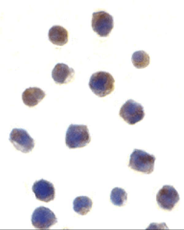
Real IHC data IHC analysis of Chk2 using anti-Chk2 antibody (PB9692). Chk2 was detected in a paraffin-embedded section of human lung cancer tissue. Heat mediated antigen retrieval was performed in EDTA buffer (pH 8.0, epitope retrieval solution). The tissue section was blocked with 10% goat serum. The tissue section was then incubated with 1 μg/ml rabbit anti-Chk2 Antibody (PB9692) overnight at 4°C. Biotinylated goat anti-rabbit IgG was used as secondary antibody and incubated for 30 minutes at 37°C. The tissue section was developed using Strepavidin-Biotin-Complex (SABC) (Catalog # SA1022) with DAB as the chromogen.
Anti-Chk2/CHEK2 Antibody ®
Cat # PB9692
Real IHC data CHEK2 Antibody (N-term) IHC analysis in formalin fixed and paraffin embedded colon carcinoma followed by peroxidase conjugation of the secondary antibody and DAB staining. This data demonstrates the use of the CHEK2 Antibody (N-term) for immunohistochemistry. Clinical relevance has not been evaluated.
Anti-CHEK2 Antibody (N-term)
Cat # A00277-1
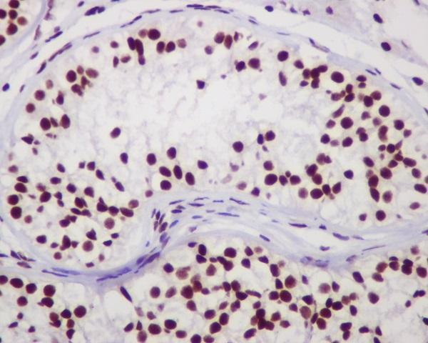
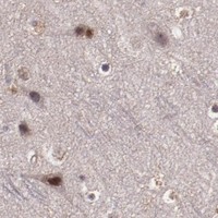
Real IHC data Immunohistochemical analysis of paraffin-embedded human testis, using Chk2 Antibody.
Anti-Chk2 CHEK2 Rabbit Monoclonal Antibody
Cat # M00277
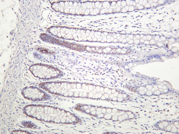
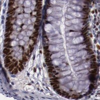
Real IHC data Human colon was stained with anti-Chk2 rabbit antibody
Anti-Chk2 Rabbit Monoclonal Antibody
Cat # M00277-3
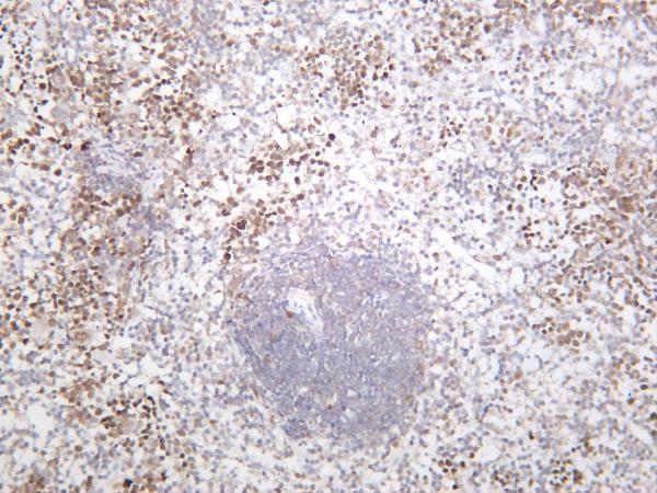
Real IHC data Mouse spleen was stained with anti-Chk2 rabbit antibody
Anti-Chk2 Rabbit Monoclonal Antibody
Cat # M00277-4
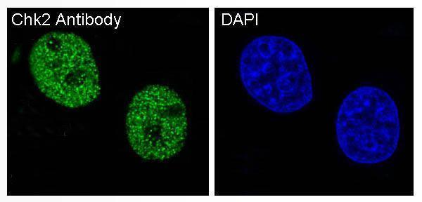
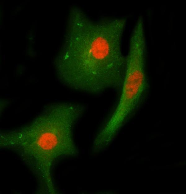
Real IF data Immunocytochemistry of Chk2 in Jurkat cells with Chk2 antibody at 1 μg/mL.
Anti-Chk2 CHEK2 Antibody
Cat # A00277

PB9692 shows paraffin-embedded human lung cancer; A00277-1 shows formalin-fixed, paraffin-embedded colon carcinoma; M00277 shows paraffin-embedded human testis (each SKU’s IHC caption). M00277-3 shows human colon, M00277-4 shows mouse spleen, and A00277 shows Jurkat cells by ICC/IF (each SKU’s image caption).

Which to pick: For tissue IHC, choose PB9692 for human paraffin sections (PB9692 IHC caption; fixative unreported) or A00277-1 when a paraffin-section, paraffin-embedded example is useful (A00277-1 IHC caption). For IF/ICC, choose A00277, which lists both applications and has a Jurkat-cell figure (A00277 catalog applications and image caption). For cross-species IHC planning, PB9692 and M00277-3 list human, mouse and rat reactivity, while M00277-4 lists human and mouse and shows mouse spleen staining; the M00277-3 and M00277-4 captions do not report fixation (catalog reactivity and each SKU’s IHC caption). The selected PB9692 tissue-IHC caption documents paraffin sections, but does not specify the fixative (selected-SKU IHC image PB9692).

Each figure is that product's own IHC / IF validation image from its datasheet.

References

  1. UniProt Consortium. UniProt entry O96017 (CHK2_HUMAN, Serine/threonine-protein kinase Chk2).
  2. Human Protein Atlas. CHEK2 tissue IHC expression (reliability: Enhanced).
  3. Human Protein Atlas. CHEK2 subcellular location (ICC-IF): Mainly localized to the nucleoplasm. In addition localized to the Golgi apparatus..
  4. Human Protein Atlas. CHEK2 antibody validation summary (2 antibodies).
  5. Status of CHEK2 and p53 in patients with early-onset and conventional gastric cancer. Oncology letters 2021 — PMC7967923.
  6. Recurrent Germline Mutations of CHEK2 as a New Susceptibility Gene in Patients with Pheochromocytomas and Paragangliomas. International journal of endocrinology 2021 — PMC8497153.
  7. Relationship between the Expression of CHK2 and p53 in Tumor Tissue and the Course of Papillary Thyroid Cancer in Patients with CHEK2 Germline Mutations. Cancers 2024 — PMC10886656.
  8. Expression of DNA doublestrand repair proteins in oral leukoplakia and the risk of malignant transformation. Oncology letters 2018 — PMC6004653.
  9. PubMed PMID:9836640 — UniProt-cited evidence.
  10. PubMed PMID:9889122 — UniProt-cited evidence.
  11. PubMed PMID:10097108 — UniProt-cited evidence.