This website uses cookies to ensure you get the best experience on our website.
- Table of Contents
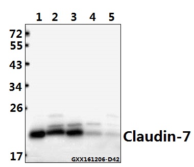
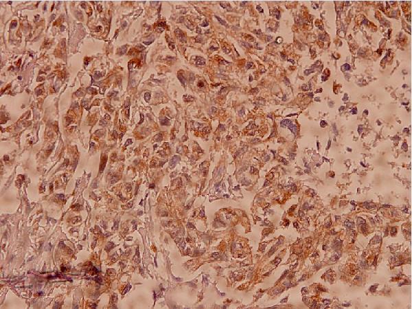
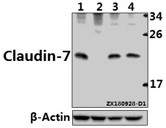
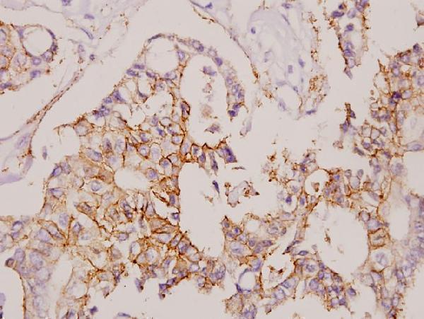
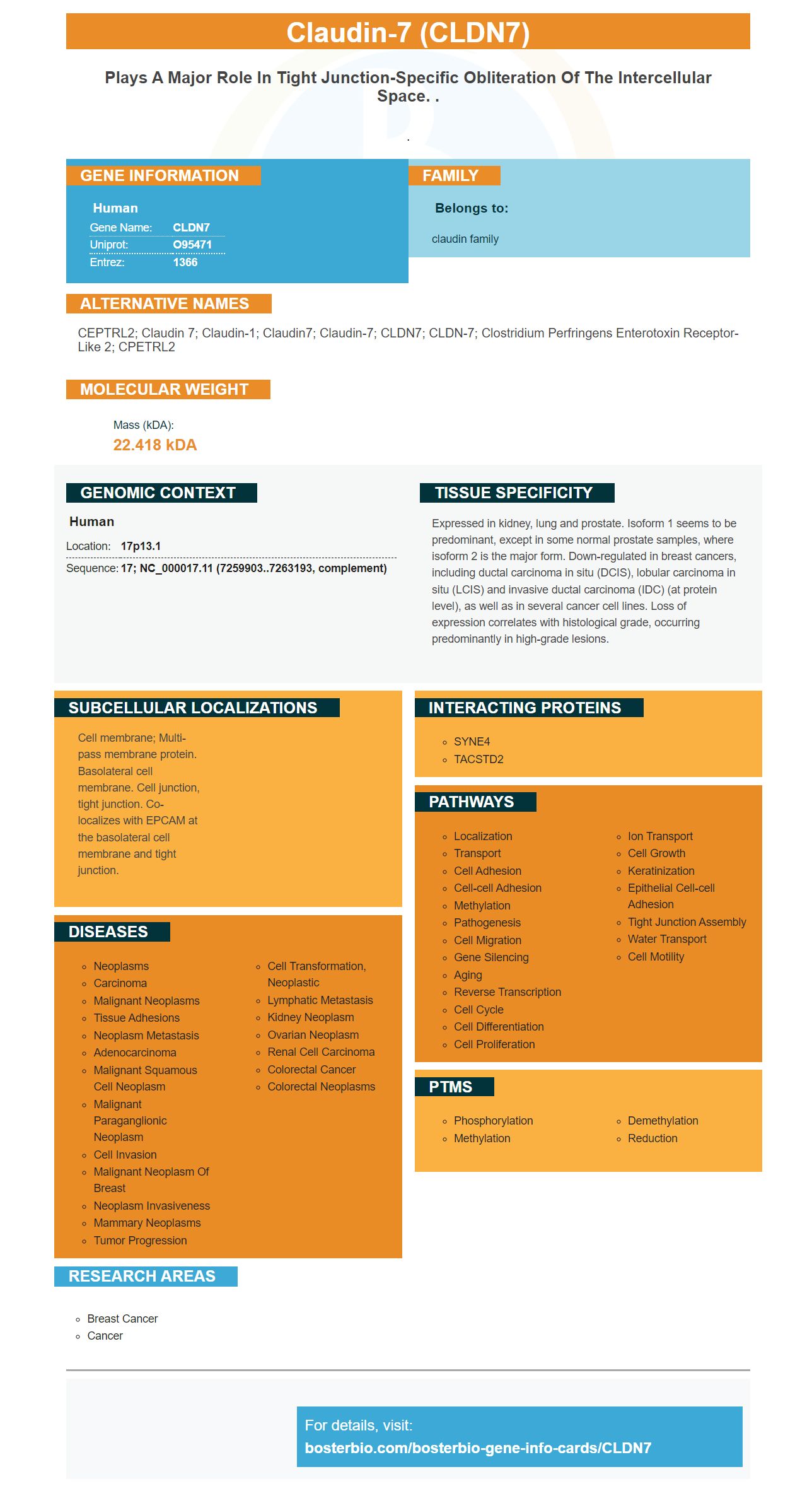
Facts about Claudin-7.
.
Human | |
---|---|
Gene Name: | CLDN7 |
Uniprot: | O95471 |
Entrez: | 1366 |
Belongs to: |
---|
claudin family |
CEPTRL2; claudin 7; claudin-1; Claudin7; Claudin-7; CLDN7; CLDN-7; clostridium perfringens enterotoxin receptor-like 2; CPETRL2
Mass (kDA):
22.418 kDA
Human | |
---|---|
Location: | 17p13.1 |
Sequence: | 17; NC_000017.11 (7259903..7263193, complement) |
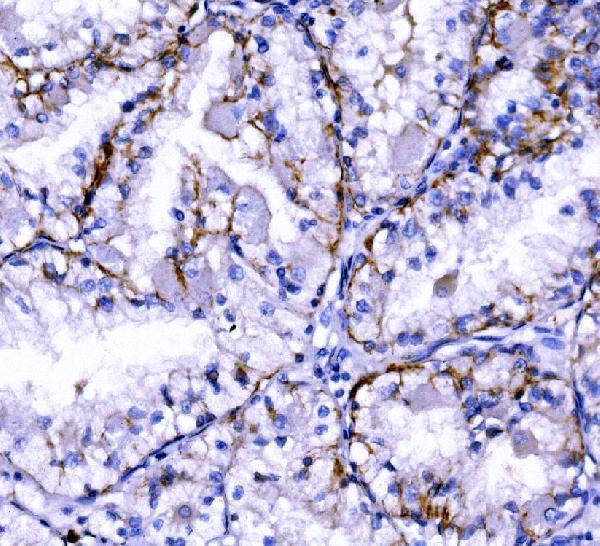
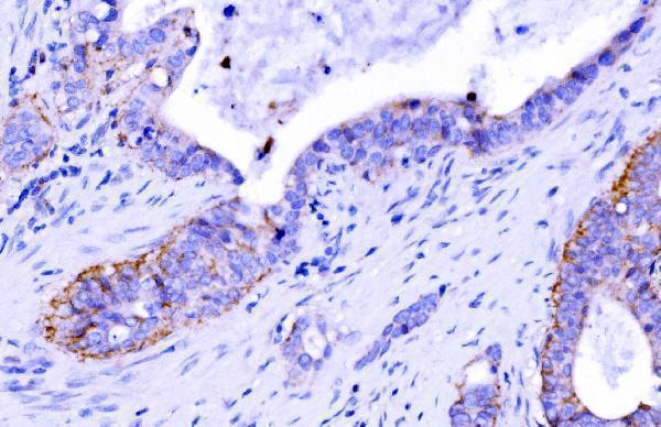
Expressed in kidney, lung and prostate. Isoform 1 seems to be predominant, except in some normal prostate samples, where isoform 2 is the major form. Down-regulated in breast cancers, including ductal carcinoma in situ (DCIS), lobular carcinoma in situ (LCIS) and invasive ductal carcinoma (IDC) (at protein level), as well as in several cancer cell lines. Loss of expression correlates with histological grade, occurring predominantly in high-grade lesions.
Cell membrane; Multi-pass membrane protein. Basolateral cell membrane. Cell junction, tight junction. Co-localizes with EPCAM at the basolateral cell membrane and tight junction.
If you're looking for the best CLDN7 marker, you've come to the right place. We've compiled information about the product, its uses, and its cost. Read on to find out more! Also, find out more about the company behind it. In the meantime, you can take advantage of our handy guide on how to use the CLDN7 marker. It's a highly useful tool for researchers, clinicians, and scientists.
The CLDN7 gene encodes a protein that is involved in the regulation of prostate-specific antigen expression. It is also down-regulated in some types of breast cancer including lobular carcinoma in situ and invasive ductal carcinoma. Loss of CLDN7 expression is related to higher histological grades and lesions. This protein co-localizes with the basolateral cell membrane and tight junctions.
A recent study found that the expression of CLDN7 varies widely across cancers, from breast cancer to colon cancer. Using the STRING database, this marker was evaluated for over and underexpression. The Pearson correlation coefficient ranged from blue to red. Its expression level is proportional to the gene rank and p-value. It has potential as a diagnostic marker in cancer and as a biomarker for disease prevention.
In gastric cancer, CLDN7 is expressed in gastric cells, but its expression is less specific than CDh27 or CDh28. However, a recent study showed that a combination of both markers can detect gastric cancer. It is possible to detect the disease using the CLDN7 marker and CDh27 together. Moreover, the two markers overlap in tissue localization, which may result in improved diagnostic accuracy.
Several studies of gastric cancers have shown that cancer cells with high CLDN7 expression had a poor prognosis. Clinical studies suggest that CLDN7 promotes gastric cancer cell proliferation and invasiveness via the YAP1 protein. Further, it has been shown to accelerate pancreatic cancer and colon cancer progression. In addition, high levels of CLDN7 can identify cancer initiation cells and detect metastases.
In a recent study, the expression of CLDN7 in patients was investigated using a double-antibody sandwich ELISA kit. A fifty-microliter sample of standard and tumor sample was added to the appropriate wells on an antibody-precoated microtiter plate. The plate was then incubated at 37degC for 45 minutes. The next step in this procedure was to add diluted biotinylated anti-Immunoglobulin G antibody to each well. After this, a chromogen solution and a stop solution were added to the wells. The reaction would stop when the color change went from blue to yellow.
The CLDN7 marker and the CLDN18 marker are two known adhesion molecules in the stomach and intestine. Combined, these markers are a powerful diagnostic tool in gastric cancer diagnosis. The two genes are co-expressed in the gastric cancer tissue and the corresponding immunostaining pattern is homogeneous. This co-detection method will provide more comprehensive detection of gastric cancer metastases and its spread beyond the primary lesion.
These studies suggest that Claudin-7 expression is strongly correlated with the presence of T-cells and dendritic cells in patients with CRC. Furthermore, CLDN15 expression is negatively correlated with neutrophil infiltration and CLDN11 expression with dendritic cell infiltration. These findings support the notion that CLDN7 expression is related to invasion and metastasis. The CLDN7 gene can be detected by ELISA and has clinical utility.
In addition to its diagnostic potential, the CLDN7 gene can also be used in prognostic analysis. It is a highly sensitive biomarker of gastric cancer and can be used to differentiate among cancer subgroups. The clinical use of this marker will be expanded in the coming years. And it is also a good biomarker for cancer research. So, why wait? Get started today and start saving lives!
The CLDN7 marker is a candidate for use in cancer research. It has been found to interact with matrix metalloproteinases and affect intercellular signal transduction. It also has possible relevance in cancer stem cells. Malignant tumour tissues contain a limited number of CSCs, which have multidirectional differentiation and immortalization potential. They are also referred to as tumour-initiating cells. The price of the CLDN7 marker depends on its availability.
The CLDN7 marker is highly relevant in tumor biology. Physiological levels of CLDNs are critical for maintaining TJ structure and function. Hence, low expression of CLDNs in tumors can enhance tumor formation and progression. It is a valuable marker for early detection of cancer. However, cost is a concern. For this reason, there are many studies that have shown a link between the CLDN7 marker and poor prognosis in various types of cancer.
PMID: 14502431 by Zheng J.-Y., et al. Regulation of the expression of the prostate-specific antigen by claudin-7.
PMID: 12673207 by Kominsky S.L., et al. Loss of the tight junction protein claudin-7 correlates with histological grade in both ductal carcinoma in situ and invasive ductal carcinoma of the breast.